Post renal aki and septicemia, Nkeck Jan
•
0 gefällt mir•63 views

This document describes a case study of a two-month-old Cameroonian male infant who presented with acute kidney injury and sepsis due to an undiagnosed case of posterior urethral valves. The infant had a history of a treated urinary tract infection as a newborn. Ultrasound revealed uretero-hydronephrosis and a distended bladder, suggesting posterior urethral valves. The infant developed Klebsiella pneumoniae sepsis and acute kidney injury with uremia and hyperkalemia due to the delayed diagnosis of the valves. He underwent a vesicostomy and was scheduled for valve resection surgery but unfortunately died three days post-operation from refractory hyperkalemia and
Empfohlen

Empfohlen
Weitere ähnliche Inhalte
Ähnlich wie Post renal aki and septicemia, Nkeck Jan
Ähnlich wie Post renal aki and septicemia, Nkeck Jan (20)
Kürzlich hochgeladen
Kürzlich hochgeladen (20)
Post renal aki and septicemia, Nkeck Jan
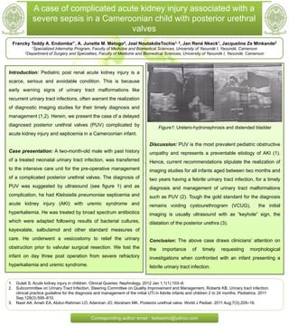
- 1. A case of complicated acute kidney injury associated with a severe sepsis in a Cameroonian child with posterior urethral valves Francky Teddy A. Endomba1*, A. Junette M. Metogo2, Joel NoutakdieTochie1, 2, Jan René Nkeck1, Jacqueline Ze Minkande2 1Specialized Internship Program, Faculty of Medicine and Biomedical Sciences, University of Yaoundé I, Yaoundé, Cameroon 2Department of Surgery and Specialties, Faculty of Medicine and Biomedical Sciences, University of Yaoundé I, Yaoundé, Cameroon Introduction: Pediatric post renal acute kidney injury is a scarce, serious and avoidable condition. This is because early warning signs of urinary tract malformations like recurrent urinary tract infections, often warrant the realization of diagnostic imaging studies for their timely diagnosis and management (1,2). Herein, we present the case of a delayed diagnosed posterior urethral valves (PUV) complicated by acute kidney injury and septicemia in a Cameroonian infant. Case presentation: A two-month-old male with past history of a treated neonatal urinary tract infection, was transferred to the intensive care unit for the pre-operative management of a complicated posterior urethral valves. The diagnosis of PUV was suggested by ultrasound (see figure 1) and as complication, he had Klebsiella pneumoniae septicemia and acute kidney injury (AKI) with uremic syndrome and hyperkaliemia. He was treated by broad spectrum antibiotics which were adapted following results of bacterial cultures, kayexalate, salbutamol and other standard measures of care. He underwent a vesicostomy to relief the urinary obstruction prior to valvular surgical resection. We lost the infant on day three post operation from severe refractory hyperkaliemia and uremic syndrome. Figure1: Uretero-hydronephrosis and distended bladder Discussion: PUV is the most prevalent pediatric obstructive uropathy and represents a preventable etiology of AKI (1). Hence, current recommendations stipulate the realization of imaging studies for all infants aged between two months and two years having a febrile urinary tract infection, for a timely diagnosis and management of urinary tract malformations such as PUV (2). Tough the gold standard for the diagnosis remains voiding cystourethrogram (VCUG), the initial imaging is usually ultrasound with as “keyhole” sign, the dilatation of the posterior urethra (3). Conclusion: The above case draws clinicians’ attention on the importance of timely requesting morphological investigations when confronted with an infant presenting a febrile urinary tract infection. 1. Gulati S. Acute kidney injury in children. Clinical Queries: Nephrology. 2012 Jan 1;1(1):103–8. 2. Subcommittee on Urinary Tract Infection, Steering Committee on Quality Improvement and Management, Roberts KB. Urinary tract infection: clinical practice guideline for the diagnosis and management of the initial UTI in febrile infants and children 2 to 24 months. Pediatrics. 2011 Sep;128(3):595–610. 3. Nasir AA, Ameh EA, Abdur-Rahman LO, Adeniran JO, Abraham MK. Posterior urethral valve. World J Pediatr. 2011 Aug;7(3):205–16. Corresponding author email : tedissimo@yahoo.com