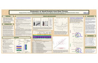
Proteomics of small proteins from plant tissues
•
1 gefällt mir•118 views

Small genes and the proteins that they encode can play important biological roles including signaling, development, and mediation of plant-microbe interactions in organisms ranging from bacteria to plants to mammals (Frith et al.; Basrai et al.; Galindo et al.; Hemm et al. 2008, 2010; Kastenmeyer et al.). However, genes that encode proteins containing <100 residues are difficult to identify reliably solely by DNA sequence analysis (Dinger et al.)
Empfohlen

Empfohlen
Weitere ähnliche Inhalte
Was ist angesagt?
Was ist angesagt? (20)
Ähnlich wie Proteomics of small proteins from plant tissues
Ähnlich wie Proteomics of small proteins from plant tissues (20)
Mehr von Expedeon
Mehr von Expedeon (20)
Kürzlich hochgeladen
Kürzlich hochgeladen (20)
Proteomics of small proteins from plant tissues
- 1. METHODS Evaluation of fractionation protocols (Figure 1) l E. coli used as a model system l ACN: removal of large proteins by acetonitrile precipitation (Aristoteli et al.) l MWCO: ultrafiltration using molecular weight cutoff filters (Aristoteli et al.) u 10 kilodalton and 30 kilodalton cutoffs evaluated l In-Gel: in-gel digestion of low-molecular-weight regions excised from SDS-PAGE gels (Shevchenko et al.) l GelFree: fractionation using the GelFree 8100 fractionation system (Protein Discovery, Knoxville TN) l Full: no fractionation (full lysate) Application to plant tissues (Figure 2) l Root and shoot tissues from small laboratory-grown A. thaliana seedlings flash frozen in liquid nitrogen l Protein extraction (Damerval et al.) l In-Gel method to enrich small proteins (see above) Each unfractionated lysate, liquid fraction, or gel slice was: l digested using trypsin l analyzed using LC-MS-MS (2D nanoLC interfaced with ThermoFinnigan LTQ) l Peptides and proteins identified using Sequest and DTASelect l Several methods for enriching small proteins were evaluated using E. coli as a model system. l Small proteins were enriched from Arabidopsis thaliana and analyzed by LC-MS-MS. l Small genes and the proteins that they encode can play important biological roles including signaling, development, and mediation of plant-microbe interactions in organisms ranging from bacteria to plants to mammals (Frith et al.; Basrai et al.; Galindo et al.; Hemm et al. 2008, 2010; Kastenmeyer et al.). However, genes that encode proteins containing <100 residues are difficult to identify reliably solely by DNA sequence analysis (Dinger et al.) l We previously described an approach to identify small- protein-encoding genes in the woody model species Populus trichocarpa that relied in part on proteomics to identify small proteins from unfractionated protein extracts (Yang et al.). l To increase the sensitivity of proteomics toward small proteins, we sought to evalute methods for enriching the low- molecular-weight proteome prior to LC-MS-MS analysis. l Using E. coli as a model system, we evaluated several methods for enriching small proteins from cell lysates. We applied the most promising to fractionation of plant root and shoot tissues from Arabidopsis thaliana in order to increase the sensitivity of LC-MS-MS analysis toward small proteins. Comparison of Small Protein Enrichment Methods Figure 3 compares SDS PAGE analyses of E. coli proteins isolated using the various methods, and from the unfractionated proteome. l All fractionation methods show depletion of large proteins. l ACN and MWCO methods appear also to entail significant losses of the small protein complement. l Low molecular weight fractions from In-Gel and GelFree methods appear to contain highest abundance of small proteins. Figure 4 shows molecular weight distributions for proteins detected by each method l Proteins identified by LC-MS-MS from each of the fractionation methods exhibited molecular mass distributions with medians significantly lower than those of unfractionated lysates l The largest numbers of small protein identifications from LC-MS-MS analysis were obtained from u In-gel digestion of the low molecular weight range of SDS-PAGE separated proteins, and u the GelFree system l Consistent with SDS-PAGE analysis, the ACN and MWCO methods provided the lowest numbers of identified small proteins. Figure 5 compares Spectrum Count values for the In-gel digestion (0-20 kDa region) to the unfractionated proteome. While 26 “small” (i.e., <= 100 amino acids in length) proteins were more abundant in the In-gel digestion, 58 small proteins were less abundant in the In-gel digestion. Table 1 summarizes results of this analysis for the various fractionation methods. Additionally, Table 1 shows that the In-Gel and GelFree methods yielded the highest fraction of Spectrum Count from small proteins. Table 2 lists selected small E. coli proteins that were more abundant in one or more enrichment samples than in the unfractionated proteome. Enrichment of small proteins appears to depend both on the particular protein, and on the fractionation method used. One factor that may affect the results is the participation of small proteins in large complexes such as the ribosome, and how such complexes survive the various fractionation protocols. Additional improvements may be gained in the future by considering consequences of the smaller numbers of distinct tryptic peptides obtained from digestion of small proteins. OVERVIEW INTRODUCTION REFERENCES Results from enrichment of E. coli proteins suggests that improved sensitivity toward small proteins is feasible with an MS-based approach, especially with further optimization of the In-Gel and Gelfree isolation protocols. Hemm et al. (2008, 2010) have employed other approaches to the detection of small proteins in E. coli, and showed that expression of a number of these proteins required subjecting the cells to various stresses. Several E. coli proteins were identified more abundantly in small protein enrichment fractions than in unfractionated proteomes. Among these proteins are several with annotations indicating that their functions are not yet characterized. Evidence for expression of these proteins from LC-MS-MS identification supports improved annotation of the corresponding small genes, and also provides candidates for further studies of the biological functions of these small proteins. Further investigation of small proteins identified from Arabidopsis thaliana will complement our ongoing research, which integrates informatics and experimental approaches for identifying genes that encode small proteins in plants (Yang et al.). CONCLUSIONSRESULTS AND DISCUSSION We thank Dr. Karuna Chourey (ORNL) for advice on protein extraction from plant tissues. Research sponsored by the Genomic Science Program, Office of Biological and Environmental Research, U.S. Department of Energy, under contract No DE-AC05- 00OR22725 with Oak Ridge National Laboratory, managed and operated by UT-Battelle, LLC. ACKNOWLEDGMENTS Aristoteli et al., J. Proteome Research 2006, 6, 571. Basrai, M.A. et al., Genome Research 1997, 7, 768. Damerval, C. et al., Electrophoresis 1986, 7, 52. Dinger, M.E. et al., Plos Comput. Biol. 2008, 4, e1000177. Frith, M.C. et al., Plos Genetics 2006, 2, 515. Galindo, M.I. et al., Plos Biology 2007, 5, 1052. Hemm, M.R. et al., Mol. Microbiol. 2008, 70, 1487. Hemm, M.R. et al., J. Bacteriology 2010, 192, 46. Kastenmayer, J.P. et al., Genome Research 2006, 16, 365. Shevchenko, A. et al., Nature Protocols 2006, 1, 2856. Yang, X. et al., Genome Research 2011, 21, 634. Figure 6. Enrichment of small proteins from Arabidopsis. Figure 4. Molecular weight distributions of LC-MS-MS protein identifications resulting from various fractionation protocols applied to the E. coli proteome. Boxplots show molecular mass distributions of proteins identified in proteomics measurements. Dark horizontal bars: median molecular mass for identified proteins. Box: 25th and 75th percentile molecular masses. Whiskers: 1.5 x interquartile range. Circles mark molecular masses of any outliers more extreme than the whiskers. 25 20 15 10 MWM Totalecolilysate Fractions from GelFree System 25 20 15 10 mwm Ecolilysate ba ACNppt(5uL) 10kDMWCO(5uL) 30kDMWCO c <30kDsmallproteinecoli mwmarkers 250 150 100 75 50 37 25 20 15 10 Figure 3. SDS PAGE analyses of (a) fractions from GelFree fractionation system, (b) supernatant from acetonitrile precipitation (ACN ppt) and flow-through from 10 kDa MWCO separation, and (c) flow-through from 30 kDa MWCO separation. Gel slice locations for the In-Gel digestion are shown approximately by the red boxes in panel b; lower box is the 0-20 kDa range, upper box is the 20-35 kDa range. MWM: molecular weight markers. Figure 5. Comparison of Spectrum Count for proteins ≤100 aa ( ) and >100 aa (x) for 0-20 kDa gel slice versus unfractionated proteome. Diagonal black line shows equal average Spectrum Count for the two methods. Data points above the diagonal are “enriched” in the gel slice relative to the unfractionated proteome, while data points below the diagonal are “depleted” in the gel slice. Spectrum Count for proteins that were not detected was replaced with a value of 0.1. Small Proteins in Arabidopsis thaliana Shoot and root tissues from Arabidopsis thaliana were separated by SDS-PAGE, and in-gel digestion performed on slices corresponding to <20 kDa and 20-35 kDa ranges. LC-MS-MS analyses of these fractions and unfractionated protein extracts were performed in duplicate. Figure 6 summarizes the results for molecular weight distributions (boxplots) and numbers of proteins identified in the various fractions (Venn diagrams.) full ACN 10kDa 30kDa <20kDa 20-35kDa Fr.2 Fr.3 Fr.4 Fr.6 050000100000150000 MW full ACN MWCO In-gel Gelfree Table 1. Comparison of Enrichment Methods for Small Proteins from E. coli Isolation Method Number of proteins that contain: Proteins Identified in Isolate (average) n** Fraction of Total Spectrum Count in Isolate From Proteins with ≤100 AA ≤100 AA >100 AA Enriched* in Isolate Depleted or Not Detected in Isolate Enriched in Isolate Depleted or Not Detected in Isolate ACN 25 64 43 1125 221 3 21% MWCO 10 kDa 9 74 9 1149 91 3 15% MWCO 30 kDa 0 81 9 1148 24 1 4% GelFree fraction 2 26 64 122 1077 449 3 16% In-Gel digestion, <20 kDa 26 58 80 1089 176 1 25% unfractionated proteome - - - - 1236 4 4% * Spectrum Count is higher (Enriched) or lower (Depleted) for isolation method compared to unfractionated proteome ** n = number of replicate LC-MS-MS measurements Table 2. Selected E. coli proteins detected more abundantly following enrichment L MW Gene Symbol Description 55 6507 rmf b0953 ribosome modulation factor 63 7281 yaiA b0389 predicted protein 63 7273 rpmC b3312 50S ribosomal subunit protein L29 66 7892 glgS b3049 predicted glycogen synthesis protein 69 7463 cspE b0623 DNA-binding transcriptional repressor 69 7402 cspC b1823 stress protein, member of the CspA-family 70 7781 cspG b0990 cold shock protein homolog, cold-inducible 70 7403 cspA b3556 RNA chaperone and anti-terminator, cold-inducible 71 8500 rpsU b3065 30S ribosomal subunit protein S21 72 8250 infA b0884 translation initiation factor IF-1 77 8639 yedF b1930 conserved protein, UPF0033 family 84 9704 rpsQ b3311 30S ribosomal subunit protein S17 85 9119 ptsH b2415 phosphohistidinoprotein-hexose phosphotransferase component of PTS system (Hpr) 90 9226 hupB b0440 HU, DNA-binding transcriptional regulator, beta subunit 90 9535 hupA b4000 HU, DNA-binding transcriptional regulator, alpha subunit 97 10387 groS b4142 Cpn10 chaperonin GroES, small subunit of GroESL 99 10776 yiiS b3922 conserved protein, UPF0381 family 1 2 1 2 1 2 0e+005e+041e+05 MW 10085(16261) 11451(19052) 136(279) 1894(2720) 3551(5867) 3304(5085) full 0-20kDa 20-35kDa 38 955 2233 0-20 kDa in-gel “full” proteome 21 86 853 235 20-35 kDa in-gel ROOTS LC-MS-MS replicate 35 917 1695 0-20 kDa in-gel “full” proteome 26 102 1015 286 20-35 kDa in-gel SHOOTS 1 2 1 2 1 2 0e+005e+041e+05 MW 9815(18057) 10033(17883) 156(332) 2038(3859) 3902(6392) 4859(8213) full 0-20kDa 20-35kDa LC-MS-MS replicate E. coli cell pellet Lyse Add cytochrome c, BSA lysate centrifuge ACN Gelfree Fractionation No enrichment (Full) supernatant MWCO In-Gel Figure 1 v Trypsin digestion v LC-MS-MS identification of peptides A. thaliana seedlings v Trypsin digestion v LC-MS-MS identification of peptides In-Gel roots shoots vGrind under liquid N2 vExtract proteins vAdd cytochrome c, BSA Noenricment(Full) Figure 2