

Empfohlen
Weitere ähnliche Inhalte
Was ist angesagt?
Was ist angesagt? (20)
Andere mochten auch
Ähnlich wie Cardiorenal syndrome
Ähnlich wie Cardiorenal syndrome (20)
Kürzlich hochgeladen
Kürzlich hochgeladen (20)
Cardiorenal syndrome
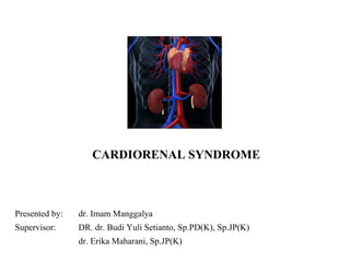
- 1. CARDIORENAL SYNDROME Presented by: dr. Imam Manggalya Supervisor: DR. dr. Budi Yuli Setianto, Sp.PD(K), Sp.JP(K) dr. Erika Maharani, Sp.JP(K)
- 3. Definition • The CRS includes a variety of acute or chronic conditions, where the primary failing organ can be either the heart or the kidney (Berl T & Herich, 2006) • A diseased heart has numerous negative effects on kidney function but, at the same time, renal insufficiency can significantly impair cardiac function (Berl T & Herich, 2006) • In acute HF, AKI appears to be more severe in patients with impaired LV ejection fraction compared with those with preserved LV function, achieving an incidence 70% in patients with cardiogenic shock (Jose P et al., 2006) • Impaired renal function is consistently found as an independent risk factor for 1-year mortality in acute HF patients, including patients with ST-segment elevation myocardial infarction (Goldberg A et al., 2005)
- 4. Incidence & Risk Factor • Reducing in eGFR related mortality in CHARM study (Candesartan in Heart Failure Assessment in Mortality and Morbidity) • Approximately 30 % of CHF patients also has CKD The Acute Decompensated Heart Failure National Registry (ADHERE) • VALIANT study: reducing of eGFR is independent risk factor (Valsartan in Acute Myocardial Infarction Trial) • Studies of Left Ventricular Dysfunction (SOLVD) elevated creatinin serum > 0,3 mg/dl: • Aging • Low EF • Creatinin serum level baseline • Low SBP • DM • Hypertension • Use of Anti platelet, Diuretics, B-Blocker, CCB
- 5. Dinna N Cruz. Cardiorenal syndrome in critical care. Am Coll Cardio J; 2013
- 6. From: Cardiorenal Syndrome J Am Coll Cardiol. 2008;52(19):1527-1539. doi:10.1016/j.jacc.2008.07.051 CRS Typ e 1 Pa thophysiologic al inte rac tions betwe n hea rt a nd kidney in type 1 ca rdiore na l syndrome (CRS) or “ acute CRS” (a brupt wors ening of cardia c func tion, e .g., ac ute ca rdioge nic shock or ac ute de compe ns ation of c hronic he art fai lure ) le ading to kidney injury. ACE = angiote nsin-c onve rting enzyme ; ANP = a tria l na triure tic peptide; BNP = B-type natriure tic pe ptide; CO = c ardia c output; GFR = glome rul ar fi ltra tion rate; KIM = kidney injury mol ec ule ; N-GAL = ne ut rophil gel atinase -a s oci ated lipoca lin; RAA = renin a ngiot ensin aldoste rone. Figure il ustration by Rob Flewel . Figure Legend: J Am Coll Cardiol. 2008;52(19):1527-1539. doi:10.1016/j.jacc.2008.07.051
- 7. Figure 1 Incidence of CRS Advances in Chronic Kidney Disease 2013: 20:56-66)
- 8. CRS type 1 • The presence of AKI sometimes inhibiting the prescription of angiotensin-converting enzyme (ACE) inhibitors, angiotensin receptor blockers (ARBs), and aldosterone inhibitors. • But the potential benefits of these interventions often outweigh their risks, even in these patients. (Verma & Solomon, 2007) • Relative or absolute tachycardia sustains the adequacy of cardiac output. Blockade of such compensatory tachycardia and sympathetic system-dependent inotropic compensation can precipitate cardiogenic shock with associated high mortality. (Chen Z et al., 2005) • An increase in creatinine is not simply a marker of illness severity, it represents the onset of AKI acting as a causative factor for cardiovascular injury acceleration (Tokuyama et al., 2007)
- 9. Figure 2 Proportion of other organ failure and CoD on AKI Advances in Chronic Kidney Disease 2013: 20:56-66)
- 10. From: Cardiorenal Syndrome J Am Coll Cardiol. 2008;52(19):1527-1539. doi:10.1016/j.jacc.2008.07.051 CRS Type 2 Pathophysiological interactions between heart and kidney in type 2 cardiorenal syndrome (CRS) or “chronic CRS” (chronic abnormalities in cardiac function, e.g., chronic heart failure) causing progressive chronic kidney disease (CKD). Figure illustration by Rob Flewell. LVH = left ventricular hypertrophy; RAA = renin angiotensin aldosterone. Figure Legend: J Am Coll Cardiol. 2008;52(19):1527-1539. doi:10.1016/j.jacc.2008.07.051
- 11. Postulated mechanisms underlying the relationship between HF and renal dysfunction. Bock J, Gottlieb S. Circulation 2010;121:2592-2600 Copyright © American Heart Association, Inc. All rights reserved.
- 12. CRS Type 3 Pathophysiological interactions between heart and kidney in type 3 CRS or “acute renocardiac syndrome” (abrupt worsening of renal function, e.g., acute kidney failure or glomerulonephritis) causing acute cardiac disorder (e.g., heart failure, arrhythmia, pulmonary edema). MPO = myeloperoxidase; other abbreviations as in Figure 1. Figure illustration by Rob Flewell. J Am Coll Cardiol. 2008;52(19):1527-1539. doi:10.1016/j.jacc.2008.07.051
- 13. CRS type 3 • When the RIFLE (risk, injury, and failure; loss; and end-stage kidney disease) consensus definition is used, AKI has been identified in close to 9% of hospital patients. (Uchino S et al., 2006) • In a large ICU database, AKI was observed in more than 35% of patients. (Bagshaw et al., 2008) • Fluid overload can contribute to the development of pulmonary edema. • Hyperkalemia can contribute to arrhythmias and may cause cardiac arrest. • Untreated uremia affects myocardial contractility through the accumulation of myocardial depressant factors (Blake et al., 1996) and pericarditis (Coresh et al., 2003). • Acidemia produces pulmonary vasoconstriction, which can significantly contribute to right-sided HF • Acidemia appears to have a negative inotropic effect (Brady & Hasbargen, 2000)
- 15. Advances in Chronic Kidney Disease 2013: 20:56-66)
- 16. Figure 3 Renoprotective in CRS Advances in Chronic Kidney Disease 2013: 20:56-66)
- 17. From: Cardiorenal Syndrome J Am Coll Cardiol. 2008;52(19):1527-1539. doi:10.1016/j.jacc.2008.07.051 Date of download: 2/18/2014 CRS Type 4 Pathophysiological interactions between heart and kidney in type 4 cardiorenal syndrome (CRS) or “chronic renocardiac syndrome” (chronic kidney disease [CKD], e.g., chronic glomerular disease, contributing to decreased cardiac function, cardiac hypertrophy, or increased risk of adverse cardiovascular events). BMI = body mass index; EPO = erythropoietin; LDL = low-density lipoprotein. Figure illustration by Rob Flewell. Figure Legend:
- 18. From: Cardiorenal Syndrome J Am Coll Cardiol. 2008;52(19):1527-1539. doi:10.1016/j.jacc.2008.07.051 CRS Type 5 Pathophysiological interactions between heart and kidney in type 5 cardiorenal syndrome (CRS) or “secondary CRS” (systemic condition, e.g., diabetes mellitus, sepsis, causing both cardiac and renal dysfunction). LPS = lipopolysaccharide (endotoxin); RVR = renal vascular resistance. Figure illustration by Rob Flewell. Figure Legend:
- 19. MANAGEMENT
- 20. Diuretics • Loop, thiazide and potassium-sparing diuretics provide diuresis and natriuresis in as quickly as 20 min after administration. • Moreover, they provide effective short-term symptomatic relief. However, the use of diuretics is not free from drawbacks, such as long-term deleterious cardiovascular effects. • A Cochrane review (Salvador D et al., 2004) examined eight trials comparing continuous infusion of a loop diuretic with bolus injections in 254 patients with CHF. The urine output (as measured in mL/24 h) was noted to be greater in patients given continuous infusion
- 21. Non-responding to diuretics • In the management of ADHF, the lack of a clinical response to diuretic therapy is commonly observed. • Diuretic therapy can worsen renal function • Diuretic resistance can be considered to be another indicator of poor prognosis in patients with CHF. • However, in the absence of definitive data, patients with volume overload should not be restricted from receiving loop or thiazide diuretics as necessary to alleviate symptoms (Geisberg & Butler, 2006) • Nesiritide can be helpful in patients with non-responding to diuretics (PROACTIVE trial) but a large meta-analysis doesn’t show benefit in long term mortality (Arora et al., 2006)
- 22. Low dose dopamine • low (renal) doses of dopamine are commonly used in conjunction with diuretic therapy. Rather than improving renal function, dopamine has been shown to impair renal oxygen kinetics, inhibit feedback systems that protect the kidney from ischemia, and possibly worsen tubular injury (Schenarts P et al., 2006) • the effect of ‘low-dose’ dopamine on renal resistance indexes concluded that low-dose dopamine can worsen renal perfusion in patients with acute renal failure (Lausckhe A et al., 2006) • Suggests a negative impact on survival, except in a very limited number of patients presenting with severe ‘low output failure’ – candidates for bridging to more definitive therapy (assist device or transplantation)
- 23. Ultrafiltration • This treatment modality is useful as a palliative measure in cases of chronic CRS when renal function is declining despite the use of loop diuretics, and when the patient is extremely edematous (The Ultrafiltration versus Intravenous Diuretics for Patients Hospitalized for Acute Decompensated Heart Failure) UNLOAD trial • CARRESS-HF trial: ultrafiltration does not provide a long-term solution to the chronic cases of CRS. These patients often continue to retain fluid
- 24. ACE-inhibitor • ACE inhibitors should be used cautiously in patients with renal insufficiency. • To reduce the incidence of renal dysfunction, ACE inhibitors should be started at a lower dose while monitoring the patient’s hydration status. • The concomitant use of nonsteroidal anti-inflammatory drugs should be avoided
- 25. Investigational study • AVP receptor antagonists • Adenosine A1 receptor antagonists • Use of hypertonic saline with diuretics
- 26. Conclusion • In both chronic and acute situations, an appreciation of the interaction between heart and kidney during dysfunction of each or both organs has practical clinical implications. The depth of knowledge and complexity of care necessary to offer best therapy to these patients demands a multidisciplinary approach, combining the expertise of cardiology, nephrology, and critical care. • The CRS has a unique and complex pathophysiology. Any degree of combination of renal insufficiency with heart failure makes patient management a great challenge and is associated with a poor prognosis. Most of the current therapies in use can have detrimental effects on renal function; hence, good clinical judgement is essential for proper patient management.
- 27. THANK YOU VERY MUCH FOR YOUR KIND ATTENTION
Hinweis der Redaktion
- Incidence of CRS type 1 in selected studies on (A) AHF and (B) ACS.
- Other organ failures seen in AKI patients (A); adapted with permission from Liano et al.31 Reported causes of death in AKI patients (B).31–32
- Figure 5. Postulated mechanisms underlying the relationship between HF and renal dysfunction. Blue arrows indicate pathways by which HF may lead to renal failure. Red arrows indicate pathways by which renal failure may lead to HF. The relative importance of these mechanisms (and additional mechanisms not discussed) is not known (ie, boxes are not drawn to scale).
- Supportive management in patients with established AKI.