A3 - Symptom Management
•Als PPTX, PDF herunterladen•
5 gefällt mir•2,084 views

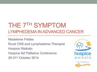
The “Seventh Symptom” – Management of Lymphoedema in advanced cancer Madeleine Fiddes

Empfohlen
Empfohlen
Weitere ähnliche Inhalte
Was ist angesagt?
Was ist angesagt? (20)
Andere mochten auch
Andere mochten auch (20)
Ähnlich wie A3 - Symptom Management
Ähnlich wie A3 - Symptom Management (20)
Mehr von HospiceNZConference
Mehr von HospiceNZConference (16)
Kürzlich hochgeladen
Kürzlich hochgeladen (20)
A3 - Symptom Management
- 1. THE 7TH SYMPTOM LYMPHEDEMA IN ADVANCED CANCER Madeleine Fiddes Rural CNS and Lymphedema Therapist Hospice Waikato Hospice NZ Palliative Conference 29-31st October 2014
- 2. Overview • Definition of lymphedema • Types of lymphedema • Stages of lymphedema • Lymphedema and Palliative Care • Treatment options • Collaboratively in the community • Case Studies • Conclusion
- 3. Lymphatic system Lymphatic system is part of circulatory system. Network of vessels and nodes throughout the body transporting lymph fluid from tissues back to bloodstream. Functions: Assist immune system Remove waste products (ALA, 2014)
- 5. Lymphedema “Lymphedema is a progressive chronic condition. It leads to excessive accumulation of protein rich lymph fluid in the tissues as a result of lymphatic failure due to either congenital abnormality or damage to the lymphatic system.” (British Lymphology Society, 2010)
- 6. Types of Lymphedema • Primary Lymphedema • Secondary Lymphedema
- 7. Primary Lymphedema Mechanical insufficiency due to unknown cause Congenital defect of lymphatic system • Milroy’s disease • Lymphedema Praecox – up to age 35 (at puberty) • Lymphedema Tardis – after age 35 (milder condition)
- 8. Secondary Lymphedema • Filariasis • Malignancy • Surgical complications • Radiation • Trauma • Infection –acute, chronic
- 10. Lymphedema and Palliative Care “Lymphedema is known to affect the physical, psychological and social well-being of the patient” (Tobin et al, 1995). “Lymphedema in palliative care is associated with worsening levels of function and dependence as well as feelings of hopelessness, disgust and social isolation” (Hewitt et al, 2010).
- 11. Oedema • Oedema: • Excess low-viscosity, protein-poor interstitial fluid that exceeds lymphatic capacity to reabsorb • Lymphedema: • Excess protein-rich interstitial fluid within skin and sub-cutaneous tissue resulting from lymphatic dysfunction
- 12. Differential diagnosis: Oedema/lymphedema - Advanced Cancer • Obstruction - DVT, compression by tumour • Hypoalbuminemia • Renal/hepatic failure • Cardiac failure • Dependant limb, immobility, neurological deficit • Effects of drugs or cytotoxic chemotherapy • Infection – e.g. Cellulitis • Malignant involvement or infiltration of lymphatic structures – e.g. SVCO (Towers et al. 2010)
- 13. Principles of Palliative management • Reverse the reversible • Patient’s priorities and goals • Benefit versus burden • Patient general condition and prognosis • Psychosocial issues Vaugn Keeley ALA conference, 2014 Explanation - Compassion - Education
- 14. Key concepts: Palliative Care and Lymphedema • Uniqueness of each patient • Interdisciplinary (team) work • Communication • Ingenuity and creativity • Good control of pain and other symptoms • Maintenance of independence • Fears and expectations • Self-care (Towers et al 2010)
- 15. We need to continually ask: Is the primary goal life prolongation or is it comfort and quality of life?
- 16. Treatment
- 17. Key components of care • Skin care • Remedial exercise • Lymphatic drainage therapy • Compression – bandaging/garments • Kinesio® Taping “The Power of Touch”
- 18. Differences Standard and Palliative CDT Standard CDT • Goal – reduce swelling, transition to garment, life-long maintenance • Four elements CDT • Two distinct phases in treatment • Definitive contra-indicators to treatment Palliative CDT • Goal – comfort, support, relief of symptoms, maintain function • CDT elements modified or omitted • Less distinction between phases • Contra-indicators relative (Towers et al, 2010)
- 19. Standard and Palliative Bandaging Standard • Full standard pressure • Multi-layer bandaging • 24-hour bandaging during the intense phase • Foam padding used • Transition to compression garments Palliative • Reduced pressure • Fewer layers required • Intensive treatment for lymphorrhea require frequent re-application • Bandaging lower leg/s • Soft padding better tolerated • May transition to lighter compression garment • (Towers et al, 2010)
- 20. Management of LE in the community Green (2010); Warilow& Jones (2012); Morgan P (n.d.) • Patient • GP • Lymphedema Therapist • Community Nurse • Interdisciplinary approach
- 21. Developing: Integrated LE patient pathway • Referral process • Central point of entry • Multidisciplinary focus • Development of study days “It is important that practitioners use standard assessment tools and documentation (Hopkins, 2010).
- 22. Case Studies
- 23. “Sally” • 66 yr. old female • Clear cell carcinoma of urethra Stage 4 • Radical Cystourethrectomy, Hysterectomy, bilateral oophorectomy, vaginectomy, vulvectomy, pelvic lymph node dissection and formation of an ileal conduit • Radiotherapy • Chemotherapy
- 24. Measurement - Leg Date 29/5/13 17/6/13 28/7/13 15/7/13 Right Left Left Left Left 10cm 20cm 25cm 25cm 23 22 20cm 28 32 28.8 29 27 30cm 35 43 40.5 38.5 38 40cm 33 40 40 37.5 37 50cm 42.5 51.3 50 48 47.5 60cm 52 62 58 57 57 70cm 58
- 26. Wendy • 56 yr. old female • R Breast Ca. – IDC Grade 2 • Extensive skin/breast changes – fungating lesion • Lymphadenopathy lower cervical, supraclavicular, axillary regions with brachial plexus involvement • Radiotherapy • Chemotherapy
- 27. Measurement – R arm Date 1 July 17 July 21 Aug Wrist 15.5 cm 16.5 cm 15 cm 10 cm 21 23.3 22 20 26.5 29 28 30 31 33 30.5 40 31 32 29
- 28. Wendy the Warrior’s supporter
- 29. “Mary” • 53 yr. old woman • Stage 4 Leiomyosarcoma – uterus • Hysterectomy, Bilateral Oophorectomy • Pleomorphic Sarcoma R arm and extensive bony metastasis
- 31. Conclusion “Although lymphedema can be challenging to manage in palliative populations, it is possible to modify existing modalities to effectively reduce oedema and improve quality of living” (Hewit et el, 2010)
- 32. 2015 National Hui NZ Lymphedema Therapists Date: 16 – 17th May 10 - 5pm Venue: Hamilton Airport Hotel 201, Airport Road, RD2, Hamilton
- 33. Palliative care today is much more than end-of-life care (Saunders, 2005).
- 34. Useful web-sites • Lymphoedema Support Network www.lymphoedema.org • Australia Lymphology Association • International Lymphoedema Framework www.lympho.org • Lymphoedema DermNet NZ • National Lymphoedema Network • Lymph notes www.lymphnotes.com • British Lymphology Society www.thebls.com • Lymphoedema Support Network www.lymphoedema.org/lsn • LymphCare
- 35. References • Towers, A. et al. (2010). Care of palliative patients with cancer related lymphedema. J. of Lymphoedema. 5(1). • Morgan. P. Health Professionals ideal roles in lymphoedema management. BJ of Community Nursing. • Green, T. (2010). Key-worker clinics: the maintenance phase of LE therapy. Chronic Oedema, October. • Hopkins, A. 2010). Mapping an integrated LE patient pathway. 5(2).
- 36. References • Todd, M. (2009). Understanding lymphoedema in advanced disease in palliative setting. IJPN. 15(10). • Hewitt, B., et el. (2010). Lymphoedema in Palliative Care. Cancer Forum, 34(2). • Cooper, G. (2012). Lymphoedema treatment in palliative care: a case study. British Journal of Nursing. 21(15). • Board, J., & Anderson, J. (2013). Treatment of lymphorrhea. British Journal of Healthcare Assistants. 7(1). • Todd, M (2010). Managing lymphoedema in palliative care patients. BJN. 18(8). • Linnitt, N. (2005). Lymphoedema: recognition, assessment and management. Woundcare. March. • Fenton, S. (2001). The power of touch in last week of life. IJPN. 17(2)
- 38. My Role • Rural CNS • Lymphedema Therapist – Casey-Smith Method • Registered ALA - member
Hinweis der Redaktion
- Lymph system is a network of vessels that form a semi-circle. Starts in tissue bed with lymphatics Fluid moves one way, starting from the interstitial tissue. Valves ensure the fluid moves one way only. No pump. Muscle movement acts as a pump. Lymph Fluid Lymph fluid carries - large proteins, dead cells, bacteria, cancer cells Originates as blood plasma. Most of the plasma / fluid returns back into the blood circulation via the small veins. A small percentage returns to blood circulation via the lymph circulation.
- National Breast and Ovarian Ca Centre Estimated 20% of breast, genituiry, Gynea or Melanoma Vulval 36-47%, Breast 20%, Cervical 24%, Melanoma 9 -29%, following SNB 4 -8 %
- Primary Primary lymphedema result from - Hypoplasia – small and ineffective vessels Hyperplasia – tangle of vessels Aplasia – absence of lymph vessels Milroy's disease - characterized by lymphedema, commonly in the legs, caused by congenital abnormalities in the lymphatic system. Disruption of the normal drainage of lymph leads to fluid accumulation and hypertrophy of soft tissues.
- Filariasis – Most common cause secondary LE – caused by a parasitic worm roundworm/larvae migrate to the lymphatics. These filarial worms are spread by a mosquito vector. Wuscheria bancrofti carry out their life cycle in two hosts. Human beings serve as the definitive host and mosquitoes as their intermediate hosts. The adult parasites reside in the lymphatics of the human host. According to WHO 2000 – 120milj – 45 milj is severely disfigured Eradication The WHO is coordinating an effort to eradicate filarisis. The mainstay of this programme is the mass use of antifilarial drugs on a regular basis for at least five years In April 2011, Sri Lanka was certified by the WHO as having eradicated this disease.
- Phases – Acute, Latent and chronic As lymphoedema develops, the protein rich fluid is gradually replaced by fibro-sclerotic tissue. This will result in ‘non-pitting’ or ‘woody’-feel to the limb Stage 0 (sub-clinical phase) No measurable evidence of swelling Heaviness/pain/pins and needles Stage 1 Some swelling evident ‘Pits’ on pressure Subsides with elevation Stage 2 Limb elevation no longer reduce swelling Fibrosis – pitting less evident Poor skin condition Stage 3 Pitting is absent Fibrotic changes
- Holistic approach is very important Managing lymphedema in PC is an important part of holistic care Palliative patients can benefit from LE Management Reduced function, immobility Pain reduction – heaviness of limb, lymphorrhea Infection minimisation – recurrent infections Skin protection Psychological support – fear and anxiety Explanation – Compassion - Education
- DVT’s are common in the hyper-co-arguable state that exist with mets cancer – the advent of low molecular heparin has simplified anti-coagulation. However use of compression and exercise remains controversial Cardiac Failure – drugs – calcium antagonists, alpha-blockers, anti-hypertensive drugs Hypoalbuminemia – anorexia, cachexia, ascitis – repeated drainage Drugs – NSIADS (non steriodal inflammatory), hormonal therapies, pregabalin, bisposphonates especially Zometa,
- Reversible – anaemia, ascitis and superior vena cave obstruction
- Understand and respect uniqueness of patient Inclusion of family in providing lymphedema care Involvement of community in providing resources/care Interdisciplinary (team) work Attention to detail – what is important to the patient? Good communication – patient, family and other providers Ingenuity/creativity in dealing with therapeutic problems Good symptom control of pain and other symptoms Maintenance if independence and function Focus of meaning of symptoms - fears/expectations Attention to therapist own emotions with limited prognosis
- The burden of the treatment must not exceed the benefit to the patient. (Honnor, 2008)
- CLD - Complex decongestive treatment MLD -Manual lymphatic drainage
- Patient – enable to take responsibility of his/her own care and management of LE Provide education programmes of information Availability of written, verbal and other information e.g. videos Develop plans of care in partnership with patient and carers Encourage and enable patients to use information technologies to learn more GP role – Early diagnosis of LE Differential diagnosis e.g. HF and DVT Diagnostic investigations Awareness and early referral to LE services LE specialist – management and supervision of LE services. Management of community based clinics LE assessments, MLD Assist and teach SLD Develop a central referral system. Auditing Education Key-worker – qualified health professional Nurses identified for LE clinics had skills in leg ulcer management and tissue viability – current skills and capacity could be used to support patients within this settings. Facilitate and develop LE support groups
- Clear pathway for management of LE Roles and skills of CNS and key worker clearly defined Treatment carried out by staff member with appropriate training Cost effective service Patient discharged from community clinics within agreed protocols Education program covered from anatomy/physiology – assessment – four key components
- Patient Summary: · 07/02/12: Clear cell carcinoma of the urethra, T4 (vagina, bladder neck) N2 (3/9) Mx (? clitoral), Stage IV. · 19/04/12: Radical cystourethrectomy, hysterectomy, bilateral oophorectomy, vaginectomy, vulvectomy, pelvic lymph node dissection and formation of ileal conduit (anterior exenteration). · 10/04/13: Restaging subcutaneous metastases, small volume pelvic disease. · 18/04/13: Lymphoedema of the left leg managed conservatively. Repeat palliative radiation to bony lesion in the left pubis. Stage IV disease (symphysis pubis involvement). · 1/9/2013: Skin lesion mons pubis slight fungation, manage conservatively
- 21/01/2008, wide local excision left breast plus modified B-reconstruction and axillary sentinel lymph node; pT1a(2.5mm) pNo cMx, Grade 2, invasive ductal carcinoma. No LVSI. ER 3+ 80%, PR 3+ 60 – 70% and HER2 2+, FISH not amplified. Adjuvant radiotherapy to the left breast, 50gy in 25 fractions. Tamoxifen discussed but not advised/started due to low risk. · October 2013, presented with extensive skin and breast changes in both breasts. · 24/10/2013, punch biopsy right breast, Grade 2/3 carcinoma, with ulceration and infiltration of the dermis. No Paget’s disease. ER 1+ (15%), PR negative, HER2 negative, cN3 cM1(skin), Stage IV disease. · 08/11/2013, commenced on AC chemotherapy. Due to toxicities, this was stopped January 2014. · March 2014, disease progression chest wall, extensive skin involvement from flank to flank. Further chemotherapy recommended but declined. · 22/07/2014, re-referred with progressive disease with fungation right breast as well as increasing right arm lymphoedema with evidence of progressive lymphadenopathy, lower cervical/supraclavicular/axillary regions with brachial plexus involvement.
- Treatment Skin care, MLD – husband – gave them some time to be intimate