Oxidative stress and mito in autism
•Als PPTX, PDF herunterladen•
1 gefällt mir•1,260 views

This PPT is a layman's summary of a brain tissue study by Chauhan and Chauhan that found oxidative stress and mitochiondrial dysfunction in autistic brains. This PPT has not been authorized or reviewed by Chauhan and Chauhan.

Empfohlen
Weitere ähnliche Inhalte
Was ist angesagt?
Was ist angesagt? (20)
Ähnlich wie Oxidative stress and mito in autism
Ähnlich wie Oxidative stress and mito in autism (20)
Kürzlich hochgeladen
Kürzlich hochgeladen (20)
Oxidative stress and mito in autism
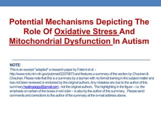
- 1. Potential Mechanisms Depicting The Role Of Oxidative Stress And Mitochondrial Dysfunction In Autism NOTE: This is an excerpt *adapted* a research paper by Fatemi et al - http://www.ncbi.nlm.nih.gov/pubmed/22370873 and features a summary of the section by Chauhan & Chauhan. Please note that this is a summary by a layman with no formal training in the subject matter and has not been reviewed or endorsed by the original authors. Any mistakes are due to the author of this summary healingsiggy@gmail.com , not the original authors. The highlighting in the figure – i.e. the emphasis on certain of the boxes in red color – is also by the author of this summary, . Please send comments and corrections to the author of the summary at the e-mail address above.
- 2. Note from healingsiggy@gmail.com • As ASD parents, many of us are aware that oxidative stress, mitochondrial dysfunction, inflammation etc. are part of the dysfunctional processes in autism. • However, many of these conclusions are based on studies done in the body, rather than on the brain. • The Chauhan and Chauhan study summarized here is remarkable in that it presents results from studies done on post-mortem brains of autistic individuals.
- 3. FIRST, LET’S LOOK AT SOME DEFINITIONS Oxidative stress Glutathione
- 4. Oxidative Stress and Why it Matters Let’s start with defining reactive oxygen species (ROS). ROS are chemically reactive molecules containing oxygen which form as a natural byproduct of the normal metabolism of oxygen. ROS have important roles in cell signaling and homeostasis. However, during times of environmental stress ROS levels can increase dramatically and may exceed the anti-oxidant capacity of a cell. Obviously, this is more likely to happen if an individual’s anti-oxidant capacity is low to begin with. When the levels of reactive oxygen species (ROS) exceed the antioxidant capacity of a cell, significant cell damage results. This is called oxidative stress. Oxidative stress can lead to inflammation, damaged cell membranes, autoimmunity, and cell death.
- 5. Glutathione and Why it Matters • Glutathione (GSH) is an anti-oxidant which prevents damage to cells caused by reactive oxygen species (ROS) such as free radicals. • Glutathione is the major free radical scavenger in the brain. • Diminished GSH levels elevate cellular vulnerability towards oxidative stress
- 7. Summary of Findings There is elevated oxidative stress in the cerebellum and frontal and temporal lobes of individuals with autism. There is reduced glutathione in brains of individuals with autism. There is mitochondrial dysfunction in brains of individuals with autism. What the Study Proposes The oxidative stress can be induced or triggered in autism by exposure to certain environmental factors, which include toxins, maternal drugs, viral and bacterial infections. The timing of this exposure to environmental factors may be pre-natal, per- natal or post-natal. Genetic factors can also influence vulnerability to oxidative stress in autism. Therefore, autism may result from genetic, environmental and immune factors, with oxidative stress as the mechanism linking these factors. See the next slide for the potential mechanisms of the role of oxidative stress and mitochondrial dysfunction in the development of autism.
- 8. Autism may result from genetic, environmental and immune factors, with oxidative stress as the mechanism linking these factors. Defect in mitochondrial Environmental risk factors – Genetic electron transport chain pre-natal, peri-natal, post-natal susceptibility factors complexes Impaired oxidative Reduced anti-oxidant defense phosphorylation, impaired Increased generation of free Glutathione redox imbalance energy (ATP) production radicals Anti-oxidant enzymes Iron/copper transport proteins Mitochondrial Dysfunction OXIDATIVE STRESS DNA methylation Lipid peroxidation, protein Epigenetic Inflammation Altered immune oxidation, DNA oxidation dysregulation response Pathogenesis and clinical development of autism
- 9. MORE DETAILS ON FINDINGS
- 10. Evidence of oxidative stress has been found in ASD brain tissues Several studies have suggested immunological abnormalities and inflammation in autism. There is also ample evidence of the presence of oxidative stress in the bodies of children with autism. We now know from studies of postmortem brain tissues that compared to age-matched control subjects, ASD individuals have elevated levels of markers of oxidative damage in the brain.
- 11. Glutathione - the body’s own powerful anti-oxidant – is reduced in autistic brains The brain is highly vulnerable to oxidative stress because of its limited antioxidant capacity, higher energy requirement, high amounts of unsaturated lipids and iron. ASD individuals have reduced antioxidant status in brain tissues compared to age-matched controls. Glutathione (GSH) is the major endogenous antioxidant produced by cells, which neutralizes ROS, and participates in detoxification and elimination of environmental toxins. A decrease in GSH, an increase in its oxidized disulfide form (GSSG), and a decrease in the redox ratio of GSH/GSSG were observed in the cerebellum and temporal cortex of individuals with autism, suggesting a glutathione redox imbalance in autism.
- 12. Evidence of mitochondrial dysfunction has been found in autistic brains Mitochondria produce ATP (energy) with the help of five electron transport chain (ETC) complexes. Emerging evidence suggests increased prevalence of mitochondrial dysfunction in autism. Chauhan and Chauhan report a deficit specific to brain regions in the levels of ETC complexes in children with autism. Healingsiggy’s note: Note that the mitochondrial dysfunction mentioned by the authors is specific to certain brain regions. Would these individuals have shown generalized mitochondrial dysfunction on biochemistry screens? Per clinical criteria? We do not know whether they did or not – but to my mind, this speaks to the fact that we may not be able to easily rule out mitochondrial dysfunction in autism just though biochemistry screens.
- 13. The level of mitochondrial dysfunction varies even within brain regions! Reduced levels of complexes III and V in the cerebellum, of complex I in the frontal cortex, and of complexes II, III, and V in the temporal cortex were observed in children with autism as compared to age-matched control subjects. Chauhan and Chauhan’s studies of different brain regions showed that oxidative stress differentially affects selective brain regions, such as cerebellum, temporal, and frontal cortices, in autism. Increased oxidative stress and mitochondrial abnormalities were not observed in the parietal and occipital cortices in autism. In other words, the level of oxidative stress and mitochondrial abnormalities varies even within brain regions.
- 14. Conclusion – there is elevated oxidative stress in autistic brains In conclusion, there is elevated oxidative stress in the cerebellum and frontal and temporal lobes of individuals with autism The oxidative stress can be induced or triggered in autism by exposure to certain environmental factors, which include toxins and toxicants, maternal drugs, viral and bacterial infections. The timing of this exposure to environmental factors may be pre-natal, per- natal or post-natal. Genetic factors can also influence vulnerability to oxidative stress in autism. Therefore, autism may result from genetic, environmental and immune factors, with oxidative stress as the mechanism linking these factors. See the next slide for the potential mechanisms of the role of oxidative stress and mitochondrial dysfunction in the development of autism.