SlideShare ist ein Scribd-Unternehmen logo
1 von 8
Epithelium stomatodeom
When the neuralcrest cells reach jaw space the
(es) ReleaseFactors initiate the reaction btwthem (Es andectoome.
Epithelial cellsof the external and internal
dental epithelium(the dental organ)
proliferatein an apical direction forminga
double layer of cells named Hertwig’s
epithelial rootsheath (RS
The Anatomy of Periodontal Tissues
Introduction:
The periodontium=(attachment apparatus)=(supportingtissueof the teeth) (peri = around,odontos = tooth) comprises the
followingtissues (Fig.1-1):(1) the root cementum(RC), (2) the periodontal ligament(PdL), (3) the alveolar bone
(AP)and (4) the gingiva(G) -The alveolar boneconsists of alveolarboneproper (ABP)=(bundle bone) and the
alveolar process
-(ABP) coming btw alveolar processand (PDL).
-Neural crest cells From neural tube
Migrate to 1st branchial branch as a result to forma band of ectomesenchymebeneath
- formation of dental lamina then processes areinitiated likecap and bud and bell stage
Cap stage :
1-condensation of ectomesenchymal cells appears in relation to the dental epithelium(dental organ (DO)) and then
2-forming the dental papilla (DP) that gives riseto the dentin and the pulp and determines the shapeand form of the tooth by
ectomesenchyme
3-forming the dental follicle (DF) that gives riseto the periodontal supportingtissues(attachmentapparatus)
Bell stage:
 The dental organ is the formative organ of enamel, ‫ستايج‬ ‫بل‬ ‫ال‬ ‫في‬ ‫ثانية‬ ‫(مر‬DP &DF) ‫ال‬ ‫ذكر‬ ‫تم‬
 The development begin with crown then root and periodontiom
 Epithelial cellsof the external and internal dental epithelium ( dental organ)
 Hertwig’s epithelial rootsheath (RS). Is a double layer cells of external and internal dental organ
1. Inner epithelial cells inducedental papilla
2. Dental papilla contain ectomesenchymal cells which differentiateto odontoblast
3. Odontoblastform dentin
at the startof dentin formation .1the inner cells of Hertwig’s epithelial rootsheath 1-synthesize and 2-secrete enamel-related
proteins,(amelogenin family)
-at the end the epithelial rootsheath becomes fenestrated and ectomesenchymal cells from the dental folliclepenetrate through
these fenestrations and contact the root surface
. The ectomesenchymal cells in contactwith the enamel-related proteins differentiate into cementoblasts and startto form
cementoid. This cementoid represents the organic matrix of the cementum and consists of a ground substanceand collagen fibers,
which intermingle with collagen fibers in the not yet fully mineralized outer layer of the dentin. It is assumed thatthe
cementum becomes firmly attached to the dentin through these fiber interactions.The formation of the cellular
cementum, which covers the apical third of the dental roots, differs from that of acellular cementum in that some of the
cementoblasts become embedded in the cementum. The remainingparts of the periodontium areformed by
ectomesenchymal cells fromthe dental folliclelateral to the cementum. Some of them differentiate into periodontal
fibroblastsand formthe fibers of the periodontal ligamentwhileothers become osteoblasts producingthe alveolar bone
proper in which the periodontal fibers areanchored. In other words, the primary alveolar wall isalso an ectomesenchymal
product. It is likely,butstill notconclusively documented, that ectomesenchymal cells remain in the mature =periodontium
and take part in the turnover of this tissue.
Gingiva:
The oral mucosa consists of
masticatory mucosa include gingivaand the covering of the hard palate
specialized mucosa covers the dorsum of the tongue
Lining mucosa The remaining part
lamina propria is underlying connective tissue layer
Gingiva consist of epithelial layer and lamina propria ( ct)
-the free gingival margin, has a scalloped outline
-In the apical direction the gingiva is continuous with the loose attached to bone, darker red alveolar mucosa (lining mucosa)
which the gingiva is separated by mucogingival junction (mgj)(arrows) or the mucogingival line.
-There is no mucogingival line present in t he palate since the
hard palate and the maxillary alveolar process are
covered by the same type of masticatory mucosa.
Two types of gingiva (fg and ag)
1- free gingiva(fg) is coral pink, has a dull surface and firm consistency
interdental papilla = interdental gingiva
Free gingival groove : is positioned at a level corresponding to the level of the cemento-enamel junction (CEJ)
free gingiva extends from the gingival margin in apical direction to the free gingival groove
2-Attached gingiva (AG)From mgj To cej or free gingival groove
gingival pocket”is not close so it is pathogenic so it is opened if it is’t so it is called gingival crevice”
the free gingival margin is located on the enamel surface approximately 1.5–2 mm coronal to the cemento-
enamel junction.
-the interdental papilla is of pyramidal form (Fig. 1-8b) while in the molar regions, the papillae are more
flattened in the buccolingual direction (Fig. 1-8a).and this depend on
-The attached gingiva is demarcated in the coronal direction, by the free gingival groove (GG)
The free gingival groove is often most pronounced on the vestibular aspect of the teeth, occurring most
frequently in the incisor and premolar regions of the mandible, and least frequently in the
mandibular molar and maxillary premolar regions.
-Stippling is smalldepression and give the appearance of orange peel. It is firmly attached to the underlying
alveolar bone and cementum by connective tissue fibers, and is, therefore, comparatively
immobile in relation to the underlying tissue.(found in 40% of adult in attached gingiva)
-the attached gingiva is of firm texture, coral pink in color, and often shows small depressions(stippling)
- the darker (am)(alveolar mucosa) is loosely bound to the underlying bone.
-the width of the gingiva tends to increase with age
-the mgj remains stable through life in relation to the lower border of the mand.
Microscopic anatomy
Oral epithelium
The epithelium covering the free gingiva may be differentiated as follows:
• Oral epithelium (OE), which faces the oral cavity
• Oral sulcular epithelium (OSE), which faces the tooth without being in contact with the tooth surface
• Junctional epithelium (JE), which provides the contact between the gingiva and the tooth not in direct with
tooth -(oe )and (ct) has wavy shape
- connective tissue papillae (CTP)is the projection of (ct) into (oe)
-rete pegs (er)are separator btw (oe) and (ct)
-contact
relationships btw
the teeth
-the width of the
approximal tooth
surrfaces
- course of cej
have cytoplasmic extensions of various size and appearance and called clear cells
- the zone around their nuclei appears lighter than that in the surrounding keratin-
producing cells
‫بمثلو‬ ‫هذول‬
‫ال‬basement
membrane
-rete pegs presents btw junctional epithelium and (ct) if inflammation is occurred
- CTP + RETE PEGS are lacking in the junctional epithelium and presence in( ors) and (oe) in non inflamed
gingiva.
- The oral epithelium is a keratinized, stratified, squamous epithelium
- the keratin-producing cells are differentiated, can be divided into the following cell layers
1. Basal layer (stratum basale or stratum germinativum)
2. Prickle cell layer (stratum spinosum)
3. Granular cell layer (stratum granulosum) contain Keratohyalin granules and clasters of collagen containing
granules
4. Keratinized cell layer (stratum corneum).
- cell nuclei are lacking in the outer cell layers. Such an epithelium is denoted orthokeratinized
-parakeratinized epithelium is the gingiva contain remnants of the nuclei like in stratum corneum
- 90% of the total cell population or oral epithelium contain
1- Melanocytes
2- Langerhans cells
3- Merkel’s cells
4- Inflammatory cells.
- merkel’s cells do not produce keratin, lack desmosomal attachment to adjacent cell And have sensory function .
-melanocytes are pigment-synthesizing cells and are responsible for the melanin pigmentation occasionally seen on the
gingiva and contains melanin granules (MG) and has no tonofilaments or hemidesmosomes.
-langerhans cells play a role in the defense mechanism of the oral mucosa and react with antigens.
- The cells in the basal layer) stratum germinativum ( and ( progenitor cell compartment) are either cylindric or cuboid, and
are in contact with the basement membrane that separates the epithelium and the connective tissue(which
mean it comes btw them ) and possess the ability to divide , so in this area the epithelium is renewed but
when it is attached to tooth surface it will be flattened
- Fig. 1-21 When two daughter cells (D) have been formed by cell division, an adjacent “older” basal cell (OB)
is pushed into the spinous cell layer and starts, as a keratinocyte, to traverse the epithelium
- there is complete equilibrium between cell renewal and cell loss so that the epithelium maintains a constant
thickness.
- Under the light microscope the basement membrane appears as a structureless zone approximately 1–2 μm
wide and act with pas stain and toappear carbohydrate (glycoprotein )in basement membrane
- extracellular substance contains proteinpolysaccharide complexes and surrounds the epithelium cells
-lamina lucida(LL) is 400 Å wide electron-lucent zone and it is beneath basal cells (bc)
- lamina densa (LD).is an electron-dense zone and it is beneath and the same thickness of (LL)
- anchoring fibers (AF) is From the lamina densa project in a fan-shaped fashion into the connective tissue and
it is approximately 1 μm in length and terminate freely in the connective tissue
-the basement membrane appears to comprise one (LL) and(LD)
- The cell membrane of the epithelial cells facing the lamina lucida harbors a number of electron-dense,
thicker zones appearing at various intervals along the cell membrane. These structures are
called hemidesmosomes (HD) are involved in the attachment of the epithelium to the
underlying basement membrane and connect the basal lamina with epithelial cells .
- The cytoplasmic tonofilaments in the cell converge towards the hemidesmosomes.
- Stratum spinosum consists of 10–20 layers of relatively large, polyhedral cells, equipped with short cytoplasmic processes
resembling spines.
- cytoplasmic processes occur at regular intervals and give the cells a prickly appearance.
- desmosomes is two hemidesmosomes facing each other and separated by a zone containing electrondense
granulated material (GM).and consist of
1-inner leaflet of the cell membrane -2-outlet leaflet of the cell membrane and the3-attached plaque(
represent granular and fibrillar material in the cytoplasm.)
- The presence of a large number of desmosomes indicates that the cohesion between the epithelial cells is
solid
-light cell not a keratinocyte but rather a “clear cell”
- there are large amount of tonofilaments in the cytoplasm of the keratinocytes.
- From (stratum basale) to stratum granulosum) both the number of tonofilaments (F) and the number of
desmosomes (D) in the cytoplasm increase.
the number of organelles, such as mitochondria (M), lamellae of rough endoplasmic reticulum (E) and Golgi
complexes (G), decrease in the keratinocytes on their way from the basal layer towards the
surface.
- the number of organelles, such as mitochondria (M), lamellae of rough endoplasmic reticulum (E) and Golgi
complexes (G), decrease in the keratinocytes on their way from the basal layer towards the
surface. In the stratum granulosum, electron-dense keratohyalin bodies (K) and clusters of
glycogen-containing granules start to occur. Such granules are believed to be related to the
synthesis of keratin.
-dento gingival epithelium
-when the tooth is fully developed the ameloblast which produces enamel become reduce in height and
produce basal lamina(epithelial attachement lamina)( lies in direct contact with the enamel)
-basal lamina +outer enamel epithelium will form reduced dental epithelium.
-The reduced enamel epithelium surrounds the crown of the tooth from the moment the enamel is properly
mineralized until the tooth starts to erupt.)then remove and come instead of it the junctional epithelu
-the mitotic activity of outer layer of (re) and and the basal layer of the oral epithelium will be increased and
start to migrate into the underlying (ct) when the tooth reach the oral epithelium
- The migrating epithelium produces an epithelial mass between the oral epithelium and the reduced dental
epithelium so that the tooth can erupt without bleeding.
-when the tooth exposed to oral cavity the whole crown covered by junctional epithelium
-the cervical region. Still covered by ameloblast and reduced dentinal epithelium later on will be junctional
epithelium
-junctional epithelium decreasing in thickness toward cej( in coronal portion (15 to 20 cell layers )but in cej (3-
4 cell )
- The cells of the oral sulcular epithelium are cuboidal and the surface of this epithelium is keratinized.
Oral epithelium Junctional epithelium
- cells volume -smaller -bigger
-intracellular space -Narrower -wider
-no. Of desmosomes - Bigger -smaller
-in the junctional epithelium which is close to tooth appears two zones 1- is electron dense which is
continuation o the lamina densa 2-electron lucent (contact to the junctional epitheliumand is
continuation o the laminalucida )
- anchoring fibers (AF) attached to the lamina densa
- (ct) is(lamina propria ) contain amophous ground substanse(matrix )
1-
Semtoblast and osteoblast produce
-matrix contain 1- collagen fibers 2- vessels and nerves 3- fibbroblast
-cells of ct are 1- fibroblast 2- mast 3- macrophage. 4- inflammatiory
1-fibrlblast
-most important
-most predominant
-spindle in shape stellateoval
-responsible of production of fibers
.cytoplasm of fibroblast contain 1- granular endoplasmic reticulaum 2- ribosomes 3- glogi 4- mitochondiria 5-
tonofilment 6- vesicle
2-mast cell
-produce vasoactive substance
-part of matrix
-control of blood flow by microvascular system
-mast cell cytoplasm contain
I. vedivcles Which contain
A-proteolytic enzyme
B- histamin
C- heparin
II. Golgi complex
III. Microvilli
3-macrophage cytoplasm contain
I. Golgi complex
II. Vesicle Contain phagosome
III. Ribosome
IV. Endoplasm trticulum
4-inflamatory cells
E.g- neutrophilic granulocytes (pmn)
-lymphocytes plasma cells
1-pmn : - neuclus is lobulate
2-lymphocytes – nucleus and ovale
3-plasma cells : mucules --> sphericles
- Golgi
- Er
- Mitochondria
- Ribosomes
Fibers in ct
1- Collagen f
2- Reticulin f
3- OxytalEan
4- Elastic f
-most predominant in gingiva and preidontium
- fibroblast produce collagen
 Traponcollagen : smallest unit of collagen mollecule
 Tropocollagen molecules consist of three polypiptyde Helix
 Polypiptyde consist of glysin ,prolin and hydroxyprolin
 Synthesis of tropocollagen occur in fibroblast and then polymarization occur out of fibroblast
 Tropocollagen is parallel to protofibrils aubsequently aggregated parallel to collagen fibrils
 tropocollagen b9eer bainhom cross linking Mature collagen fibers , less soluble
The alvoelar mucosa wain ma bet7o6a btmashe from non keratinized to karitinizid
PERIODONTAL LIGAMENT:
is soft, richly vascular and cellular connective tissue and is continuos with lamina propria(ct)of the gingiva in
coronal direction
There are two types of alveolar bone
1-lamina dura(is a abp but in x-ray) is alveolar bone which covers the alveolus
2-spongy bone has the appearance of a meshwork
-
-Alveolar crest is the coronal border of the bone
- The alveolar bone surrounds the tooth to a level approximately 1mm apical to the cemento-enamel junction
(CEJ).
- The periodontal ligament space has the shape of an hourglass and is narrowest at the mid-root level. And its
width is 0.25 mm . And its function is elicited the forces during masticatory and mobility .
The periodontal ligament divided into groups according to their arrangement
1. Alveolar crest fibers (ACF)
2. Horizontal fibers (HF)
3. Oblique fibers (OF) most common
4. Apical fibers (APF)
- The periodontal ligament and the root cementum develop from the loose connective tissue which surrounds
the tooth bud and contain fibroblast
- The tooth bud is formed in a crypt of the bone.
- fig-159a -These fiber bundles oriented towards the coronal portion of the bone crypt will later form the
dento-gingival fiber group, the dento-periosteal fiber group and the trans septal fiber group
which belong to the oriented fibers of the gingiva and it is apical to cej
fig 159 b- -the true PDL fibers =principle fibers
- First, fibers can be identified entering the most marginal portion of the alveolar bone.
159 c- Later, more apically positioned bundles of oriented collagen fibers are seen.
159-d - when the tooth has reached contact in occlusion and is functioning properly, the fibers of the
periodontal ligament associate into groups of well oriented dento-alveolar collagen fibers
demonstrated in Fig. 1-58. These collagen structures undergo constant remodeling (i.e.
resorption of old fibers and formation of new ones).
-160a – root cementum has small fine brush like fibrils found in pl space
- bone has small number of radiating , thin collagen fibrils and At this stage the surface of the bone is
covered by osteoblasts
160b- root cementum still short
-bone the number and thickness of fibers entering the bone increase. These fibers radiate towards the
loose connective tissue in the mid-portion of the periodontal ligament area (PL), which contains
more or less randomly oriented collagen fiber and the fibers entering the bone become longer
160-c The fi bers originating from the cementum subsequently increase in length and thickness and fuse in
the periodontal ligament space with the fi bers originating from the alveolar bone. When the
tooth, following eruption, reaches contact in occlusion and starts to function, the principal fi
bers become organized in bundles and run continuously from the bone to the cementum.
- The principal fi bers embedded in the cementum (Sharpey’s fi bers) have a smaller diameter but are more
numerous than those embedded in the alveolar bone proper (Sharpey’s fi bers)
- The periodontal ligament also contains a few elastic f i bers associated with the blood vessels ,oxytalan fibers
are also present in the PDL
- are located in the ligament closer to the tooth than to the alveolar bone. Very often they insert into the
cementum.
- The cells of the periodontal ligament are: fi broblasts, osteoblasts, cementoblasts, osteoclasts, as well as
epithelial cells and nerve fi bers. The fi broblasts are aligned along the principal fi bers, while
cementoblasts line the surface of the cementum, and the osteoblasts line the bone surface
-epithelial cell rests of mallasse: are clusters of epithelial cells and represent remnants of the hertwigs
epithelial root sheath and surrounded by a basement membrane (bm has desmosome and
menidesmosome
- The epithelial cell rests are situated in the periodontal ligament at a distance of 15–75 μm from the
cementum (C) on the root surface
- The epithelial cells contain only few mitochondria and have a poorly developed endoplasmic reticulum. This
means that they are vital, but resting, cells with minute metabolism
Root cementum:
Blood supply of the periodontium
-superior or inferior alveolar artery(a.a.i) gives dental artery (a.d.) Which enters the root canal
- the intraseptal artery (a.i.) before it enters the tooth socket and gives (rami perforantes, rr.p.)which
penetrate the alveolar bone proper in canals(this canals called volkmanns canals) at all levels of
the socket and anastomose in the periodontal ligament space to form polyhedral network
which surround the root .
-supraperiosteal blood vessels which is mainly supply of gingiva
-internal maxillary artery Ascending palatine artery Greater palatine artery which runs
though the greater palatine canal and puts out branches which supply the gingiva and the
masticatory mucosa of the palate
-there are anastomoses like facial artery and the blood vessels of the mandible
-free gingiva have blood supply
-subepithelial plexus located beneath oral epithelial of free and attached gingiva 40 μm thickness which
means that they are mainly venules and ,
yields thin capillary loops (7 μm )in (ct) papilla
-the no. Of capillary loops is constant
superior alveolar n.
Inferior alveolar n.
-dento-gingival plexus (dp) is consists of a fi ne-meshed network of blood vessels and it is beneath junction
epithelium
-In healthy gingiva, no capillary loops occur in the dento-gingival plexus.
-Note that the free gingiva receives its blood supply from (1) supraperiosteal blood vessels, (2) the blood
vessels of the periodontal ligament, and (3) the blood vessels of the alveolar bone.
-Lymphatic system of the periodontium
-the lymph capillaries, form an extensive network in the connective tissue. The wall of the lymph capillary
consists of a single layer of endothelial cells and is the smallest lymph vessels
1. Lymph absorbed from tissue fluid to lymph capillary
2. From capillaries to lymph vessels(Have valves )
3. It passes through one or more lymph nodes (the lymph node function is flittering and supply the
lymph with lymphocytes)
4. Then to blood stream
-sub mental lymph nodes : The labial and lingual gingiva of the mandibular incisor region and the teeth
-deep cervical lymph nodes : the palatal gingiva of the maxilla
-submandibular lymph: The buccal gingiva of the maxilla and the buccal and lingual gingiva in the mandibular
premolar–molar region
jugulodigastric lymph node :The third molars
-Except for the third molars and mandibular incisors, all teeth with their adjacent periodontal tissues are
drained to the submandibular lymph
Nerves of the periodontium
-periodontium contains nociceptors and mechanoreceptors and sensory recepter
-Nerves recording pain, touch, and pressure have their trophic center in the semilunar ganglion and are
brought to the periodontium via the trigeminal nerve and its end branche
-The nerves enter the periodontal ligament through the perforations (Volkmann’s canals) in the socket wall
-nerves join larger bundles which take a course parallel to the long axis of the tooth
-Ruffi ni’s corpuscles have been identifi ed in the periodontal ligament.
Maxilla:have 4 nerves
1-superior labial which comes from infraorbital n. ––––>from premolar to premolar from labial
aspect
2- post.supr.dental n. –––>buccal gingiva of molars
3-greater palatine –––>lingual aspect for all teeth except
Incisors which innervated by long sphenopalatine n.
Mandible :have 3 n.
1-mental –––>from canine to canine
2-buccal n. –––>from premolar to molar
3-sublingual from lingual n.–––> all teeth from lingual aspect

Weitere ähnliche Inhalte

Was ist angesagt? (20)

Pdl
PdlPdl
Pdl
 
Periodontium characteristics and parts
Periodontium characteristics and partsPeriodontium characteristics and parts
Periodontium characteristics and parts
 
262333569 cementum-in-health-and-disease-ppt
262333569 cementum-in-health-and-disease-ppt262333569 cementum-in-health-and-disease-ppt
262333569 cementum-in-health-and-disease-ppt
 
Periodontal ligaments
Periodontal ligamentsPeriodontal ligaments
Periodontal ligaments
 
Periodontium brian
Periodontium brianPeriodontium brian
Periodontium brian
 
Cementum ^_^ Emad
Cementum ^_^ Emad Cementum ^_^ Emad
Cementum ^_^ Emad
 
Cementum seminar
Cementum seminarCementum seminar
Cementum seminar
 
periodontal ligament turnover - Dr Harshavardhan Patwal
periodontal ligament turnover - Dr Harshavardhan Patwalperiodontal ligament turnover - Dr Harshavardhan Patwal
periodontal ligament turnover - Dr Harshavardhan Patwal
 
Periodontal ligament
Periodontal ligamentPeriodontal ligament
Periodontal ligament
 
Periodontal ligament
Periodontal ligamentPeriodontal ligament
Periodontal ligament
 
Molecular Biology of Cementum and Alveolar Bone
Molecular Biology of Cementum and Alveolar BoneMolecular Biology of Cementum and Alveolar Bone
Molecular Biology of Cementum and Alveolar Bone
 
Cementum
Cementum Cementum
Cementum
 
Cellular and molecular biology of cementum
Cellular and molecular biology of cementum   Cellular and molecular biology of cementum
Cellular and molecular biology of cementum
 
CEMENTUM
CEMENTUM  CEMENTUM
CEMENTUM
 
Cementum /certified fixed orthodontic courses by Indian dental academy
Cementum  /certified fixed orthodontic courses by Indian dental academy Cementum  /certified fixed orthodontic courses by Indian dental academy
Cementum /certified fixed orthodontic courses by Indian dental academy
 
Cementum
CementumCementum
Cementum
 
Cementum.
Cementum.Cementum.
Cementum.
 
Cementum ppt
Cementum pptCementum ppt
Cementum ppt
 
Cementum
CementumCementum
Cementum
 
Oral Histology - Cementum
Oral Histology - CementumOral Histology - Cementum
Oral Histology - Cementum
 

Andere mochten auch

Clinical features ofgingivitis. periodontics
Clinical features ofgingivitis. periodonticsClinical features ofgingivitis. periodontics
Clinical features ofgingivitis. periodonticsGururam MDS
 
Radiograpy in pediatric dental patient
Radiograpy in pediatric dental patientRadiograpy in pediatric dental patient
Radiograpy in pediatric dental patientPurna Patel
 
Chronic gingivitis
Chronic gingivitisChronic gingivitis
Chronic gingivitisIAU Dent
 
PACs in West Africa - Achievements and Challenges
PACs in West Africa - Achievements and ChallengesPACs in West Africa - Achievements and Challenges
PACs in West Africa - Achievements and Challengesparliaments.cluster
 
Romeo and Juliet Summary- Review sheet
Romeo and Juliet Summary- Review sheetRomeo and Juliet Summary- Review sheet
Romeo and Juliet Summary- Review sheetCatherine Yaeger
 
WBG - Discussion Note on Network and CoP Learning for Pacific PACs
WBG - Discussion Note on Network and CoP Learning for Pacific PACsWBG - Discussion Note on Network and CoP Learning for Pacific PACs
WBG - Discussion Note on Network and CoP Learning for Pacific PACsparliaments.cluster
 
OSCARS 2016|BEST DRESSED CELEBRITIES
OSCARS 2016|BEST DRESSED CELEBRITIESOSCARS 2016|BEST DRESSED CELEBRITIES
OSCARS 2016|BEST DRESSED CELEBRITIESFacesOf Ony
 
XXL MAGAZINE FRONT COVER ANALYSIS
XXL MAGAZINE FRONT COVER ANALYSISXXL MAGAZINE FRONT COVER ANALYSIS
XXL MAGAZINE FRONT COVER ANALYSISkeshialavallsmith
 
Mравославно вероучение за деца
Mравославно вероучение за децаMравославно вероучение за деца
Mравославно вероучение за децаPetar Vasilev
 
XXL MAGAZINE CONTENTS PAGE ANALYSIS
XXL MAGAZINE CONTENTS PAGE ANALYSISXXL MAGAZINE CONTENTS PAGE ANALYSIS
XXL MAGAZINE CONTENTS PAGE ANALYSISkeshialavallsmith
 

Andere mochten auch (20)

100275
100275100275
100275
 
Clinical features ofgingivitis. periodontics
Clinical features ofgingivitis. periodonticsClinical features ofgingivitis. periodontics
Clinical features ofgingivitis. periodontics
 
Radiograpy in pediatric dental patient
Radiograpy in pediatric dental patientRadiograpy in pediatric dental patient
Radiograpy in pediatric dental patient
 
Chronic gingivitis
Chronic gingivitisChronic gingivitis
Chronic gingivitis
 
протокол №1
протокол №1протокол №1
протокол №1
 
SNA 2015
SNA 2015SNA 2015
SNA 2015
 
PACs in West Africa - Achievements and Challenges
PACs in West Africa - Achievements and ChallengesPACs in West Africa - Achievements and Challenges
PACs in West Africa - Achievements and Challenges
 
Will robots replace teachers?
Will robots replace teachers?Will robots replace teachers?
Will robots replace teachers?
 
Romeo and Juliet Summary- Review sheet
Romeo and Juliet Summary- Review sheetRomeo and Juliet Summary- Review sheet
Romeo and Juliet Summary- Review sheet
 
WBG - Discussion Note on Network and CoP Learning for Pacific PACs
WBG - Discussion Note on Network and CoP Learning for Pacific PACsWBG - Discussion Note on Network and CoP Learning for Pacific PACs
WBG - Discussion Note on Network and CoP Learning for Pacific PACs
 
OSCARS 2016|BEST DRESSED CELEBRITIES
OSCARS 2016|BEST DRESSED CELEBRITIESOSCARS 2016|BEST DRESSED CELEBRITIES
OSCARS 2016|BEST DRESSED CELEBRITIES
 
Anasazi
AnasaziAnasazi
Anasazi
 
Data Dimensions NWCD 2015
Data Dimensions NWCD 2015Data Dimensions NWCD 2015
Data Dimensions NWCD 2015
 
XXL MAGAZINE FRONT COVER ANALYSIS
XXL MAGAZINE FRONT COVER ANALYSISXXL MAGAZINE FRONT COVER ANALYSIS
XXL MAGAZINE FRONT COVER ANALYSIS
 
Denny's Campaign Book
Denny's Campaign BookDenny's Campaign Book
Denny's Campaign Book
 
Tokyopop Final
Tokyopop FinalTokyopop Final
Tokyopop Final
 
About me
About meAbout me
About me
 
Mравославно вероучение за деца
Mравославно вероучение за децаMравославно вероучение за деца
Mравославно вероучение за деца
 
NABCE 2015
NABCE 2015NABCE 2015
NABCE 2015
 
XXL MAGAZINE CONTENTS PAGE ANALYSIS
XXL MAGAZINE CONTENTS PAGE ANALYSISXXL MAGAZINE CONTENTS PAGE ANALYSIS
XXL MAGAZINE CONTENTS PAGE ANALYSIS
 

Ähnlich wie Periodontics dentistry Histology By Hassan Ayyad

2000 biologia celular y molecular de la encia
2000 biologia celular y molecular de la encia2000 biologia celular y molecular de la encia
2000 biologia celular y molecular de la enciakenigal
 
Oral cavity anatomy and histology
Oral cavity anatomy and histologyOral cavity anatomy and histology
Oral cavity anatomy and histologyAliLaith5230
 
Gingiva (Macroscopic features)
Gingiva (Macroscopic features)Gingiva (Macroscopic features)
Gingiva (Macroscopic features)PremKumar2314
 
Chronology of dental development and development of occlusion
Chronology  of  dental  development and  development  of  occlusionChronology  of  dental  development and  development  of  occlusion
Chronology of dental development and development of occlusionshilpathaklotra
 
Anatomy of the Periodontium
Anatomy of the PeriodontiumAnatomy of the Periodontium
Anatomy of the PeriodontiumEnas Elgendy
 
development of root, Root formation and periodontal ligament
development of root, Root formation and periodontal ligamentdevelopment of root, Root formation and periodontal ligament
development of root, Root formation and periodontal ligamentAkram bhuiyan
 
Root formation and periodontal ligament
Root formation and periodontal ligamentRoot formation and periodontal ligament
Root formation and periodontal ligamentHamzeh AlBattikhi
 
Periodontium
PeriodontiumPeriodontium
PeriodontiumStom Vnmu
 
Development of periodontium
Development of periodontium Development of periodontium
Development of periodontium Mehul Shinde
 
Development of periodontium 1
Development of periodontium 1Development of periodontium 1
Development of periodontium 1Mehul Shinde
 
Dentinogingival junction
Dentinogingival junctionDentinogingival junction
Dentinogingival junctionBanMaraby
 
GINGIVA SEMINAR .pptx
GINGIVA SEMINAR .pptxGINGIVA SEMINAR .pptx
GINGIVA SEMINAR .pptxANIL KUMAR
 

Ähnlich wie Periodontics dentistry Histology By Hassan Ayyad (20)

junctional epithelium
junctional epitheliumjunctional epithelium
junctional epithelium
 
2000 biologia celular y molecular de la encia
2000 biologia celular y molecular de la encia2000 biologia celular y molecular de la encia
2000 biologia celular y molecular de la encia
 
Junctionalepitheliumlecture
JunctionalepitheliumlectureJunctionalepitheliumlecture
Junctionalepitheliumlecture
 
Junctional epithelium
Junctional epitheliumJunctional epithelium
Junctional epithelium
 
Gingiva
Gingiva Gingiva
Gingiva
 
Hist 9
Hist 9Hist 9
Hist 9
 
Enamel
EnamelEnamel
Enamel
 
Oral cavity anatomy and histology
Oral cavity anatomy and histologyOral cavity anatomy and histology
Oral cavity anatomy and histology
 
Gingiva
GingivaGingiva
Gingiva
 
Gingiva (Macroscopic features)
Gingiva (Macroscopic features)Gingiva (Macroscopic features)
Gingiva (Macroscopic features)
 
Chronology of dental development and development of occlusion
Chronology  of  dental  development and  development  of  occlusionChronology  of  dental  development and  development  of  occlusion
Chronology of dental development and development of occlusion
 
Anatomy of the Periodontium
Anatomy of the PeriodontiumAnatomy of the Periodontium
Anatomy of the Periodontium
 
Gingiva
Gingiva Gingiva
Gingiva
 
development of root, Root formation and periodontal ligament
development of root, Root formation and periodontal ligamentdevelopment of root, Root formation and periodontal ligament
development of root, Root formation and periodontal ligament
 
Root formation and periodontal ligament
Root formation and periodontal ligamentRoot formation and periodontal ligament
Root formation and periodontal ligament
 
Periodontium
PeriodontiumPeriodontium
Periodontium
 
Development of periodontium
Development of periodontium Development of periodontium
Development of periodontium
 
Development of periodontium 1
Development of periodontium 1Development of periodontium 1
Development of periodontium 1
 
Dentinogingival junction
Dentinogingival junctionDentinogingival junction
Dentinogingival junction
 
GINGIVA SEMINAR .pptx
GINGIVA SEMINAR .pptxGINGIVA SEMINAR .pptx
GINGIVA SEMINAR .pptx
 

Kürzlich hochgeladen

Paris 2024 Olympic Geographies - an activity
Paris 2024 Olympic Geographies - an activityParis 2024 Olympic Geographies - an activity
Paris 2024 Olympic Geographies - an activityGeoBlogs
 
Alper Gobel In Media Res Media Component
Alper Gobel In Media Res Media ComponentAlper Gobel In Media Res Media Component
Alper Gobel In Media Res Media ComponentInMediaRes1
 
URLs and Routing in the Odoo 17 Website App
URLs and Routing in the Odoo 17 Website AppURLs and Routing in the Odoo 17 Website App
URLs and Routing in the Odoo 17 Website AppCeline George
 
Employee wellbeing at the workplace.pptx
Employee wellbeing at the workplace.pptxEmployee wellbeing at the workplace.pptx
Employee wellbeing at the workplace.pptxNirmalaLoungPoorunde1
 
SOCIAL AND HISTORICAL CONTEXT - LFTVD.pptx
SOCIAL AND HISTORICAL CONTEXT - LFTVD.pptxSOCIAL AND HISTORICAL CONTEXT - LFTVD.pptx
SOCIAL AND HISTORICAL CONTEXT - LFTVD.pptxiammrhaywood
 
How to Make a Pirate ship Primary Education.pptx
How to Make a Pirate ship Primary Education.pptxHow to Make a Pirate ship Primary Education.pptx
How to Make a Pirate ship Primary Education.pptxmanuelaromero2013
 
A Critique of the Proposed National Education Policy Reform
A Critique of the Proposed National Education Policy ReformA Critique of the Proposed National Education Policy Reform
A Critique of the Proposed National Education Policy ReformChameera Dedduwage
 
Contemporary philippine arts from the regions_PPT_Module_12 [Autosaved] (1).pptx
Contemporary philippine arts from the regions_PPT_Module_12 [Autosaved] (1).pptxContemporary philippine arts from the regions_PPT_Module_12 [Autosaved] (1).pptx
Contemporary philippine arts from the regions_PPT_Module_12 [Autosaved] (1).pptxRoyAbrique
 
Grant Readiness 101 TechSoup and Remy Consulting
Grant Readiness 101 TechSoup and Remy ConsultingGrant Readiness 101 TechSoup and Remy Consulting
Grant Readiness 101 TechSoup and Remy ConsultingTechSoup
 
PSYCHIATRIC History collection FORMAT.pptx
PSYCHIATRIC   History collection FORMAT.pptxPSYCHIATRIC   History collection FORMAT.pptx
PSYCHIATRIC History collection FORMAT.pptxPoojaSen20
 
MENTAL STATUS EXAMINATION format.docx
MENTAL     STATUS EXAMINATION format.docxMENTAL     STATUS EXAMINATION format.docx
MENTAL STATUS EXAMINATION format.docxPoojaSen20
 
Organic Name Reactions for the students and aspirants of Chemistry12th.pptx
Organic Name Reactions  for the students and aspirants of Chemistry12th.pptxOrganic Name Reactions  for the students and aspirants of Chemistry12th.pptx
Organic Name Reactions for the students and aspirants of Chemistry12th.pptxVS Mahajan Coaching Centre
 
“Oh GOSH! Reflecting on Hackteria's Collaborative Practices in a Global Do-It...
“Oh GOSH! Reflecting on Hackteria's Collaborative Practices in a Global Do-It...“Oh GOSH! Reflecting on Hackteria's Collaborative Practices in a Global Do-It...
“Oh GOSH! Reflecting on Hackteria's Collaborative Practices in a Global Do-It...Marc Dusseiller Dusjagr
 
Hybridoma Technology ( Production , Purification , and Application )
Hybridoma Technology  ( Production , Purification , and Application  ) Hybridoma Technology  ( Production , Purification , and Application  )
Hybridoma Technology ( Production , Purification , and Application ) Sakshi Ghasle
 
Introduction to ArtificiaI Intelligence in Higher Education
Introduction to ArtificiaI Intelligence in Higher EducationIntroduction to ArtificiaI Intelligence in Higher Education
Introduction to ArtificiaI Intelligence in Higher Educationpboyjonauth
 
_Math 4-Q4 Week 5.pptx Steps in Collecting Data
_Math 4-Q4 Week 5.pptx Steps in Collecting Data_Math 4-Q4 Week 5.pptx Steps in Collecting Data
_Math 4-Q4 Week 5.pptx Steps in Collecting DataJhengPantaleon
 
Crayon Activity Handout For the Crayon A
Crayon Activity Handout For the Crayon ACrayon Activity Handout For the Crayon A
Crayon Activity Handout For the Crayon AUnboundStockton
 
Separation of Lanthanides/ Lanthanides and Actinides
Separation of Lanthanides/ Lanthanides and ActinidesSeparation of Lanthanides/ Lanthanides and Actinides
Separation of Lanthanides/ Lanthanides and ActinidesFatimaKhan178732
 
Software Engineering Methodologies (overview)
Software Engineering Methodologies (overview)Software Engineering Methodologies (overview)
Software Engineering Methodologies (overview)eniolaolutunde
 
Presiding Officer Training module 2024 lok sabha elections
Presiding Officer Training module 2024 lok sabha electionsPresiding Officer Training module 2024 lok sabha elections
Presiding Officer Training module 2024 lok sabha electionsanshu789521
 

Kürzlich hochgeladen (20)

Paris 2024 Olympic Geographies - an activity
Paris 2024 Olympic Geographies - an activityParis 2024 Olympic Geographies - an activity
Paris 2024 Olympic Geographies - an activity
 
Alper Gobel In Media Res Media Component
Alper Gobel In Media Res Media ComponentAlper Gobel In Media Res Media Component
Alper Gobel In Media Res Media Component
 
URLs and Routing in the Odoo 17 Website App
URLs and Routing in the Odoo 17 Website AppURLs and Routing in the Odoo 17 Website App
URLs and Routing in the Odoo 17 Website App
 
Employee wellbeing at the workplace.pptx
Employee wellbeing at the workplace.pptxEmployee wellbeing at the workplace.pptx
Employee wellbeing at the workplace.pptx
 
SOCIAL AND HISTORICAL CONTEXT - LFTVD.pptx
SOCIAL AND HISTORICAL CONTEXT - LFTVD.pptxSOCIAL AND HISTORICAL CONTEXT - LFTVD.pptx
SOCIAL AND HISTORICAL CONTEXT - LFTVD.pptx
 
How to Make a Pirate ship Primary Education.pptx
How to Make a Pirate ship Primary Education.pptxHow to Make a Pirate ship Primary Education.pptx
How to Make a Pirate ship Primary Education.pptx
 
A Critique of the Proposed National Education Policy Reform
A Critique of the Proposed National Education Policy ReformA Critique of the Proposed National Education Policy Reform
A Critique of the Proposed National Education Policy Reform
 
Contemporary philippine arts from the regions_PPT_Module_12 [Autosaved] (1).pptx
Contemporary philippine arts from the regions_PPT_Module_12 [Autosaved] (1).pptxContemporary philippine arts from the regions_PPT_Module_12 [Autosaved] (1).pptx
Contemporary philippine arts from the regions_PPT_Module_12 [Autosaved] (1).pptx
 
Grant Readiness 101 TechSoup and Remy Consulting
Grant Readiness 101 TechSoup and Remy ConsultingGrant Readiness 101 TechSoup and Remy Consulting
Grant Readiness 101 TechSoup and Remy Consulting
 
PSYCHIATRIC History collection FORMAT.pptx
PSYCHIATRIC   History collection FORMAT.pptxPSYCHIATRIC   History collection FORMAT.pptx
PSYCHIATRIC History collection FORMAT.pptx
 
MENTAL STATUS EXAMINATION format.docx
MENTAL     STATUS EXAMINATION format.docxMENTAL     STATUS EXAMINATION format.docx
MENTAL STATUS EXAMINATION format.docx
 
Organic Name Reactions for the students and aspirants of Chemistry12th.pptx
Organic Name Reactions  for the students and aspirants of Chemistry12th.pptxOrganic Name Reactions  for the students and aspirants of Chemistry12th.pptx
Organic Name Reactions for the students and aspirants of Chemistry12th.pptx
 
“Oh GOSH! Reflecting on Hackteria's Collaborative Practices in a Global Do-It...
“Oh GOSH! Reflecting on Hackteria's Collaborative Practices in a Global Do-It...“Oh GOSH! Reflecting on Hackteria's Collaborative Practices in a Global Do-It...
“Oh GOSH! Reflecting on Hackteria's Collaborative Practices in a Global Do-It...
 
Hybridoma Technology ( Production , Purification , and Application )
Hybridoma Technology  ( Production , Purification , and Application  ) Hybridoma Technology  ( Production , Purification , and Application  )
Hybridoma Technology ( Production , Purification , and Application )
 
Introduction to ArtificiaI Intelligence in Higher Education
Introduction to ArtificiaI Intelligence in Higher EducationIntroduction to ArtificiaI Intelligence in Higher Education
Introduction to ArtificiaI Intelligence in Higher Education
 
_Math 4-Q4 Week 5.pptx Steps in Collecting Data
_Math 4-Q4 Week 5.pptx Steps in Collecting Data_Math 4-Q4 Week 5.pptx Steps in Collecting Data
_Math 4-Q4 Week 5.pptx Steps in Collecting Data
 
Crayon Activity Handout For the Crayon A
Crayon Activity Handout For the Crayon ACrayon Activity Handout For the Crayon A
Crayon Activity Handout For the Crayon A
 
Separation of Lanthanides/ Lanthanides and Actinides
Separation of Lanthanides/ Lanthanides and ActinidesSeparation of Lanthanides/ Lanthanides and Actinides
Separation of Lanthanides/ Lanthanides and Actinides
 
Software Engineering Methodologies (overview)
Software Engineering Methodologies (overview)Software Engineering Methodologies (overview)
Software Engineering Methodologies (overview)
 
Presiding Officer Training module 2024 lok sabha elections
Presiding Officer Training module 2024 lok sabha electionsPresiding Officer Training module 2024 lok sabha elections
Presiding Officer Training module 2024 lok sabha elections
 

Periodontics dentistry Histology By Hassan Ayyad

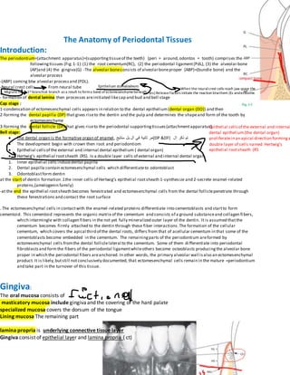
  • 1. Epithelium stomatodeom When the neuralcrest cells reach jaw space the (es) ReleaseFactors initiate the reaction btwthem (Es andectoome. Epithelial cellsof the external and internal dental epithelium(the dental organ) proliferatein an apical direction forminga double layer of cells named Hertwig’s epithelial rootsheath (RS The Anatomy of Periodontal Tissues Introduction: The periodontium=(attachment apparatus)=(supportingtissueof the teeth) (peri = around,odontos = tooth) comprises the followingtissues (Fig.1-1):(1) the root cementum(RC), (2) the periodontal ligament(PdL), (3) the alveolar bone (AP)and (4) the gingiva(G) -The alveolar boneconsists of alveolarboneproper (ABP)=(bundle bone) and the alveolar process -(ABP) coming btw alveolar processand (PDL). -Neural crest cells From neural tube Migrate to 1st branchial branch as a result to forma band of ectomesenchymebeneath - formation of dental lamina then processes areinitiated likecap and bud and bell stage Cap stage : 1-condensation of ectomesenchymal cells appears in relation to the dental epithelium(dental organ (DO)) and then 2-forming the dental papilla (DP) that gives riseto the dentin and the pulp and determines the shapeand form of the tooth by ectomesenchyme 3-forming the dental follicle (DF) that gives riseto the periodontal supportingtissues(attachmentapparatus) Bell stage:  The dental organ is the formative organ of enamel, ‫ستايج‬ ‫بل‬ ‫ال‬ ‫في‬ ‫ثانية‬ ‫(مر‬DP &DF) ‫ال‬ ‫ذكر‬ ‫تم‬  The development begin with crown then root and periodontiom  Epithelial cellsof the external and internal dental epithelium ( dental organ)  Hertwig’s epithelial rootsheath (RS). Is a double layer cells of external and internal dental organ 1. Inner epithelial cells inducedental papilla 2. Dental papilla contain ectomesenchymal cells which differentiateto odontoblast 3. Odontoblastform dentin at the startof dentin formation .1the inner cells of Hertwig’s epithelial rootsheath 1-synthesize and 2-secrete enamel-related proteins,(amelogenin family) -at the end the epithelial rootsheath becomes fenestrated and ectomesenchymal cells from the dental folliclepenetrate through these fenestrations and contact the root surface . The ectomesenchymal cells in contactwith the enamel-related proteins differentiate into cementoblasts and startto form cementoid. This cementoid represents the organic matrix of the cementum and consists of a ground substanceand collagen fibers, which intermingle with collagen fibers in the not yet fully mineralized outer layer of the dentin. It is assumed thatthe cementum becomes firmly attached to the dentin through these fiber interactions.The formation of the cellular cementum, which covers the apical third of the dental roots, differs from that of acellular cementum in that some of the cementoblasts become embedded in the cementum. The remainingparts of the periodontium areformed by ectomesenchymal cells fromthe dental folliclelateral to the cementum. Some of them differentiate into periodontal fibroblastsand formthe fibers of the periodontal ligamentwhileothers become osteoblasts producingthe alveolar bone proper in which the periodontal fibers areanchored. In other words, the primary alveolar wall isalso an ectomesenchymal product. It is likely,butstill notconclusively documented, that ectomesenchymal cells remain in the mature =periodontium and take part in the turnover of this tissue. Gingiva: The oral mucosa consists of masticatory mucosa include gingivaand the covering of the hard palate specialized mucosa covers the dorsum of the tongue Lining mucosa The remaining part lamina propria is underlying connective tissue layer Gingiva consist of epithelial layer and lamina propria ( ct)
  • 2. -the free gingival margin, has a scalloped outline -In the apical direction the gingiva is continuous with the loose attached to bone, darker red alveolar mucosa (lining mucosa) which the gingiva is separated by mucogingival junction (mgj)(arrows) or the mucogingival line. -There is no mucogingival line present in t he palate since the hard palate and the maxillary alveolar process are covered by the same type of masticatory mucosa. Two types of gingiva (fg and ag) 1- free gingiva(fg) is coral pink, has a dull surface and firm consistency interdental papilla = interdental gingiva Free gingival groove : is positioned at a level corresponding to the level of the cemento-enamel junction (CEJ) free gingiva extends from the gingival margin in apical direction to the free gingival groove 2-Attached gingiva (AG)From mgj To cej or free gingival groove gingival pocket”is not close so it is pathogenic so it is opened if it is’t so it is called gingival crevice” the free gingival margin is located on the enamel surface approximately 1.5–2 mm coronal to the cemento- enamel junction. -the interdental papilla is of pyramidal form (Fig. 1-8b) while in the molar regions, the papillae are more flattened in the buccolingual direction (Fig. 1-8a).and this depend on -The attached gingiva is demarcated in the coronal direction, by the free gingival groove (GG) The free gingival groove is often most pronounced on the vestibular aspect of the teeth, occurring most frequently in the incisor and premolar regions of the mandible, and least frequently in the mandibular molar and maxillary premolar regions. -Stippling is smalldepression and give the appearance of orange peel. It is firmly attached to the underlying alveolar bone and cementum by connective tissue fibers, and is, therefore, comparatively immobile in relation to the underlying tissue.(found in 40% of adult in attached gingiva) -the attached gingiva is of firm texture, coral pink in color, and often shows small depressions(stippling) - the darker (am)(alveolar mucosa) is loosely bound to the underlying bone. -the width of the gingiva tends to increase with age -the mgj remains stable through life in relation to the lower border of the mand. Microscopic anatomy Oral epithelium The epithelium covering the free gingiva may be differentiated as follows: • Oral epithelium (OE), which faces the oral cavity • Oral sulcular epithelium (OSE), which faces the tooth without being in contact with the tooth surface • Junctional epithelium (JE), which provides the contact between the gingiva and the tooth not in direct with tooth -(oe )and (ct) has wavy shape - connective tissue papillae (CTP)is the projection of (ct) into (oe) -rete pegs (er)are separator btw (oe) and (ct) -contact relationships btw the teeth -the width of the approximal tooth surrfaces - course of cej
  • 3. have cytoplasmic extensions of various size and appearance and called clear cells - the zone around their nuclei appears lighter than that in the surrounding keratin- producing cells ‫بمثلو‬ ‫هذول‬ ‫ال‬basement membrane -rete pegs presents btw junctional epithelium and (ct) if inflammation is occurred - CTP + RETE PEGS are lacking in the junctional epithelium and presence in( ors) and (oe) in non inflamed gingiva. - The oral epithelium is a keratinized, stratified, squamous epithelium - the keratin-producing cells are differentiated, can be divided into the following cell layers 1. Basal layer (stratum basale or stratum germinativum) 2. Prickle cell layer (stratum spinosum) 3. Granular cell layer (stratum granulosum) contain Keratohyalin granules and clasters of collagen containing granules 4. Keratinized cell layer (stratum corneum). - cell nuclei are lacking in the outer cell layers. Such an epithelium is denoted orthokeratinized -parakeratinized epithelium is the gingiva contain remnants of the nuclei like in stratum corneum - 90% of the total cell population or oral epithelium contain 1- Melanocytes 2- Langerhans cells 3- Merkel’s cells 4- Inflammatory cells. - merkel’s cells do not produce keratin, lack desmosomal attachment to adjacent cell And have sensory function . -melanocytes are pigment-synthesizing cells and are responsible for the melanin pigmentation occasionally seen on the gingiva and contains melanin granules (MG) and has no tonofilaments or hemidesmosomes. -langerhans cells play a role in the defense mechanism of the oral mucosa and react with antigens. - The cells in the basal layer) stratum germinativum ( and ( progenitor cell compartment) are either cylindric or cuboid, and are in contact with the basement membrane that separates the epithelium and the connective tissue(which mean it comes btw them ) and possess the ability to divide , so in this area the epithelium is renewed but when it is attached to tooth surface it will be flattened - Fig. 1-21 When two daughter cells (D) have been formed by cell division, an adjacent “older” basal cell (OB) is pushed into the spinous cell layer and starts, as a keratinocyte, to traverse the epithelium - there is complete equilibrium between cell renewal and cell loss so that the epithelium maintains a constant thickness. - Under the light microscope the basement membrane appears as a structureless zone approximately 1–2 μm wide and act with pas stain and toappear carbohydrate (glycoprotein )in basement membrane - extracellular substance contains proteinpolysaccharide complexes and surrounds the epithelium cells -lamina lucida(LL) is 400 Å wide electron-lucent zone and it is beneath basal cells (bc) - lamina densa (LD).is an electron-dense zone and it is beneath and the same thickness of (LL) - anchoring fibers (AF) is From the lamina densa project in a fan-shaped fashion into the connective tissue and it is approximately 1 μm in length and terminate freely in the connective tissue -the basement membrane appears to comprise one (LL) and(LD) - The cell membrane of the epithelial cells facing the lamina lucida harbors a number of electron-dense, thicker zones appearing at various intervals along the cell membrane. These structures are called hemidesmosomes (HD) are involved in the attachment of the epithelium to the underlying basement membrane and connect the basal lamina with epithelial cells . - The cytoplasmic tonofilaments in the cell converge towards the hemidesmosomes. - Stratum spinosum consists of 10–20 layers of relatively large, polyhedral cells, equipped with short cytoplasmic processes resembling spines.
  • 4. - cytoplasmic processes occur at regular intervals and give the cells a prickly appearance. - desmosomes is two hemidesmosomes facing each other and separated by a zone containing electrondense granulated material (GM).and consist of 1-inner leaflet of the cell membrane -2-outlet leaflet of the cell membrane and the3-attached plaque( represent granular and fibrillar material in the cytoplasm.) - The presence of a large number of desmosomes indicates that the cohesion between the epithelial cells is solid -light cell not a keratinocyte but rather a “clear cell” - there are large amount of tonofilaments in the cytoplasm of the keratinocytes. - From (stratum basale) to stratum granulosum) both the number of tonofilaments (F) and the number of desmosomes (D) in the cytoplasm increase. the number of organelles, such as mitochondria (M), lamellae of rough endoplasmic reticulum (E) and Golgi complexes (G), decrease in the keratinocytes on their way from the basal layer towards the surface. - the number of organelles, such as mitochondria (M), lamellae of rough endoplasmic reticulum (E) and Golgi complexes (G), decrease in the keratinocytes on their way from the basal layer towards the surface. In the stratum granulosum, electron-dense keratohyalin bodies (K) and clusters of glycogen-containing granules start to occur. Such granules are believed to be related to the synthesis of keratin. -dento gingival epithelium -when the tooth is fully developed the ameloblast which produces enamel become reduce in height and produce basal lamina(epithelial attachement lamina)( lies in direct contact with the enamel) -basal lamina +outer enamel epithelium will form reduced dental epithelium. -The reduced enamel epithelium surrounds the crown of the tooth from the moment the enamel is properly mineralized until the tooth starts to erupt.)then remove and come instead of it the junctional epithelu -the mitotic activity of outer layer of (re) and and the basal layer of the oral epithelium will be increased and start to migrate into the underlying (ct) when the tooth reach the oral epithelium - The migrating epithelium produces an epithelial mass between the oral epithelium and the reduced dental epithelium so that the tooth can erupt without bleeding. -when the tooth exposed to oral cavity the whole crown covered by junctional epithelium -the cervical region. Still covered by ameloblast and reduced dentinal epithelium later on will be junctional epithelium -junctional epithelium decreasing in thickness toward cej( in coronal portion (15 to 20 cell layers )but in cej (3- 4 cell ) - The cells of the oral sulcular epithelium are cuboidal and the surface of this epithelium is keratinized. Oral epithelium Junctional epithelium - cells volume -smaller -bigger -intracellular space -Narrower -wider -no. Of desmosomes - Bigger -smaller -in the junctional epithelium which is close to tooth appears two zones 1- is electron dense which is continuation o the lamina densa 2-electron lucent (contact to the junctional epitheliumand is continuation o the laminalucida ) - anchoring fibers (AF) attached to the lamina densa - (ct) is(lamina propria ) contain amophous ground substanse(matrix )
  • 5. 1- Semtoblast and osteoblast produce -matrix contain 1- collagen fibers 2- vessels and nerves 3- fibbroblast -cells of ct are 1- fibroblast 2- mast 3- macrophage. 4- inflammatiory 1-fibrlblast -most important -most predominant -spindle in shape stellateoval -responsible of production of fibers .cytoplasm of fibroblast contain 1- granular endoplasmic reticulaum 2- ribosomes 3- glogi 4- mitochondiria 5- tonofilment 6- vesicle 2-mast cell -produce vasoactive substance -part of matrix -control of blood flow by microvascular system -mast cell cytoplasm contain I. vedivcles Which contain A-proteolytic enzyme B- histamin C- heparin II. Golgi complex III. Microvilli 3-macrophage cytoplasm contain I. Golgi complex II. Vesicle Contain phagosome III. Ribosome IV. Endoplasm trticulum 4-inflamatory cells E.g- neutrophilic granulocytes (pmn) -lymphocytes plasma cells 1-pmn : - neuclus is lobulate 2-lymphocytes – nucleus and ovale 3-plasma cells : mucules --> sphericles - Golgi - Er - Mitochondria - Ribosomes Fibers in ct 1- Collagen f 2- Reticulin f 3- OxytalEan 4- Elastic f -most predominant in gingiva and preidontium - fibroblast produce collagen
  • 6.  Traponcollagen : smallest unit of collagen mollecule  Tropocollagen molecules consist of three polypiptyde Helix  Polypiptyde consist of glysin ,prolin and hydroxyprolin  Synthesis of tropocollagen occur in fibroblast and then polymarization occur out of fibroblast  Tropocollagen is parallel to protofibrils aubsequently aggregated parallel to collagen fibrils  tropocollagen b9eer bainhom cross linking Mature collagen fibers , less soluble The alvoelar mucosa wain ma bet7o6a btmashe from non keratinized to karitinizid PERIODONTAL LIGAMENT: is soft, richly vascular and cellular connective tissue and is continuos with lamina propria(ct)of the gingiva in coronal direction There are two types of alveolar bone 1-lamina dura(is a abp but in x-ray) is alveolar bone which covers the alveolus 2-spongy bone has the appearance of a meshwork - -Alveolar crest is the coronal border of the bone - The alveolar bone surrounds the tooth to a level approximately 1mm apical to the cemento-enamel junction (CEJ). - The periodontal ligament space has the shape of an hourglass and is narrowest at the mid-root level. And its width is 0.25 mm . And its function is elicited the forces during masticatory and mobility . The periodontal ligament divided into groups according to their arrangement 1. Alveolar crest fibers (ACF) 2. Horizontal fibers (HF) 3. Oblique fibers (OF) most common 4. Apical fibers (APF) - The periodontal ligament and the root cementum develop from the loose connective tissue which surrounds the tooth bud and contain fibroblast - The tooth bud is formed in a crypt of the bone. - fig-159a -These fiber bundles oriented towards the coronal portion of the bone crypt will later form the dento-gingival fiber group, the dento-periosteal fiber group and the trans septal fiber group which belong to the oriented fibers of the gingiva and it is apical to cej fig 159 b- -the true PDL fibers =principle fibers - First, fibers can be identified entering the most marginal portion of the alveolar bone. 159 c- Later, more apically positioned bundles of oriented collagen fibers are seen. 159-d - when the tooth has reached contact in occlusion and is functioning properly, the fibers of the periodontal ligament associate into groups of well oriented dento-alveolar collagen fibers demonstrated in Fig. 1-58. These collagen structures undergo constant remodeling (i.e. resorption of old fibers and formation of new ones). -160a – root cementum has small fine brush like fibrils found in pl space - bone has small number of radiating , thin collagen fibrils and At this stage the surface of the bone is covered by osteoblasts 160b- root cementum still short
  • 7. -bone the number and thickness of fibers entering the bone increase. These fibers radiate towards the loose connective tissue in the mid-portion of the periodontal ligament area (PL), which contains more or less randomly oriented collagen fiber and the fibers entering the bone become longer 160-c The fi bers originating from the cementum subsequently increase in length and thickness and fuse in the periodontal ligament space with the fi bers originating from the alveolar bone. When the tooth, following eruption, reaches contact in occlusion and starts to function, the principal fi bers become organized in bundles and run continuously from the bone to the cementum. - The principal fi bers embedded in the cementum (Sharpey’s fi bers) have a smaller diameter but are more numerous than those embedded in the alveolar bone proper (Sharpey’s fi bers) - The periodontal ligament also contains a few elastic f i bers associated with the blood vessels ,oxytalan fibers are also present in the PDL - are located in the ligament closer to the tooth than to the alveolar bone. Very often they insert into the cementum. - The cells of the periodontal ligament are: fi broblasts, osteoblasts, cementoblasts, osteoclasts, as well as epithelial cells and nerve fi bers. The fi broblasts are aligned along the principal fi bers, while cementoblasts line the surface of the cementum, and the osteoblasts line the bone surface -epithelial cell rests of mallasse: are clusters of epithelial cells and represent remnants of the hertwigs epithelial root sheath and surrounded by a basement membrane (bm has desmosome and menidesmosome - The epithelial cell rests are situated in the periodontal ligament at a distance of 15–75 μm from the cementum (C) on the root surface - The epithelial cells contain only few mitochondria and have a poorly developed endoplasmic reticulum. This means that they are vital, but resting, cells with minute metabolism Root cementum: Blood supply of the periodontium -superior or inferior alveolar artery(a.a.i) gives dental artery (a.d.) Which enters the root canal - the intraseptal artery (a.i.) before it enters the tooth socket and gives (rami perforantes, rr.p.)which penetrate the alveolar bone proper in canals(this canals called volkmanns canals) at all levels of the socket and anastomose in the periodontal ligament space to form polyhedral network which surround the root . -supraperiosteal blood vessels which is mainly supply of gingiva -internal maxillary artery Ascending palatine artery Greater palatine artery which runs though the greater palatine canal and puts out branches which supply the gingiva and the masticatory mucosa of the palate -there are anastomoses like facial artery and the blood vessels of the mandible -free gingiva have blood supply -subepithelial plexus located beneath oral epithelial of free and attached gingiva 40 μm thickness which means that they are mainly venules and , yields thin capillary loops (7 μm )in (ct) papilla -the no. Of capillary loops is constant
  • 8. superior alveolar n. Inferior alveolar n. -dento-gingival plexus (dp) is consists of a fi ne-meshed network of blood vessels and it is beneath junction epithelium -In healthy gingiva, no capillary loops occur in the dento-gingival plexus. -Note that the free gingiva receives its blood supply from (1) supraperiosteal blood vessels, (2) the blood vessels of the periodontal ligament, and (3) the blood vessels of the alveolar bone. -Lymphatic system of the periodontium -the lymph capillaries, form an extensive network in the connective tissue. The wall of the lymph capillary consists of a single layer of endothelial cells and is the smallest lymph vessels 1. Lymph absorbed from tissue fluid to lymph capillary 2. From capillaries to lymph vessels(Have valves ) 3. It passes through one or more lymph nodes (the lymph node function is flittering and supply the lymph with lymphocytes) 4. Then to blood stream -sub mental lymph nodes : The labial and lingual gingiva of the mandibular incisor region and the teeth -deep cervical lymph nodes : the palatal gingiva of the maxilla -submandibular lymph: The buccal gingiva of the maxilla and the buccal and lingual gingiva in the mandibular premolar–molar region jugulodigastric lymph node :The third molars -Except for the third molars and mandibular incisors, all teeth with their adjacent periodontal tissues are drained to the submandibular lymph Nerves of the periodontium -periodontium contains nociceptors and mechanoreceptors and sensory recepter -Nerves recording pain, touch, and pressure have their trophic center in the semilunar ganglion and are brought to the periodontium via the trigeminal nerve and its end branche -The nerves enter the periodontal ligament through the perforations (Volkmann’s canals) in the socket wall -nerves join larger bundles which take a course parallel to the long axis of the tooth -Ruffi ni’s corpuscles have been identifi ed in the periodontal ligament. Maxilla:have 4 nerves 1-superior labial which comes from infraorbital n. ––––>from premolar to premolar from labial aspect 2- post.supr.dental n. –––>buccal gingiva of molars 3-greater palatine –––>lingual aspect for all teeth except Incisors which innervated by long sphenopalatine n. Mandible :have 3 n. 1-mental –––>from canine to canine 2-buccal n. –––>from premolar to molar 3-sublingual from lingual n.–––> all teeth from lingual aspect