
Empfohlen
Empfohlen
Weitere ähnliche Inhalte
Was ist angesagt?
Was ist angesagt? (20)
Ähnlich wie Plasma membrane : cell biology
Ähnlich wie Plasma membrane : cell biology (20)
Mehr von Gauri Haval
Mehr von Gauri Haval (17)
Kürzlich hochgeladen
Kürzlich hochgeladen (20)
Plasma membrane : cell biology
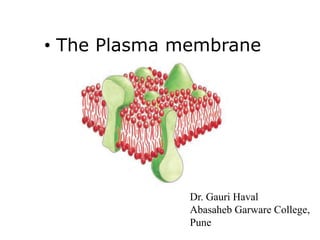
- 1. • The Plasma membrane Dr. Gauri Haval Abasaheb Garware College, Pune
- 2. • Plasma membrane encloses the cell, defines boundaries and maintains the essential differences in the cytosol and external environment. • All biological membranes have a common structure of thin film of lipid and protein molecules. • The cell membranes are fluid, dynamic structures.
- 3. Danielli & Davson model: • This model was prepared in 1935 called as Sandwich model. It says membrane has two lipid layers coated on its either side with hydrated proteins therefore trilamellar structure. Phospholipid molecules have their non polar ends i.e. tails extending into the centre of the membrane while hydrophilic heads are in contact with surface proteins
- 4. • The Danielli and Davson model got support from electron microscope studies. It showed that the plasma membrane contains two dark layers (electron dense granular protein layers) both separated by a light area. The total thickness turned out to be 7.5nm. • The stability of the structure is maintained by mutual attraction between hydrocarbon chains of the lipid molecule and electrostatic forces between the proteins and the head of the lipid molecule. Danielli and Davson have also predicted the lipid bilayer be about 6.0 nm in thickness and each of protein about 1nm thick. The total thickness of the plasma membrane comes to about 8.0nm
- 5. Unit Membrane • Robertson (1960) proposed the unit membrane hypothesis. He used the evidence from electron microscope. This hypothesis states that all cellular membranes have an identical trilamellar structure- dark-light-dark. However, the thickness of the unit membrane has been found to be greater in plasma membrane (10 nm) than in intracellular membranes of E.R. and Golgi apparatus i.e. 5- 7nm.
- 6. Fluid Mosaic model: • S.J. Singer and G.L. Nicholson suggested this model which is widely accepted. • The plasma membrane contains a bimolecular lipid layer. Both the surfaces are interrupted by protein molecules. The proteins are located here and there in a mosaic pattern. Some proteins are attached at the polar surface of the lipid (Extrinsic proteins) while other proteins partially penetrate the bilayer or span the membrane entirely. These are called Trans membrane proteins
- 7. • The extrinsic proteins or ectoproteins frequently contain chains of sugar or oligosaccharides to form glycoprotein. Some oligosaccharides are also attached to lipids to form glycolipids. As there is variety of chemicals required to form a membrane it is called mosaic i.e. mosaic of chemicals • The fluid mosaic model suggests that plasma membrane is not rigid. It has a fluid consistency. Also there are movements of molecules in the lipid and protein. This model represents the fluidity of the membrane and mosaic of chemicals hence it is called fluid mosaic model.
- 8. • The lipid bilayer is formed spontaneously by lipid molecules as they are amphipathic. Lipid molecules are insoluble in water but dissolve readily in organic solvent. The three major types of lipids in plasma membrane are phospholipids (most abundant), cholesterol and glycolipids. All the three are amphipathic. i.e. they have a hydrophilic end and a hydrophobic end. In the structure tails are hydrophobic and heads are hydrophilic. The length of the tail varies. One tail is saturated and other one is unsaturated. Differences in length of the tail and in saturation are important because they influence the ability of Phospholipid molecule to pack against one another and thereby affect the fluidity of the membrane. Thus the fluidity of the plasma membrane depends on its composition.
- 9. • Osmosis (Greek, osmos “to push”) – Movement of water down its concentration gradient • Hydrostatic pressure – Movement of water causes fluid mechanical pressure – Pressure gradient across a semi-permeable membrane Osmotic Properties of Cells
- 10. Erythrocyte Cell Equilibrium •No osmotic pressure - cell is in an isotonic solution - Water does not cross membrane •Increased [Osmotic] in cytoplasm - cell is in an hypotonic solution - Water enters cell, swelling •Decreased [Osmotic] in cytoplasm - cell is in an hypotonic solution - Water leaves cell, shrinking
- 11. Cell Permeability • Passive transport is carrier mediated – Facilitated diffusion – Solute molecule combines with a “carrier” or transporter – Electrochemical gradients determines the direction – Integral membrane proteins form channels
- 12. Crossing the Membrane • Simple or passive diffusion • Passive transport – Channels or pores • Facilitated transport – Assisted by membrane-floating proteins • Active transport pumps and carriers – ATP is required – Enzymes and reactions may be required
- 13. Cellular Transport • Passive transport – no energy is needed to move particles. –Facilitated diffusion – embedded proteins act as tunnels allowing particles to “fall” through.
- 16. Channel Mediated Transport • Proteins form aqueous pores allowing specific solutes to pass across the membrane • Allow much faster transport than carrier proteins
- 17. Coupled Transport • Some solutes “go along for the ride” with a carrier protien or an ionophore Can also be a Channel coupled transport
- 18. Cellular Transport • Active transport – energy is needed to move particles. – Carrier proteins – embedded proteins change shape to open and close passages across the membrane. – Endocytosis – taking something into the cell. – Exocytosis – expelling something from the cell.
- 19. Active Transport • Energy is required
- 20. •Against their electrochemical gradients •For every 3 ATP, 3 Na+ out, 2 K+ in Na+/K+ Pump • Actively transport Na+ out of the cell and K+ into the cell
- 21. • Na+ exchange (symport) is also used in epithelial cells in the gut to drive the absorption of glucose from the lumen, and eventually into the bloodstream (by passive transport) Na+/K+ Pump
- 22. Endocytosis and Exocytosis • Exocytosis - membrane vesicle fuses with cell membrane, releases enclosed material to extracellular space. • Endocytosis - cell membrane invaginates, pinches in, creates vesicle enclosing contents
- 24. Membrane Receptors • Converting chemical signal into electrical one. • These are fast acting receptors, a typical ex. of nervous system. Chemical signals in the form of neurotransmitters are transduced by ion channel ion channel linked receptors directly into an electrical signal in the form of voltage difference across the plasma membrane. Ion Gated Channels
- 25. G-Protein membrane receptors Importance of protein phosphorylation (kinases, conformation change, enzyme activation)
- 26. • This is the largest family of cell surface receptors. The binding of a signal to this receptor results in the switching ‘on’ of a G protein on the internal face of the membrane. Once activated this G protein will initiate a process that will alter cellular behaviour. •
- 30. microvilli finger-like projections of cell cytoplasm lined by cell membrane
- 31. cilia actively motile processes structure: 9 pairs of microtubules + 1 central pair associated proteins: dynein nexin basal body of cilia: 9 triplets of microtubules
- 32. desmosome transmembrane proteins: desmosomal cadherin, desmocolin, desmoglein proteins of plaque: plakoglobin, desmoplakin, plakophilin, plektin cytokeratin intermediate filaments anchored to plaque
- 33. hemidesmosome, a half of desmosom connecting net of intermediary filaments of cell to the basal part of an extracellular matrix ultrastructural dense plaque composed of intracellular protein (desmoplakin, plektin, BP 230), cytokeratin intermediary filaments are anchored to that binding to the extracellular matrix is provided by the set of transmembrane link proteins (α, a β, integriny)
- 34. Plasmodesmata • Found in plants, but are very similar to gap junctions • Extension of the plasma membrane • Desmotubule links ER's of both cells and transports hydrophobic molecules • Neck region contains proteins that regulate permeability