
Empfohlen
Weitere ähnliche Inhalte
Was ist angesagt?
Was ist angesagt? (20)
Ähnlich wie Pleural effusion
Ähnlich wie Pleural effusion (20)
Mehr von GAMANDEEP
Mehr von GAMANDEEP (20)
Kürzlich hochgeladen
Kürzlich hochgeladen (20)
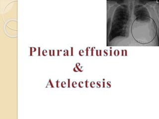
Pleural effusion
- 2. Pleural effusion Pleural effusion, a collection of fluid in the pleural space, is rarely a primary disease process but is usually secondary to other diseases.
- 3. Causes Pleural effusion may be a complication of :- Heart failure, Tuberculosis, Pneumonia, Pulmonary infections, Nephritic syndrome, Connective tissue disease, Pulmonary embolism, and Neoplasm (tumor).
- 4. Pathophysiology The effusion can be composed of a relatively clear fluid, or it can be bloody or purulent. An effusion of clear fluid may be a transudate or an exudate. (Transudate is fluid pushed through the capillary due to high pressure within the capillary. Exudate is fluid that leaks around the cells of the capillaries caused by inflammation.) A transudate (filtrates of plasma that move across intact capillary walls) occurs when factors influencing the formation and re- absorption of pleural fluid are altered, usually by imbalances in hydrostatic or oncotic pressures.
- 5. Continued.. The finding of a transudative effusion generally implies that the pleural membranes are not diseased. The most common cause of a transudative effusion is heart failure. An exudates (extravasation of fluid into tissues or a cavity) usually results from inflammation by bacterial products or tumors involving the pleural surfaces.
- 6. Clinical Manifestations Pneumonia causes fever, chills, Pleuritic chest pain, Malignant effusion may result in dyspnea and coughing. A large pleural effusion causes shortness of breath.
- 7. Assessment and Diagnostic Findings Assessment of the area of the pleural effusion reveals decreased or absent breath sounds, decreased fremitus, and a dull, flat sound when percussed. In an extremely large pleural effusion, the assessment reveals a patient in acute respiratory distress.
- 8. Continued.. Tracheal deviation away from the affected side may also be noted. Physical examination, chest x-ray, chest CT scan, and thoracentesis confirm the presence of fluid. In some instances, a lateral decubitus x-ray is obtained. A pleural effusion can be diagnosed because this position allows for the “layering out” of the fluid, and an air–fluid line is visible
- 9. Continued.. Pleural fluid analysis by bacterial culture, Gram stain, acid fast bacillus stain (for TB), red and white blood cell counts, chemistry studies (glucose, amylase, lactic dehydrogenase, protein), cytologic analysis for malignant cells, and pH.
- 10. Medical management The objectives of treatment : discover the underlying cause to prevent re accumulation of fluid, and to relieve discomfort, dyspnea, and respiratory compromise. Specific treatment is directed at the underlying cause (eg, heart failure, pneumonia, lung cancer, cirrhosis).
- 11. Thoracentesis is performed to remove fluid, to obtain a specimen for analysis, and to relieve dyspnea and respiratory compromise.
- 12. CONTINUED.. Repeated thoracenteses result in pain, depletion of protein and electrolytes, and sometimes pneumothorax. Once the pleural space is adequately drained, a chemical pleurodesis may be performed to obliterate the pleural space and prevent reaccumulation of fluid
- 13. Other treatments Surgical pleurectomy Insertion of a small catheter attached to a drainage bottle for outpatient management Implantation of a Pleura peritoneal shunt
- 14. PP shunt A pleura peritoneal shunt consists of two catheters connected by a pump chamber containing two one- way valves. Fluid moves from the pleural space to the pump chamber and then to the peritoneal cavity. The patient manually pumps on the reservoir daily to move fluid from the pleural space to the peritoneal space.
- 15. Atelectesis It refers to closure or collapse of alveoli and often is described in relation to x-ray findings and clinical signs and symptoms. May be acute or chronic and may cover a broad range of pathophysiologic changes, Microatelectasis /Macroatelectasis
- 16. Continued.. Excess secretions or mucus plugs may also cause obstruction of airflow and result in atelectasis in an area of the lung. Atelectasis also is observed in patients with a chronic airway obstruction that impedes or blocks air flow to an area of the lung Atelectasis resulting from bronchial obstruction by secretions may occur in patients with impaired cough mechanisms (eg, postoperative, musculoskeletal or neurologic disorders) or in debilitated, bedridden patients.
- 17. Atelectasis may also result from excessive pressure on the lung tissue, which restricts normal lung expansion on inspiration. Such pressure may be produced by fluid accumulating within the pleural space (pleural effusion), air in the pleural space (pneumothorax), or blood in the pleural space (hemothorax).
- 18. Pathophysiology Following obstruction of bronchus the blood circulating absorbs gas from alveoli Lead to retraction of lungs Blood perfuses unventillated lung Results in arterial hypoximea Uninvolved surrounding lung distends displacing surrounding structures Mediastinal shift
- 19. Clinical manifestations Fever is universally cited as a clinical sign of atelectasis, marked respiratory distress in In acute atelectasis involving a large amount of lung tissue (lobar atelectasis), Dyspnea, tachycardia, tachypnea, pleural pain, and central cyanosis
- 20. Assessment and Diagnostic Findings Decreased breath sounds and crackles are heard over the affected area. In addition, chest x-ray findings may reveal patchy infiltrates or consolidated areas. Pulse oximetry (SpO2) may demonstrate a low saturation of hemoglobin with oxygen (less than 90%) or a lower-than-normal partial pressure of arterial oxygen (PaO2).
- 21. CONTINUED.. Cough, Sputum Production, And Low-grade Fever. Dyspnea, Tachycardia,tachypnea Pleural Pain Central Cyanosis
- 22. Management First line measures: frequent turning, early ambulation, lung volume expansion maneuvers (eg, deep- breathing exercises, incentive spirometry), and coughing
- 23. Other treatments such as positive expiratory pressure or PEP therapy (a simple mask and one way valve system that provides varying amounts of expiratory resistance [usually 5 to 15 cm H2O]) Continuous or intermittent positive pressure- breathing (IPPB)
- 24. Bronchoscopy
- 25. Continued.. Chest physical therapy to mobilize secretions. Nebulizer treatments with a bronchodilator medication or sodium bicarbonate. Endotracheal intubation and mechanical ventilation may be necessary. With a large pleural effusion that is compressing lung tissue and causing alveolar collapse, treatment may include thoracentesis, removal of the fluid by needle aspiration, or insertion of a chest tube.
- 26. Continued.. Management of chronic atelectasis focuses on removing the cause of the obstruction of the airways or the compression of the lung tissue. Bronchoscopy may be used to open an airway obstructed by lung cancer or a nonmalignant lesion, and the procedure may involve cryotherapy or laser therapy.
- 27. Nursing management Nursing Assessment Obtain history of previous pulmonary condition. Assess patient for dyspnea and tachypnea. Auscultate and percuss lungs for abnormalities Presence of respiratory secretions
- 28. Nursing diagnosis 1. Ineffective Breathing Pattern related to collection of fluid in pleural space 2. Ineffective tissue perfusion related to decreased lung compliance secondary to disease condition as evidenced by decreased SPO2. 3. Altered body temperature related to infection secondary to disease condition as evidenced by patient’s body temperature (38.6 degree Celsius). 4. Self care deficit related to critical condition of health and mechanical ventilator secondary to sedation. 5. Anxiety related to unknown outcome.
- 29. Continued.. 6. Knowledge deficient regarding condition, treatment and self-care. 7. Risk for impaired skin integrity related to prolonged immobility secondary to disease condition. 8. Risk for infection related to invasive procedures as well as invasive and non invasive lines. 9. Anticipatory grieving related to perceived impending death of the patient.
- 30. Maintaining Normal Breathing Pattern • Observe patient's breathing pattern, oxygen saturation, and other vital signs, for evidence of improvement or deterioration • Administered oxygen as indicated by dyspnea and hypoxemia. • Mechanical ventilator parameters were monitored • Patient was positioned in propped up position.
- 31. Maintain effective and patent airway Interventions The patient’s respiratory status was assessed Auscultation of breath sounds done. Signs of cyanosis, dyspnea and hypoxia observed. Patient’s ABG levels monitored regularly. Patient kept in a propped up or Fowler’s position.
- 32. Continued.. Humidification provided. Chest physiotherapy was provided to the patient Suctioning was done under aseptic techniques.
- 33. Maintaining effective tissue perfusion The patient’s respiratory status was assessed. Auscultation of breath sounds was done. Signs of cyanosis, dyspnea and hypoxia observed. Patient’s ABG levels were monitored regularly. Vital signs monitored regularly. Patient kept in a propped up position. Ventilator circuits checked for any leakage. Adequate hyper-oxygenation done prior to and after suctioning.
- 34. Reduce fever Vital signs monitored regularly. Blood investigations (TLC, Blood CS and ET aspirate) were sent as prescribed. Sponging done. Cool environment was maintained Administered antipyretic and antibiotics as prescribed.
- 35. Continued.. I.V. tubing/lines were changed according to hospital protocol. Hydration of the patient was maintained via feed Temperature reassessed every 2 hourly.
- 36. To reduce anxiety Anxiety level along with previous coping mechanism of the family was assessed Established trusting relationship with family Family encouraged to ask questions & clarify doubts Family provided explanation in simple words Importance and rationale for ventilation and ET insertion was explained. Family members involved in plan of care. Diversional activities encouraged.
- 37. To maintain skin integrity Assessment of the patient’s skin was done carefully for signs of infection, inflammation, and breakdown. Patient’s position changed every 2 hours. Skin care and back care provided. Soiled linen and clothes were changed accordingly Nutritional status of the patient was maintained
- 38. References Woods S.L.et al. Cardiac nursing . Lippincott Williams & Wilkins. 2000 Lippincott Manual of Nursing Practice. 8th edition. Lippincott Williams & Wilkins Publishers; 2010. p. 68, 328-332, 361. Smeltznner SC, Bare BG, Hinkle JL, Cheever KH. Brunner & Suddarth’s Textbook of Medical-Surgical Nursing. 11th edition. Lippincott Williams & Wilkins Publishers; 2008. p. 789-805. Longo et al.Harrisons principles of internal medicine.18(2):2012 Chintamoni Lewis LS et al.Lewis’s medical surgical nursing:assessment and management of clinical problems.7. New delhi.ELSEVIER;2011
Hinweis der Redaktion
- For this x-ray, the patient lies on the affected side in a side-lying position.