Weitere ähnliche Inhalte
Ähnlich wie Whitesides 2006 (20)
Kürzlich hochgeladen (20)
Whitesides 2006
- 1. What is microfluidics? It is the science and technology of systems that
process or manipulate small (10–9
to 10–18
litres) amounts of fluids,
using channels with dimensions of tens to hundreds of micrometres.
The first applications of microfluidic technologies have been in analy-
sis, for which they offer a number of useful capabilities: the ability to
use very small quantities of samples and reagents, and to carry out
separations and detections with high resolution and sensitivity; low
cost; short times for analysis; and small footprints for the analytical
devices1
. Microfluidics exploits both its most obvious characteristic
— small size — and less obvious characteristics of fluids in microchan-
nels, such as laminar flow. It offers fundamentally new capabilities in
the control of concentrations of molecules in space and time.
As a technology, microfluidics seems almost too good to be true: it
offerssomanyadvantagesandsofewdisadvantages(atleastinitsmajor
applicationsinanalysis).Butithasnotyetbecomewidelyused.Whynot?
Why is every biochemistry laboratory not littered with ‘labs on chips’?
Why does every patient not monitor his or her condition using micro-
fluidichome-testsystems?Theanswersarenotyetclear.Iamconvinced
that microfluidictechnologywill becomeamajorthemeintheanalysis,
andperhapssynthesis,ofmolecules:theadvantagesitoffersaretoocom-
pellingtoletpass.Havingsaidthat,theanswerstoquestionsconcerning
the time and circumstances required for microfluidics to develop into a
major new technology are important not just for this field, but also for
other new technologies struggling to make it into the big time.
The origins and the future of microfluidics
George M. Whitesides1
The manipulation of fluids in channels with dimensions of tens of micrometres — microfluidics — has emerged
as a distinct new field. Microfluidics has the potential to influence subject areas from chemical synthesis and
biological analysis to optics and information technology. But the field is still at an early stage of development.
Even as the basic science and technological demonstrations develop, other problems must be addressed:
choosing and focusing on initial applications, and developing strategies to complete the cycle of development,
including commercialization. The solutions to these problems will require imagination and ingenuity.
1
Department of Chemistry and Chemical Biology, Harvard University, Cambridge, Massachusetts 02138, USA.
The field of microfluidics has four parents: molecular analysis,
biodefence,molecularbiologyandmicroelectronics.Firstcameanalysis.
The distant origins of microfluidics lie in microanalytical methods —
gas-phasechromatography(GPC),high-pressureliquidchromatography
(HPLC)andcapillaryelectrophoresis(CE)—which,incapillaryformat,
revolutionized chemical analysis. These methods (combined with the
powerofthelaserinopticaldetection)madeitpossibletosimultaneously
achievehighsensitivityandhighresolutionusingverysmallamountsof
sample. With the successes of these microanalytical methods, it seemed
obvious to develop new, more compact and more versatile formats
for them, and to look for other applications of microscale methods in
chemistry and biochemistry.
A second, different, motivation for the development of microfluidic
systems came with the realization — after the end of the cold war —
that chemical and biological weapons posed major military and
terrorist threats. To counter these threats, the Defense Advanced
Research Projects Agency (DARPA) of the US Department of Defense
supported a series of programmes in the 1990s aimed at developing
field-deployable microfluidic systems designed to serve as detec-
tors for chemical and biological threats. These programmes were
the main stimulus for the rapid growth of academic microfluidic
technology.
Thethirdmotivationalforcecamefromthefieldofmolecularbiology.
The explosion of genomics in the 1980s, followed by the advent of
other areas of microanalysis related to molecular biology, such as
high-throughput DNA sequencing, required analytical methods with
much greater throughput, and higher sensitivity and resolution than
had previously been contemplated in biology. Microfluidics offered
approaches to overcome these problems.
The fourth contribution was from microelectronics. The origi-
nal hope of microfluidics was that photolithography and associated
technologies that had been so successful in silicon microelectronics,
and in microelectromechanical systems (MEMS), would be directly
applicable to microfluidics. Some of the earliest work in fluidic micro-
systems did, in fact, use silicon and glass, but these materials have
largely been displaced by plastics. For analyses of biological samples
in water, devices fabricated in glass and silicon are usually unneces-
sary or inappropriate. Silicon, in particular, is expensive, and opaque
to visible and ultraviolet light, so cannot be used with conventional
optical methods of detection. It is easier to fabricate the components
required for microanalytical systems — especially pumps and valves
— in elastomers than in rigid materials. Neither glass nor silicon has
all the properties (especially permeability to gases) required for work
with living mammalian cells.
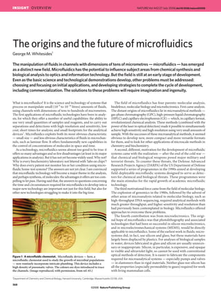
Figure 1 | A microfluidic chemostat. Microfluidic devices — here, a
microfluidic chemostat used to study the growth of microbial populations
— now routinely incorporate intricate plumbing. This device includes a
high density of pneumatic valves. The colours are dyes introduced to trace
the channels. (Image reproduced, with permission, from ref. 65.)
368
INSIGHT OVERVIEW NATURE|Vol 442|27 July 2006|doi:10.1038/nature05058
NaturePublishing Group©2006
- 2. Thus, microfluidic devices have not developed as clones of silicon
microelectronicdevices.Muchoftheexploratoryresearchinmicrofluidic
systems has been carried out in a polymer — poly(dimethylsiloxane),
or PDMS — the properties of which are entirely distinct from those of
silicon2,3
. PDMS is an optically transparent, soft elastomer. Whether
the microfluidic devices that ultimately become widely used will use
PDMS or one of the engineering polymers (such as polycarbonate or
polyolefin) remains to be seen. The ease with which new concepts can
be testedinPDMS,however, anditsabilityto supportcertainvery useful
components (such as pneumatic valves), have made it the key material
for exploratory research and research engineering at the early stages
of development. Microelectronic technologies have, however, been
indispensable for the development of microfluidics, and as the field has
developed, glass, steel and silicon have again emerged as materials with
which to build specialized systems that require chemical and thermal
stability. The mechanical stability of silicon and glass are also useful in
the nascent field of nanofluidics (the study of fluids in channels with
nanometre-scale — ideally less than 50 nm — dimensions), in which
channels with rigid walls can be useful4,5
.
Microfluidics has seen the rapid development of new methods of
fabrication, and of the components — the microchannels that serve
as pipes, and other structures that form valves6,7
, mixers8–10
and
pumps11
— that are essential elements of microchemical ‘factories’ on
a chip. However, its impact on science has not yet been revolutionary.
Revolutions in technology require both a broad range of different types
of component and subsystem, and their integration into complete, func-
tional systems. The field of microfluidics is in early adolescence, and
still lacks both these essential requirements, in addition to the integra-
tion of components into systems that can be used by non-experts. As a
field, it is a combination of unlimited promise, pimples and incomplete
commitment. This is a very exciting time for the field, but we still do
not know exactly what it will be when it grows up.
The present
A microfluidic system must have a series of generic components:
a method of introducing reagents and samples (probably as fluids,
although ideally with the option to use powders); methods for moving
these fluids around on the chip, and for combining and mixing them;
and various other devices (such as detectors for most microanalytical
work, and components for purification of products for systems used in
synthesis). The field has, so far, centred on demonstrating concepts for
these components. Two particularly important contributions have been
the development of soft lithography in PDMS as a method for fabricating
prototype devices12
; and the development of a simple method of fabri-
cating pneumatically activated valves, mixers and pumps on the basis
of soft-lithographic procedures13
. These methods have made it possible
to fabricate prototype devices that test new ideas in a time period much
shorter (typically less than 2 days from design to working device) than
that which could be achieved using silicon technology (typically, for non-
specialists, a month or more). Quake’s pneumatic valves are particularly
important as components that have enabled the design and examination
of complicated devices, and these have opened up a number of areas of
application (Fig. 1). ‘Quake valves’ use the restriction of a fluidic channel
by an adjacent channel under pressure; their operation depends on the
fact that PDMS is an elastomer, and no corresponding devices exist (or
can exist) in rigid materials such as silicon and glass (or rigid engineering
polymers such as polycarbonate).
Togetherwithnewmethodsoffabrication,microfluidicshasbeenable
to exploit certain fundamental differences between the physical prop-
erties of fluids moving in large channels and those travelling though
micrometre-scale channels14–16
. Janasek et al. describe scaling relations
that relate (or differentiate) macroscopic and microfluidic systems, with
special emphasis on lab-on-a-chip devices (see page 374). Of these dif-
ferences,themostimportantisturbulence(oritsabsence:laminarflow).
On large scales, fluids mix convectively: for example, the mixing of milk
when it is swirled into coffee, or smoke, leaving a chimney, with air. This
type of mixing reflects the fact that in macroscopic fluids, inertia is often
moreimportantthanviscosity.Inmicrosystems,withwaterasafluid,the
oppositeistrue:fluidsdonotmixconvectively—whentwofluidstreams
cometogetherinamicrochannel,theyflowinparallel,withouteddiesor
turbulence, and the only mixing that occurs is the result of diffusion of
molecules across the interface between the fluids. Although this type of
flow — known as laminar flow — requires the development of specific
devicesorcomponentstoaccomplishmixing(whenmixingisrequired),
it has proved an advantage (and one that is characteristic of microfluidic
systems)inmanycircumstances.Theratioofinertialtoviscousforceson
fluids is characterized by the Reynolds number (Re) — one of the many
dimensionless parameters used in studying fluids15
.
Fluidsflowinginmicrosystemshavemanyotherinterestinganduseful
characteristics,onlysomeofwhichhavebeenexploited.Oneparticularly
usefulcharacteristiciselectro-osmoticflow(EOF)17
.Whenanion-con-
taining fluid (for example, water) is placed in a microchannel that has
fixed charges on its surface (such as silicon dioxide or surface-oxidized
PDMS)andanelectricalpotentialisappliedalongthechannel,thefluid
moves as a plug, rather than with the parabolic-flow profile observed
when pumping is accomplished by applying pressure to the fluid. EOF
minimizesthebroadeningofplugsofsamplethatoccurswithmanypres-
sure-drivensystems,andallowsveryhighresolutionseparationsofionic
species. It is a key contributor to electrophoretic separations of DNA in
microchannels18
. A second potentially useful characteristic is the ability
of nanofluidics to manipulate water in channels whose dimensions are
similar to those of the Debye layer19
: we do not, in fact, understand the
characteristics of fluids at those scales, and nanofluidic systems offer
windows into new phenomena in fluid physics.
a
5 mm
500 µm
200 µm
b
c
Figure 2 | Efficient screening for optimal protein crystallization conditions.
Microfluidic devices are well suited for screening conditions under
which proteins crystallize, as demonstrated initially by Quake20
.
a, A device in which droplets containing proteins are trapped in wells in
the microchannels21
. The droplets are then subjected to many different
conditions for crystallization. b, An optical micrograph of dyed droplets
in the wells of the device. (Images courtesy of S. Fraden and J.-u. Shim,
Brandeis University, USA.) c, Droplets containing crystallized proteins.
The droplets are produced in a microfluidic device, then collected in a
glass capillary. (Image adapted, with permission, from ref. 22.)
369
NATURE|Vol 442|27 July 2006 INSIGHT OVERVIEW
NaturePublishing Group©2006
- 3. Current applications
There are now enough methods of fabrication, and a sufficient range of
components, to make it possible to begin to apply microfluidic systems
to the resolution of problems (rather than simply to the demonstration
of principles). The most highly developed of their applications is prob-
ably their use to screen conditions (such as pH, ionic strength and com-
position, cosolvents, and concentration) for protein crystallization20–22
(Fig. 2); these procedures offer the potential to screen large numbers
of conditions, to separate nucleation and growth of crystals, and to
minimize the damage to crystals by handling once they have formed.
Some of this technology is now commercially available. Other applica-
tions for which there are laboratory demonstrations include separations
coupled to mass spectroscopy23
, high-throughput screening in drug
development24,25
, bioanalyses26
, examination and manipulation of sam-
ples consisting of a single cell27,28
or a single molecule29,30
, and synthesis
of 18
F-labelled organic compounds for positron emission tomography
(PET)31
. The area of single-molecule studies is discussed in this issue
by Craighead (see page 387).
Themanipulationofmultiphaseflowsisanotherstrengthofmicrofluidic
systems.Theyenablethegenerationandmanipulationofmonodisperse
bubbles32,33
(Fig. 3) or droplets34–37
of a dispersed gas or liquid phase in
a continuous liquid stream; these dispersions suggest new routes to the
production of polymer particles, emulsions and foams38
. Droplets can
also serve as compartments in which to study fast organic reactions.
Fluids in microchannels form the basis of new optical systems: a range
of systems — from waveguides comprising a liquid with a high index of
refractionflowinglaminarlybetweentwostreamsoflow-index‘cladding’,
to applications of fluids in lenses and Bragg mirrors — are based on
microfluidics39–44
.Psaltis,YangandQuakepaintadetailedpictureofthis
new field, and of some of its potentials, in this issue (see page 381).
Cellbiologyisanareaofresearchintowhichmicrofluidicsystemsbring
anewcapability.Jensenetal.(seepage403)describetypesofsystemthat
seemcertaintobecomeusefulnewtoolsforcellbiologists,aswellascapa-
bilities that are still needed. Eukaryotic cells, when attached and spread,
have linear dimensions of 10–100 μm; these dimensions are well suited
for current microfluidic devices, and PDMS — with its excellent optical
transparency,lowtoxicityandhighpermeabilitytodioxygenandcarbon
dioxide—isamaterialthatisprobablyuniquelysuitableasamediumfor
thefabricationofmicrochambersinwhichtogrowandobservecells45–48
(Fig. 4). PDMS microfluidic systems have applications in the extensive
studyofmanyareasofcellbiology,includingthecytoskeleton49
,theforces
exerted by cells on the substrate to which they are attached50
, the con-
tents of cells (down to the single-cell level)27,51
, separations of motile and
non-motile cells (for example, sperm)52
, and embryos53–55
.
Chemicalsynthesis(especiallyinorganicandmedicinalchemistry)—an
area in which microfluidic systems would seem to fit naturally — has
been slow to adopt microfluidic structures as a strategy for the develop-
mentofnewcapabilities.(Someofthecharacteristicsofchemicalreactions
in microsystems are discussed in this issue by deMello, page 394.) Two
factors contribute to this slow adoption. First, the flexibility of conven-
tional apparatus has, so far, not been equalled in microfluidic systems.
Second,PDMS—thematerialmostcommonlyusedinacademicstudies
of microfluidics — dissolves in, or is swelled by, many common organic
solvents56
.Theuseofsilicon,glassorsteel57–59
,orperhapspolymersother
than PDMS60
, may both solve this problem and allow reactions at high
temperatures and pressures, but the fabrication of devices with any of
thesematerialsismoredifficultthanwithPDMS.Pumpingandvalvingin
rigidmaterialssuchassteelmustbeaccomplishedusingentirelydifferent
strategies from those used in PDMS.
Thedevelopmentofpracticalmicroanalyticalsystems61–64
—especially
those for bioanalysis — continues rapidly, although, given its early focus,
this area has been slower than expected to reach widespread routine
use. Part of the problem is that there is limited technology in two parts
of the cycle of analysis: sample preparation and detection. Biological
samples — particularly clinical samples (such as blood or faeces), or
those obtained by environmental sampling (such as soil) — are often
dilute or complicated. Before these samples can be analysed by micro-
fluidic devices, they must be converted to a form that is compatible with
the intended analysis, and then introduced into the analytical device.
The procedures required to complete these tasks are surprisingly sam-
ple-dependent, and not necessarily ‘micro’ in scale. After a sample has
been prepared, introduced into the analytical device and processed, it
must then be detected. This detection is still commonly accomplished
by a microscope located off-chip. Having the microfluidic chip as just a
small part of a system in which sample introduction and detection are
much more complicated than the chip’s operation may be appropriate
in some circumstances, but does detract from the potential advantages
of microfluidic devices. Other standard problems, such as pumping,
valving and on-chip reagent storage, also require better solutions than
those available so far.
The future
What requirements must be fulfilled for microfluidics to become a
major new technology? Will it live up to the hopes experienced at its
conception? As a field, the problems it faces are those faced by most
fields as they develop. The fact that microfluidics has not yet lived up
to its early advertising is not a surprise, and the reasons for the rate at
which it has developed are both characteristic of new technologies, and
suggestive of areas in which to focus work in the future.
Liquid
Liquid
Gas
a
b
c
500 µm
100 µm
Liquid
Liquid
Gas
Figure 3 | Creating and using bubbles in microfluidic devices. In a
microfluidic ‘flow-focusing’ device, streams of liquid pinch off a gaseous
thread to produce bubbles that are remarkably monodisperse33
. The flow
rate of the liquid and the pressure applied to the gas control the size of the
bubbles, and the frequency with which they form. a, A schematic diagram
of a flow-focusing system. b, An optical micrograph of the production of
a foam comprising monodisperse bubbles. c, An optical micrograph of
bubbles enhancing the mixing of an aqueous solution of ink (black) and an
aqueous stream containing a surfactant (white).
370
NATURE|Vol 442|27 July 2006INSIGHT OVERVIEW
NaturePublishing Group©2006
- 4. Generalissuesinintroducingnewtechnologies
The original hope for microfluidics, and that which still motivates many
of us working in the field, is that it will be a practical technology — one
widely used in a number of different types of application. I am confident
that, ultimately, it will be, but there are several problems that it — in
common with other new technologies — must first solve. Above all,
it must become successful commercially, rather than remain a field
based on proof-of-concept demonstrations and academic papers. The
impact of microfluidic systems — as with other tools such as lasers,
NMR (nuclear magnetic resonance) spectroscopy and scanning probe
microscopes — will only become apparent when everyone is using
them. Microfluidics must be able to solve problems for users who are
not experts in fluid physics or nanolithography, such as clinicians, cell
biologists, police officers or public health officials. For these applica-
tions, corporations must take on the task of making appropriate systems
widely and inexpensively available.
As with all technology in transition from university laboratories to
industry,thequestionof‘Whoownswhat?’—theproblemofintellectual
property — is one that must be resolved. For technology with very high
value, such as biopharmaceuticals and information-processing systems,
issues of intellectual property can usually be resolved by compromising
on royalties, up-front payments or equity. However, some of the most
interesting applications of microfluidics are those that would demand
large volumes but low prices — for example, in public heath monitor-
ing, environmental monitoring, and for use in the medical systems of
developing economies. In these areas, the historical differences in the
valuations placed by universities and industry on university-based tech-
nology can become a serious issue: if the university places the value of an
invention too high, it is simply not worthwhile to develop a commercial
technology from it.
Thereisalsotheissueoftheso-called‘first-userpremium’.Intheintro-
ductionofanewtechnology,thefirstcommercialuserofthattechnology
paysadisproportionateshareofthecostsofitsdevelopment,andaccepts
a disproportionate share of the risk for that development. If the applica-
tionofsuchadevelopmentisaveryappealing—ifitisofpotentiallyhigh
value (the ‘killer application’, or ‘killer app’) — these costs and risks are
more acceptable. The high-value killer app for microfluidics has not yet
emerged,althoughmarketsinresearchbiologyarecertainlydeveloping.
High-valueapplications
There are, in principle, high-value applications for microfluidic systems,
although developing these applications requires innovations in both
microfluidics and in biomedicine; doing two things at once is always
difficult. The development of new types of bioassay for monitoring
patient response to therapy is one such application; development of
assays for home testing, or for use in doctors’ offices at early stages of
disease (early detection of ‘biomarkers’), is a second. Both are plau-
sible developments in biomedicine, but will require both an under-
standing of biomarkers of disease and microfluidic systems that are
highly developed. In the future, it is certainly possible that healthcare
will move from treating to anticipating disease. Widespread, sensitive,
frequent screening or testing will be a necessity for such anticipatory
healthcare, and microfluidic systems are the most plausible technology
for such testing.
Toolsforthepharmaceuticalindustry
The pharmaceutical industry is technically sophisticated, and capable,
in principle, of adopting sophisticated new technologies. The industry
is also suffering from a crisis in productivity, and desperately needs
new tools to guide the development of new drugs — especially to help
predict the behaviour of potential new drugs in humans from perform-
ance in animals and cells. Some analytical applications of microfluidic
systems in the production and use of biopharmaceuticals seem straight-
forward (for example, analytical systems to monitor and optimize the
production of protein drugs such as therapeutic antibodies); others
(such as assays based on primary human cells that could predict per-
formance in human clinical trials) are technically more complicated,
but also feasible, at least in some instances. In either case, the assays
must package microsystems (almost certainly microfluidic systems) in
a highly reproducible and easily manipulated format that could be used
routinely by technicians.
Research
The introduction and development of new technologies is often facili-
tated by large, relatively cost-insensitive uses in research: equipment
for processing and analysing DNA and RNA are recent examples. The
development of new microfluidic tools for genomics, proteomics and
metabolomics is proceeding rapidly in research laboratories, and will
provide a stimulus for large-scale production.
Large-volumemicroanalyticaltools
Among the most interesting and important of the potential applications
of microfluidic systems are those toward which it was originally tar-
geted: biomedical and related applications requiring small amounts of
sample, routine operation by untrained personnel, and low cost (Fig. 5).
a
b c
Figure 4 | A new platform for cellular and developmental biology. Laminar
flow in microchannels, together with the biocompatibility of PDMS,
enable new methods of studying cellular and developmental biology. One
system examines the effect of temperature on the development of a fruitfly
embryo55
. The embryo (the large oval in a) is immobilized in the middle
of a microchannel. Aqueous streams of two different temperatures flow
over the halves of the embryo (b shows the cold half, c the warm half); the
differences in the embryo are reflected in the density of cells (marked by
the light-blue nuclei). The embryo is ~500 μm wide.
Figure 5 | A simple, inexpensive microfluidic diagnostic device.
Components of microfluidic devices can be designed to be inexpensive and
easy to operate. This device7
performs sandwich immunoassays — tests
that are used widely in medicine and biological research. The screws in this
system (marked with dashed circles) act as simple, manually operated valves.
Green-dyed water marks the channels. Low-cost, portable, easy-to-operate
microfluidic devices such as this one may find applications in resource-poor
environments. (Image adapted, with permission, from ref. 7.)
371
NATURE|Vol 442|27 July 2006 INSIGHT OVERVIEW
NaturePublishing Group©2006
- 5. There is an appealing commonality in a number of these applications,
and the volumes of appropriate analyses could, in principle, be very
large (hundreds of millions of tests per year). This group of applica-
tions would include healthcare delivery and monitoring in developing
economies, home healthcare and use in doctors’ offices in developed
economies, uses in homeland security and counterterrorism, use by first
responders (police, paramedics and fire departments), applications in
veterinary medicine, and incorporation into environmental and food-
safety monitoring. Diagnostic systems for developing economies will
require low-cost, adaptable microfluidic technology for its success; this
rapidly developing field is described in this issue by Yager et al. (see
page 412).
These applications suffer from the problem of chicken and egg: the
volume of use will only be large if the cost of the analysis is low and the
state of development of the assay is high; and the cost will only be low
if the volume is large.
Newscienceandtechnology
The development of microfluidics has just begun. A number of factors
suggest that there are many early-stage applications of microsystems
containing fluids, including the exploration of fluidic optics and cells,
the development of new types of organic synthesis in small-channel
systems, the continuing development of technologies based on large
arrays of detectors and on high-throughput screening, the fabrication of
microrobotic systems using hydraulic systems based on microfluidics,
other fluidic versions of MEMS, and work on biomimetic systems with
microfluidic components. The extension of microfluidic systems into
nanofluidics — in which the dimensions of the channels and the thick-
ness of the layer of structured fluid at the walls of the device become
comparable — will make possible the exploration of the properties of
near-surface water, and of ion and electrolyte transport at this inter-
face. The biocompatibility of PDMS suggests that it might ultimately
be possible to embed microfluidic devices in vivo for certain types of
biomedically relevant analysis. Single-cell and single-molecule analysis
require technologies that can work with small volumes of sample, which
might allow the testing of fundamental assumptions of cell biology and
molecular chemistry and biology.
Designandmanufacturingsystemsformicrofluidicdevices
An important aspect of the commercial development of microfluidics
— crucial to many of these applications — is the development of the
technology for manufacturing microfluidic devices. Ultimately, there will
probablybeseveralsuchtechnologies,butintheearlystagesthedefinition
of a single set of materials and processes needed to convert laboratory
demonstrations into working commercial devices is an important step.
Should devices be developed in Mylar, or PDMS, or polycarbonate?
What will be the specifications for user interfaces? How important will
very-large-volume technologies, such as roll-to-roll processing, be? And
what about technologies for sealing and packaging?
Conclusion
So, what next for microfluidics? It is both a science and a technology.
It offers great — perhaps even revolutionary — new capabilities for
the future. It is also in its infancy, and a great deal of work needs to be
done before it can be claimed to be more than an active field of aca-
demic research. However, the fundamentals of the field are very strong:
much of the world’s technology requires the manipulation of fluids, and
extending those manipulations to small volumes, with precise dynamic
control over concentrations, while discovering and exploiting new phe-
nomena occurring in fluids at the microscale level, must, ultimately, be
very important. ■
1. Manz, A. etal. Planar chips technology for miniaturization and integration of separation
techniques into monitoring systems — capillary electrophoresis on a chip. J.Chromatog.
593, 253–258 (1992).
2. Ng, J. M. K., Gitlin, I., Stroock, A. D. & Whitesides, G. M. Components for integrated
poly(dimethylsiloxane) microfluidic systems. Electrophoresis 23, 3461–3473 (2002).
3. Whitesides, G. M. & Stroock, A. D. Flexible methods for microfluidics.Phys.Today 54,
42–48 (2001).
4. Mijatovic, D., Eijkel, J. C. T. & van den Berg, A. Technologies for nanofluidic systems: top-
down vs. bottom-up — a review. LabChip 5, 492–500 (2005).
5. Czaplewski, D. A., Kameoka, J., Mathers, R., Coates, G. W. & Craighead, H. G. Nanofluidic
channels with elliptical cross sections formed using a nonlithographic process.Appl.Phys.
Lett. 83, 4836–4838 (2003).
6. Hong, J. W. & Quake, S. R. Integrated nanoliter systems.NatureBiotechnol. 21, 1179–1183
(2003).
7. Weibel, D. B. etal. Torque-actuated valves for microfluidics. Anal.Chem. 77, 4726–4733
(2005).
8. Nguyen, N. T. & Wu, Z. G. Micromixers — a review. J.Micromech.Microeng. 15, R1–R16
(2005).
9. Gunther, A., Jhunjhunwala, M., Thalmann, M., Schmidt, M. A. & Jensen, K. F. Micromixing
of miscible liquids in segmented gas-liquid flow. Langmuir 21, 1547–1555 (2005).
10. Garstecki, P., Fischbach, M. A. & Whitesides, G. M. Design for mixing using bubbles in
branched microfluidic channels. Appl.Phys.Lett. 86, 244108 (2005).
11. Laser, D. J. & Santiago, J. G. A review of micropumps. J.Micromech.Microeng. 14, R35–R64
(2004).
12. McDonald, J. C. etal. Fabrication of microfluidic systems in poly(dimethylsiloxane).
Electrophoresis 21, 27–40 (2000).
13. Thorsen, T., Maerkl, S. J. & Quake, S. R. Microfluidic large-scale integration. Science 298,
580–584 (2002).
14. Stone, H. A., Stroock, A. D. & Ajdari, A. Engineering flows in small devices: microfluidics
toward a lab-on-a-chip. Annu.Rev.FluidMech. 36, 381–411 (2004).
15. Squires, T. M. & Quake, S. R. Microfluidics: fluid physics at the nanoliter scale.Rev.Mod.
Phys. 77, 977–1026 (2005).
16. Beebe, D. J., Mensing, G. A. & Walker, G. M. Physics and applications of microfluidics in
biology. Annu.Rev.Biomed.Eng. 4, 261–286 (2002).
17. Santiago, J. G. Electroosmotic flows in microchannels with finite inertial and pressure
forces. Anal.Chem. 73, 2353–2365 (2001).
18. Wainright, A., Nguyen, U. T., Bjornson, T. & Boone, T. D. Preconcentration and separation
of double-stranded DNA fragments by electrophoresis in plastic microfluidic devices.
Electrophoresis 24, 3784–3792 (2003).
19. Karnik, R., Castelino, K. & Majumdar, A. Field-effect control of protein transport in a
nanofluidic transistor circuit.Appl.Phys.Lett. 88, 123114 (2006).
20. Hansen, C. L., Skordalakes, E., Berger, J. M. & Quake, S. R. A robust and scalable microfluidic
metering method that allows protein crystal growth by free interface diffusion. Proc.Natl
Acad.Sci.USA 99, 16531–16536 (2002).
21. Shim, J.-u., Cristobal, G., Link, D. R., Thorsen, T. & Fraden, S. Using microfluidics to decouple
nucleation and growth of protein crystals.J.Amer.Chem.Soc. (submitted).
22. Zheng, B., Tice, J. D., Roach, L. S. & Ismagilov, R. F. A droplet-based, composite PDMS/
glass capillary microfluidic system for evaluating protein crystallization conditions by
microbatch and vapor-diffusion methods with on-chip X-ray diffraction. Angew.Chem.Int.
Ed. 43, 2508–2511 (2004).
23. Ramsey, R. S. & Ramsey, J. M. Generating electrospray from microchip devices using
electroosmotic pumping. Anal.Chem. 69, 1174–1178 (1997).
24. Dittrich, P. S. & Manz, A. Lab-on-a-chip: microfluidics in drug discovery. NatureRev.Drug
Discov. 5, 210–218 (2006).
25. Pihl, J., Karlsson, M. & Chiu, D. T. Microfluidic technologies in drug discovery.DrugDiscov.
Today 10, 1377–1383 (2005).
26. Sia, S. K. & Whitesides, G. M. Microfluidic devices fabricated in poly(dimethylsiloxane) for
biological studies. Electrophoresis 24, 3563–3576 (2003).
27. Wheeler, A. R. etal. Microfluidic device for single-cell analysis. Anal.Chem. 75, 3581–3586
(2003).
28. Werdich, A. A. etal. A microfluidic device to confine a single cardiac myocyte in a sub-
nanoliter volume on planar microelectrodes for extracellular potential recordings.LabChip
4, 357–362 (2004).
29. Dittrich, P. S. & Manz, A. Single-molecule fluorescence detection in microfluidic channels
— the Holy Grail in μTAS? Anal.Bioanal.Chem. 382, 1771–1782 (2005).
30. Stavis, S. M., Edel, J. B., Samiee, K. T. & Craighead, H. G. Single molecule studies of quantum
dot conjugates in a submicrometer fluidic channel. LabChip 5, 337–343 (2005).
31. Lee, C. C. etal. Multistep synthesis of a radiolabeled imaging probe using integrated
microfluidics. Science 310, 1793–1796 (2005).
32. Ganan-Calvo, A. M. & Gordillo, J. M. Perfectly monodisperse microbubbling by capillary
flow focusing. Phys.Rev.Lett. 87, 274501 (2001).
33. Garstecki, P. etal. Formation of monodisperse bubbles in a microfluidic flow-focusing
device. Appl.Phys.Lett. 85, 2649–2651 (2004).
34. Thorsen, T., Roberts, R. W., Arnold, F. H. & Quake, S. R. Dynamic pattern formation in a
vesicle-generating microfluidic device. Phys.Rev.Lett. 86, 4163–4166 (2001).
35. Link, D. R., Anna, S. L., Weitz, D. A. & Stone, H. A. Geometrically mediated breakup of drops
in microfluidic devices. Phys.Rev.Lett. 92, 054503 (2004).
36. Anna, S. L., Bontoux, N. & Stone, H. A. Formation of dispersions using “flow focusing” in
microchannels. Appl.Phys.Lett. 82, 364–366 (2003).
37. Tan, Y. C., Fisher, J. S., Lee, A. I., Cristini, V. & Lee, A. P. Design of microfluidic channel
geometries for the control of droplet volume, chemical concentration, and sorting. LabChip
4, 292–298 (2004).
38. Xu, S. Q. etal. Generation of monodisperse particles by using microfluidics: control over
size, shape, and composition. Angew.Chem.Int.Ed. 44, 724–728 (2005).
39. Wolfe, D. B. etal. Dynamic control of liquid-core/liquid-cladding optical waveguides. Proc.
NatlAcad.Sci.USA 101, 12434–12438 (2004).
40. Kerbage, C. & Eggleton, B. J. Tunable microfluidic optical fiber gratings.Appl.Phys.Lett. 82,
1338–1340 (2003).
41. Datta, A. etal. Microfabrication and characterization of Teflon AF-coated liquid core
waveguide channels in silicon. IEEESens.J. 3, 788–795 (2003).
42. Balslev, S. & Kristensen, A. Microfuidic single-mode laser using high-order Bragg grating
and antiguiding segments. Opt.Express 13, 344–351 (2005).
43. Campbell, K. et al. A microfluidic 2×2 optical switch. Appl. Phys. Lett. 85, 6119–6121
(2004).
372
NATURE|Vol 442|27 July 2006INSIGHT OVERVIEW
NaturePublishing Group©2006
- 6. 44. Vezenov, D. V. etal. A low-threshold, high-efficiency microfluidic waveguide laser. J.Am.
Chem.Soc. 127, 8952–8953 (2005).
45. Hung, P. J., Lee, P. J., Sabounchi, P., Lin, R. & Lee, L. P. Continuous perfusion microfluidic cell
culture array for high-throughput cell-based assays.Biotechnol.Bioeng. 89, 1–8 (2005).
46. Chung, B. G. etal. Human neural stem cell growth and differentiation in a gradient-
generating microfluidic device. LabChip 5, 401–406 (2005).
47. Taylor, A. M. etal. Microfluidic multicompartment device for neuroscience research.
Langmuir 19, 1551–1556 (2003).
48. Walker, G. M. etal. Effects of flow and diffusion on chemotaxis studies in a microfabricated
gradient generator. LabChip 5, 611–618 (2005).
49. Takayama, S. etal. Selective chemical treatment of cellular microdomains using multiple
laminar streams. Chem.Biol. 10, 123–130 (2003).
50. Lu, H. etal. Microfluidic shear devices for quantitative analysis of cell adhesion. Anal.Chem.
76, 5257–5264 (2004).
51. McClain, M. A. etal. Microfluidic devices for the high-throughput chemical analysis of
cells. Anal.Chem. 75, 5646–5655 (2003).
52. Cho, B. S. etal. Passively driven integrated microfluidic system for separation of motile
sperm. Anal.Chem. 75, 1671–1675 (2003).
53. Walters, E. M., Clark, S. G., Beebe, D. J. & Wheeler, M. B. Mammalian embryo culture in a
microfluidic device. MethodsMol.Biol. 254, 375–382 (2004).
54. Glasgow, I. K. etal. Handling individual mammalian embryos using microfluidics. IEEE
Trans.Biomed.Eng. 48, 570–578 (2001).
55. Lucchetta, E. M., Lee, J. H., Fu, L. A., Patel, N. H. & Ismagilov, R. F. Dynamics of Drosophila
embryonic patterning network perturbed in space and time using microfluidics. Nature
434, 1134–1138 (2005).
56. Lee, J. N., Park, C. & Whitesides, G. M. Solvent compatibility of poly(dimethylsiloxane)-
based microfluidic devices. Anal.Chem. 75, 6544–6554 (2003).
57. Jensen, K. F. Silicon-based microchemical systems: characteristics and applications.MRS
Bull. 31, 101–107 (2006).
58. Lowe, H. & Ehrfeld, W. State-of-the-art in microreaction technology: concepts,
manufacturing and applications. Electrochim.Acta 44, 3679–3689 (1999).
59. Snyder, D. A. etal. Modular microreaction systems for homogeneously and
heterogeneously catalyzed chemical synthesis.Helv.Chim.Acta 88, 1–9 (2005).
60. Rolland, J. P., Van Dam, R. M., Schorzman, D. A., Quake, S. R. & DeSimone, J. M. Solvent-
resistant photocurable “liquid Teflon” for microfluidic device fabrication. J.Am.Chem.Soc.
126, 2322–2323 (2004).
61. Auroux, P. A., Koc, Y., deMello, A., Manz, A. & Day, P. J. R. Miniaturised nucleic acid
analysis. LabChip 4, 534–546 (2004).
62. Breslauer, D. N., Lee, P. J. & Lee, L. P. Microfluidics-based systems biology.Mol.Biosys. 2,
97–112 (2006).
63. Huh, D., Gu, W., Kamotani, Y., Grotberg, J. B. & Takayama, S. Microfluidics for flow
cytometric analysis of cells and particles.Physiol.Meas. 26, R73–R98 (2005).
64. Suh, R., Takayama, S. & Smith, G. D. Microfluidic applications for andrology. J.Androl. 26,
664–670 (2005).
65. Balagaddé, F. K., You, L., Hansen, C. L., Arnold, F. H. & Quake, S. R. Long-term monitoring of
bacteria undergoing programmed population control in a microchemostat.Science 309,
137–140 (2005).
Acknowledgements I thank M. Fuerstman for extensive help in preparing this
manuscript. This work was supported by a grant from the National Institutes of
Health.
Author Information Reprints and permissions information is available at
npg.nature.com/reprintsandpermissions. The author declares no competing
financial interests. Correspondence should be addressed to the author
(gwhitesides@gmwgroup.harvard.edu).
373
NATURE|Vol 442|27 July 2006 INSIGHT OVERVIEW
NaturePublishing Group©2006