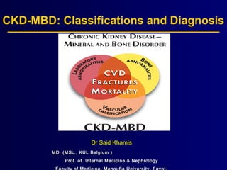
1. CKD-MBD: Classifications and Diagnosis
Dr Said KhamisDr Said Khamis
MD, (MSc., KUL Belgium )MD, (MSc., KUL Belgium )
Prof. of Internal Medicine & NephrologyProf. of Internal Medicine & Nephrology
2. Agenda
The size of the problem (CKD-MBD)
Definitions & Classifications
Bone disease in CKD (B)
Vascular calcifications/CVD (V/C)
Role of Calcium and Phosphorus (L)
Importance of Vitamin D in CKD-MBD (L)
CKD & SHPT (L)
Frequency of Monitoring
CKD-MBD 2017 Guidelines (Diagnosis of CKD-MBD & VC)
3. Disorders of mineral metabolism are major risk
factors in CKD-MBD
Moe SM, Chertow GMMoe SM, Chertow GM. Clin J Am Soc Nephrol. Clin J Am Soc Nephrol 2006.2006.
The risk associated with disorders of mineral metabolism is higher than that associated with inefficient dialysisThe risk associated with disorders of mineral metabolism is higher than that associated with inefficient dialysisThe risk associated with disorders of mineral metabolism is higher than that associated with inefficient dialysisThe risk associated with disorders of mineral metabolism is higher than that associated with inefficient dialysis
0
2
4
6
8
10
12
14
16
18
20
Low URR Anaemia High Pi High Ca High PTH All
mineral
Attributablemortalityrisk(%)
(5.1%)(5.1%)
(11.3%)(11.3%)
(6%)(6%)
(17.5%)(17.5%)
7. • However, we must keep in mind that all these
CKD-MBD players have a close connection & any
change in one could certainly affect the other
• Therefore, only an integrative approach for CKD-
MBD therapy will succeed
8. 1970 1980 1990 2000 2010 2017
Aluminum
Binders
PO Calcitriol
Calcium
Binders
Vitamin D
Analogues
K/DOQI
IV Calcitriol
History of Treatment Strategies for
Secondary Hyperparathyroidism
Focus: Bone Disease,
Systemic effects of PTH,
High Ca was
thought to be good
Sevelamer
Cinacalcet HCl
KIDGO
Focus: Fractures, Mortality,
& Vascular Calcification
KIDGO
VIT K2
9. Definition of CKD-MBD And Renal
Osteodystrophy
Definition of CKD-MBD
– A systemic disorder of mineral and bone metabolism due to CKD
manifested by either one or the combination of the following:
L/Abnormalities of calcium, Phosphorus, PTH, or Vitamin D
metabolism
B/ Abnormalities in bone turnover, mineralization, volume, linear growth, or
strength.
V/C: Vascular or other soft-tissue calcification
Definition of Renal Osteodystrophy
– Renal osteodystrophy is an alteration of bone morphology in patients
with CKD.
– It is one measure of the skeletal component of the systemic disorder of
CKD-MBD that is quantifiable by histomorphometry of bone biopsy.
– Uhlig et al, AJKD Vol 55, No 5, May 2010.
– KDIGO classification of CKD-MBD 2009
11. Framework for classification of CKD-MBD
MBD parameter Laboratory
abnormalities
Bone disease Calcification of
Vascular or other
soft tissue
L + - -
LB + + -
LC + - +
LBC/V + + +
Moe S et al. Kidney Int 2006;69:1945–
53.
L= laboratory abnormalities; B = bone disease; C/V = calcification of vascular or other soft tissue.L= laboratory abnormalities; B = bone disease; C/V = calcification of vascular or other soft tissue.
12. Definition of CKD-MBD And Renal
Osteodystrophy
Definition of CKD-MBD
– A systemic disorder of mineral and bone metabolism due to CKD
manifested by either one or the combination of the following:
L/Abnormalities of calcium, Phosphorus, PTH, or Vitamin D
metabolism
B/ Abnormalities in bone turnover, mineralization, volume, linear growth, or
strength.
V/C: Vascular or other soft-tissue calcification
Definition of Renal Osteodystrophy
– Renal osteodystrophy is an alteration of bone morphology in patiens
with CKD.
– It is one measure of the skeletal component of the systemic disorder of
CKD-MBD that is quantifiable by histomorphometry of bone biopsy.
– Uhlig et al, AJKD Vol 55, No 5, May 2010.
– KDIGO classification of CKD-MBD 2009
16. Spectrum of ROD
Mixed LesionMixed Lesion
Osteitis FibrosaOsteitis Fibrosa
Normal BoneNormal Bone
FormationFormation
HighHigh
Low turnoverLow turnover High turnoverHigh turnover
AdynamicAdynamic
OsteomalaciaOsteomalacia
LowLow
PTHPTH
< 150 pg/ml< 150 pg/ml 150 – 300 pg/ml150 – 300 pg/ml > 300 pg/ml> 300 pg/ml
17.
18.
19.
20.
21.
22.
23.
24.
25. Imaging in Chronic Kidney Disease Metabolic Bone Disease‐
Seminars in Dialysis, Volume: 30, Issue: 4, Pages: 361-368, First published: 05 April 2017, DOI: (10.1111/sdi.12598)
26 year old male with hand radiograph‐ ‐
demonstrating mild periosteal reaction
along the metacarpal bones (arrows).
26. Imaging in Chronic Kidney Disease Metabolic Bone Disease‐
Seminars in Dialysis, Volume: 30, Issue: 4, Pages: 361-368, First published: 05 April 2017, DOI: (10.1111/sdi.12598)
(A,B) 39 year old male with lateral and AP radiographs of the elbow demonstrating well defined, lobular,‐ ‐
calcified mass in the soft tissues about the elbow (arrow) consistent with periarticular calcification. (C) 42‐
year old male with right hip radiograph demonstrating periarticular “tumoral” calcification adjacent to the right‐
femoral neck (arrow).
27. Amino terminal and carboxy-Amino terminal and carboxy-
terminal propeptides of type Iterminal propeptides of type I
collagen (PINP, PICP).collagen (PINP, PICP).
33. Definition of CKD-MBD And Renal
Osteodystrophy
Definition of CKD-MBD
– A systemic disorder of mineral and bone metabolism due to CKD
manifested by either one or the combination of the following:
L/Abnormalities of calcium, Phosphorus, PTH, or Vitamin D
metabolism
B/ Abnormalities in bone turnover, mineralization, volume, linear growth, or
strength.
V/C: Vascular or other soft-tissue calcification
Definition of Renal Osteodystrophy
– Renal osteodystrophy is an alteration of bone morphology in patiens
with CKD.
– It is one measure of the skeletal component of the systemic disorder of
CKD-MBD that is quantifiable by histomorphometry of bone biopsy.
– Uhlig et al, AJKD Vol 55, No 5, May 2010.
– KDIGO classification of CKD-MBD 2009
34. Abnormal boneAbnormal bone
AgeAge
Oxidation (OxLDL)Oxidation (OxLDL)
DiabetesDiabetes
HTNHTN
Advanced glycationAdvanced glycation
end-productsend-products
SmokingSmoking
GeneticsGenetics
DyslipidemiaDyslipidemia
Carbonyl stressCarbonyl stress
Low fetuin-ALow fetuin-A
Traditional Risk FactorsTraditional Risk Factors Non-traditional Risk FactorsNon-traditional Risk Factors
Elevated IL-1, Il-6, TNFElevated IL-1, Il-6, TNFαα
HomocysteineHomocysteine
Abnormal mineral metabolismAbnormal mineral metabolism
FracturesFractures
Cardiovascular disease in CKDCardiovascular disease in CKD
36. •Two types of vascular calcification
• Intimal calcification leads to calcific plaques or circumferentially calcified atherosclerosis
• Medial calcification is nonocclusive and leads to vascular stiffening; it can cause local ischemia and also affect
the capacity of the vasculature to dampen increases in arterial pressure with each ventricular systole, leading to
left ventricular hypertrophy
•Traditionally, the CAC score obtained by electron beam CT is used to quantify calcification burden
•Other available techniques can provide semi-quantitative evidence of calcification, including duplex ultrasonography,
37. Types of Vascular Calcification in CKD
Uremic arteriopathyUremic arteriopathy AtherosclerosisAtherosclerosis
41. Imaging techniques for V/C
The best diagnostic technique is unknown
Which bed?
– Coronary vs central vs peripheral
Which technique?
– Simplicity and accuracy
Which golden standard?
– CAC vs hard clinical outcomes
42. Ideally
Appropriate vascular bed
Simple, available, interpretable
Detects disease with reasonable sensitivity/specificty
Detects progression
Detects effect of treatment
Results correlate with hard clinical outcomesResults correlate with hard clinical outcomes
46. Definition of CKD-MBD And Renal
Osteodystrophy
Definition of CKD-MBD
– A systemic disorder of mineral and bone metabolism due to CKD
manifested by either one or the combination of the following:
L/Abnormalities of calcium, Phosphorus, PTH, or Vitamin D
metabolism
B/ Abnormalities in bone turnover, mineralization, volume, linear growth, or
strength.
V/C: Vascular or other soft-tissue calcification
Definition of Renal Osteodystrophy
– Renal osteodystrophy is an alteration of bone morphology in patiens
with CKD.
– It is one measure of the skeletal component of the systemic disorder of
CKD-MBD that is quantifiable by histomorphometry of bone biopsy.
– Uhlig et al, AJKD Vol 55, No 5, May 2010.
– KDIGO classification of CKD-MBD 2009
53. Serum Phosphorus Levels and Mortality in
CKD Non-Dialysis Patients
Mortality risk increases as phosphorus levels rise, even within normal range
Each 0.5 mg/dL increase in serum phosphorus was associated with increased mortality
Statistically significant increases in mortality were noted when phosphorus levels reached 3.5 mg/dL
or above
Adapted from Kestenbaum B, Sampson JN, Rudser KD, et al. J Am Soc Nephrol. 2005;16:520-528.
1.00
1.15
1.32 1.34
1.83
1.90
1.00
1.20
1.40
1.60
1.80
2.00
2.5-2.99 3.0-3.49 3.5-3.99 4.0-4.49 4.5-4.99 >5.0
Phosphorus (mg/dL)
Adjustedhazardratio(HR)
Mortality rates by phosphate category
72% of patients
(n=3,289)
54. *Multivariable adjusted
With permission from Block GA, Klassen PS, Lazarus JM, et al. J Am Soc Nephrol. 2004;15:2208-2218.
Elevated Serum Phosphorus
and Mortality Risk in Dialysis Patients
Relativeriskofdeath*
<3 3-4 4-5 5-6 6-7 7-8 8-9 >9
Serum phosphorous concentration (mg/dL)
0.00
1.0
1.4
1.6
2.0
2.2
0.08
1.2
1.8
N = 40,538
Referent
Range
55. KDIGO Focus: Normal Treatment Target
Ranges for Phosphorous and Calcium
Stage Target PO4
1,2
Target Ca1,2
3
KDIGO: Maintain Normal
KDOQI: 2.7-4.6 mg/dL
KDIGO: Maintain Normal
KDOQI: Normal for Lab
4-5
KDIGO: Maintain Normal
KDOQI: 2.7-4.6 mg/dL
KDIGO: Maintain Normal
KDOQI: Normal for Lab
5D
KDIGO: Towards Normal
KDOQI: 3.5-5.5 mg/dL
KDIGO: Maintain Normal
KDOQI: 8.4-9.5 mg/dL
Emphasis on individual levels of serum calcium and phosphorus
rather than Ca x P product
1. Kidney Disease: Improving Global Outcomes (KDIGO) CKD-MBD Work Group. KDIGO clinical practice guideline
for the diagnosis, evaluation, prevention, and treatment of chronic kidney disease-mineral and bone disorder (CKD-MBD)
Kidney Int. 2009;76(suppl 113):S1-S130.
2. National Kidney Foundation (NKF). KDOQI clinical practice guidelines for bone metabolism and disease in chronic
kidney disease. Am J Kidney Dis. 2003;42(4 suppl 3):S1-S201.
56. Summary Slide
Despite early phosphate retention, many patients have
serum phosphorus levels within normal range
Mortality risk increases as phosphorus levels rise,
even when levels remain within normal range
Increased serum phosphorus levels are associated
with CV events and mortality
Adequate dialysis may have an additive role in
controlling hyperphosphatemia
58. Calcium and
Phosphorus
Homeostasis
Bone Health
Cardiovascular
Effects
Renin-Angiotensin
Regulation
Decreased Risk for:
Hypertension
Type II Diabetes
(via stimulation of pancreatic
insulin production)
Heart Failure
Immunomodulatory
Effects
Multiple Sclerosis
Type 1 Diabetes
(via ß-islet cell
destruction)
Psoriasis
Rheumatoid Arthritis
Inflammatory Bowel
Disease
Periodontal Disease
Growth & Regulation
Antiproliferation
Prodifferentiation
Apoptotic
Anti-angiogenic
Prostate,
Colon,
Breast Cancers etc.
Neuromuscular
Effects
Muscle Mass
Muscle Strength
Better Balance
25(OH)D
Major Circulating Metabolite
1,25(OH)2D
Biologically Active
Keratinocytes
M
acrophages
Colon
Prostate
Breast, etc.
Kidney
Physiologic Effects of Vitamin D
Throughout the Body
Adapted from: Holick MF. Mayo Clin Proc. 2006;81:353-373.
59. 1,25-dihydroxyvitamin D levels and 90-day all-cause (A) and CV mortality (B) in hemodialysis
patients according to whether patients received active vitamin D therapy
With permission from Wolf M, et al. Kidney Int. 2007;72:1004-1013.
1,25(OH)2D3 Levels and
Mortality in Dialysis Patients
1,25-dihydroxyvitamin D (pg/mL)
Oddsratioofall-causemortality
10
8
6
4
2
0
<5 6–13 >13
*
*
1,25-dihydroxyvitamin D (pg/mL)
OddsratioofCVmortality
10
8
6
4
2
0
<5 6–13 >13
*
A B
RR
No active vitamin D therapy
Active vitamin D therapy
*P<0.05 for the comparison of the individual vitamin D level―vitamin D treatment groups with the corresponding referent groups.
R=subjects treated with active vitamin D and 1,25-dihydroxyvitamin D levels ≥13 pg/mL.
No active vitamin D therapy
Active vitamin D therapy
61. Summary
Patients with CKD have a high prevalence of both
25(OH)D and 1,25(OH)2D deficiency
As kidney function declines, patients lose the ability
to convert 25(OH)D to 1,25(OH)D
Patients with lower levels of 1,25(OH)2D had a higher
risk of mortality
63. Compensatory Mechanism of PTH During
Disturbances in Mineral Metabolism
With permission from Levin A, et al. Kidney Int. 2007;71:31-38.
1.25-dihydroxyvitaminD(pg/mL)
25HydroxyvitaminD(ng/mL)
IntactPTH(pg/mL)
Median values of 1,25-dihydroxyvitamin D, 25 Hydroxyvitamin D,
and intact PTH by GFR levels
GFR level (mL/min)
50
45
40
35
30
25
20
15
10
5
0
>80 79-70 69-60 59-50 49-40 39-30 29-20 <20
50
0
100
150
1,25-dihydroxyvitamin D (pg/mL)
25 Hydroxyvitamin D (ng/mL)
Intact PTH (pg/mL)
64. Elevated PTH Levels and Cardiovascular Disease
in Patients with CKD Stages 3 and 4
37
41
49
30
35
40
45
50
<35 35-70 >70
Rateofcardiovasculardisease,%
Parathyroid hormone level, pg/mL
With permission from Bhuriya R, et al. Am J Kidney Dis. 2009;53 (4 suppl 4):S3-S10.
P<0.001
65. Clin J Am Soc Nephrol 2018, in press
PTH in Focus
66.
67. PTH Assays
•First-generation assays
• Radioimmunoassay using an antibody against the mid-region or carboxy-terminal end
• Detects full-length PTH as well as the multiple carboxy- and amino-terminal fragments
• Unreliable
•Second-generation assay/intact PTH assays/two-step first generation immunoradiometric assays
(IRMA)
• Involve two antibodies, one that detects the amino terminus and the other the carboxy terminus
• Most commonly used assay in clinical practice
• However, in addition to detecting full-length PTH, it also detects fragments commonly referred to
as 7-84 PTH
• This 7-84 PTH may have antagonistic effects to full-length PTH on bone
•Third generation assays/whole PTH assays/Biointact PTH assays only detect 1-84 PTH
•Poor correlation between any of the PTH assays and bone histology in CKD
71. KDIGO Focus: Consider Normal
Limit for PTH
Stage Treatment Target Range
3
KDIGO: Upper Limit of Normal* (2C)
KDOQI: 35-70 pg/mL
4
KDIGO: Upper Limit of Normal* (2C)
KDOQI: 70-110 pg/mL
5
KDIGO: Upper Limit of Normal* (2C)
KDOQI: 150-300 pg/mL
5D
KDIGO: 2 to 9 times Upper Limit of Normal (2C)
KDOQI: 150-300 pg/mL
1. Kidney Disease: Improving Global Outcomes (KDIGO) CKD-MBD Work Group. KDIGO clinical practice guideline
for the diagnosis, evaluation, prevention, and treatment of chronic kidney disease-mineral and bone disorder (CKD-MBD)
Kidney Int. 2009;76(suppl 113):S1-S130.
2. Adapted from National Kidney Foundation (NKF). KDOQI clinical practice guidelines for bone metabolism and disease in
chronic kidney disease. Am J Kidney Dis. 2003;42(4 suppl 3):S1-S201.
*In patients with CKD stages 3-5*In patients with CKD stages 3-5 not on dialysis,not on dialysis, in whom serum PTH is progressively rising and remainsin whom serum PTH is progressively rising and remains
persistently above the upper limit of normal for the assay despite correction of modifiable factors, treatment withpersistently above the upper limit of normal for the assay despite correction of modifiable factors, treatment with
calcitriol or vitamin D analogs is suggested.calcitriol or vitamin D analogs is suggested. (2C)(2C)
72. Summary
Elevated PTH levels are a compensatory mechanism
for 1,25D depletion — both are prominent and
progressive across the CKD continuum
Elevated PTH levels and 1,25D depletion have each
been independently associated with higher mortality
Vitamin D when appropriate without inducing what
is overtly oversuppressed parathyroid hormone
High calcium and high phosphorus with or without
vitamin D is a required objective
73.
74. Conclusions:
After switching the frequency of routine blood work from 4- to 6-week intervals, performance on
anemia and CKD-MBD targets did not change and the reduction in blood work was associated
with laboratory cost savings.
76. Kidney Disease: Improving Global Outcomes
CHAPTER 3.1:
DIAGNOSIS OF CKD-MBD:
BIOCHEMICAL
ABNORMALITIES
77. Kidney Disease: Improving Global Outcomes
ASSESSMENT
3.1.1: We recommend monitoring serum levels of calcium,
phosphate, PTH, and alkaline phosphatase activity beginning in
CKD G3a (1C). In children, we suggest such monitoring beginning
in CKD G2 (2D).
3.1.2: In patients with CKD G3a–G5D, it is reasonable to base the
frequency of monitoring serum calcium, phosphate, and PTH on
the presence and magnitude of abnormalities, and the rate of
progression of CKD (Not Graded).
78. Kidney Disease: Improving Global Outcomes
ASSESSMENT
3.1.2 (cont’d.): Reasonable monitoring intervals would be:
o In CKD G3a–G3b: for serum calcium and phosphate, every 6–12 months; and
for PTH, based on baseline level and CKD progression.
o In CKD G4: for serum calcium and phosphate, every 3–6 months; and for PTH,
every 6–12 months.
o In CKD G5, including G5D: for serum calcium and phosphate, every 1–3
months; and for PTH, every 3–6 months.
o In CKD G4–G5D: for alkaline phosphatase activity, every 12 months, or more
frequently in the presence of elevated PTH (see Chapter 3.2).
In CKD patients receiving treatments for CKD-MBD, or in whom
biochemical abnormalities are identified, it is reasonable to increase the
frequency of measurements to monitor for trends and treatment efficacy
and side effects (Not Graded).
79. Kidney Disease: Improving Global Outcomes
ASSESSMENT
3.1.3: In patients with CKD G3a–G5D, we suggest that 25(OH)D
(calcidiol) levels might be measured, and repeated testing
determined by baseline values and therapeutic interventions
(2C).
We suggest that vitamin D deficiency and insufficiency be
corrected using treatment strategies recommended for the
general population (2C).
3.1.4: In patients with CKD G3a–G5D, we recommend that
therapeutic decisions be based on trends rather than on a single
laboratory value, taking into account all available CKD-MBD
assessments (1C).
80. Kidney Disease: Improving Global Outcomes
ASSESSMENT
3.1.5: In patients with CKD G3a–G5D, we suggest that individual
values of serum calcium and phosphate, evaluated together, be
used to guide clinical practice rather than the mathematical
construct of calcium-phosphate product (Ca x P) (2D).
3.1.6: In reports of laboratory tests for patients with CKD G3a–
G5D, we recommend that clinical laboratories inform clinicians
of the actual assay method in use and report any change in
methods, sample source (plasma or serum), or handling
specifications to facilitate the appropriate interpretation of
biochemistry data (1B).
82. Kidney Disease: Improving Global Outcomes
TESTING FOR CKD-MBD
3.2.1: In patients with CKD G3a-G5D with evidence of CKD-MBD
and/or risk factors for osteoporosis, we suggest bone mineral
density (BMD) testing to assess fracture risk if results will impact
treatment decisions (2B).
3.2.2: In patients with CKD G3a-G5D, it is reasonable to perform a
bone biopsy if knowledge of the type of renal osteodystrophy will
impact treatment decisions (Not Graded).
83. Kidney Disease: Improving Global Outcomes
ASSESSMENT
3.2.3: In patients with CKD G3a–G5D, we suggest that measurements of
serum PTH or bone-specific alkaline phosphatase can be used to
evaluate bone disease because markedly high or low values predict
underlying bone turnover (2B).
3.2.4: In patients with CKD G3a–G5D, we suggest not routinely
measuring bone-derived turnover markers of collagen synthesis (such as
procollagen type I C-terminal propeptide) and breakdown (such as
type I collagen cross-linked telopeptide, cross-laps, pyridinoline, or
deoxypyridinoline) (2C).
3.2.5: We recommend that infants with CKD G2–G5D have their length
measured at least quarterly, while children with CKD G2–G5D should be
assessed for linear growth at least annually (1B).
85. Kidney Disease: Improving Global Outcomes
ASSESSMENT
3.3.1: In patients with CKD G3a–G5D, we suggest that a lateral
abdominal radiograph can be used to detect the presence or
absence of vascular calcification, and an echocardiogram can be
used to detect the presence or absence of valvular calcification,
as reasonable alternatives to computed tomography-based
imaging (2C).
3.3.2: We suggest that patients with CKD G3a–G5D with known
vascular or valvular calcification be considered at highest
cardiovascular risk (2A).
It is reasonable to use this information to guide the management
of CKD-MBD (Not Graded).
87. Chronic kidney disease-mineral and bone disorder
(CKD-MBD) patients have a huge morbidity and
mortality.
Only relatively minor progress in therapeutic
strategies has been made in the past decades.
This is at least partially due to a lack of predictive
diagnostic tools allowing personalized treatment of
CKD-MBD patients
Without precise diagnostic tools a personalized
therapy is not possible. This, however, was part of the
success story in oncology.
88. However, there is hope; Researchers highlighted key
recommendations in areas of controversy or conjecture in the
management of (CKD-MBD) in a recent synopsis of the
(KDIGO) 2017 .
The original update, published in Kidney International (2017;7(Suppl 1):1-
59), resulted in 15 revised recommendations based on evidence of
varying strengths accumulated since the 2009 KDIGO guidelines
They described the recent progress in the diagnosis of
disturbances of the PTH, calcium & phosphate & metabolism
in patients with CKD plus the currently available imaging
modalities to Dx. different subcategories of CKD-MBD.
These new tools may have the potential of allowing
personalized therapy for the treatment of CKD-MBD and
hence improving outcome.
91. Study of Cardiovascular Calcifications among Regular Hemodialysis Patients with
and Without Chronic Hepatitis C Virus Infection
Abdel-hady Hassan*, Reda S.B*, Walaa F.**, Ehab A A.*, Ashraf G D* and Dina M A*.
Internal Medicine* and Cardiology** Departments, Faculty of Medicine, Menofiya University Hospitals,
Egypt. ( in press.)
92. Thank you for your attention
Phramongkutklao Hospital and
College of Medicine
93. Elias MR et al., Current Osteoporosis report. October 2018
Hinweis der Redaktion
The population-attributable risk for mortality in a large (39,530) cohort of haemodialysis patients for several laboratory abnormalities was determined.
Several variables were considered: hyperphosphataemia (high Pi, defined as serum Pi ≥5.0 mg/dL), hypercalcaemia (high Ca, defined as serum Ca &gt;10 mg/dL), and moderate to severe hyperparathyroidism (high PTH, defined as PTH &gt;600 pg/mL) individually and in combination.
Comparison of population-attributable risk percentages for inefficient dialysis (low urea reduction ratio [URR], defined as URR &lt;65%) and anaemia (defined as haemoglobin &lt;11 g/dL) showed that the attributable risk associated with disorders of mineral metabolism (17.5%) was higher than that associated with inefficient dialysis (5.1%) or anaemia (11.3%). (6%)
Reference
Moe SM, Chertow GM. The case against calcium-based phosphate binders. Clin J Am Soc Nephrol 2006;1:697–703.
For patients with CKD stages 3-5 with biochemical abnormalities of CKD-MBD and low BMD, KDIGO guidelines recommend bone biopsy before the initiation of treatment with antiresorptive agents
Biochemical laboratory and imaging tests do not adequately predict the underlying bone histology. Thus, although bone biopsy is invasive and cannot be performed easily in all patients, it is the gold standard for the diagnosis of renal osteodystrophy. Bone biopsy should be considered in patients for whom the cause of clinical symptoms and biochemical abnormalities is not certain and for whom the effect of treatment on bone needs to be assessed
Bone biopsy is the most accurate test for the diagnosis of adynamic bone disease, and the presence of adynamic bone disease is a contraindication to bisphosphonate treatment
In patients with CKD stages 3-5D with evidence of CKD-MBD, we suggest that BMD testing not be performed routinely because BMD does not predict fracture risk as it does in the general population, and BMD does not predict the type of renal osteodystrophy
Use of teriparatide is contraindicated in patients with CKD with SHPT, although it might be a useful therapy in patients after parathyroidectomy with low BMD and/or fragility fractures. No information is available regarding the use of teriparatide in patients with CKD with adynamic bone disease.
The term CKD-mineral and bone disorder (CKD-MBD) be used to describe the broader clinical syndrome that develops as a systemic disorder of mineral and bone metabolism due to CKD, which is manifested by any one or a combination of the following:
1. Laboratory abnormalities of Ca, Pi, PTH or vitamin D metabolism.
2. Bone abnormalities in turnover, mineralization, volume, linear growth or strength.
3. Calcification of the vasculature or other soft tissues, and classified based on the presence or absence of any combination of these three primary components (L,B,C, Table).
References
Moe S, Drueke T, Cunningham J et al. Definition, evaluation, and classification of renal osteodystrophy: A position statement from Kidney Disease: Improving Global Outcomes (KDIGO). Kidney Int 2006;69:1945–53.
For patients with CKD stages 3-5 with biochemical abnormalities of CKD-MBD and low BMD, KDIGO guidelines recommend bone biopsy before the initiation of treatment with antiresorptive agents
Biochemical laboratory and imaging tests do not adequately predict the underlying bone histology. Thus, although bone biopsy is invasive and cannot be performed easily in all patients, it is the gold standard for the diagnosis of renal osteodystrophy. Bone biopsy should be considered in patients for whom the cause of clinical symptoms and biochemical abnormalities is not certain and for whom the effect of treatment on bone needs to be assessed
Bone biopsy is the most accurate test for the diagnosis of adynamic bone disease, and the presence of adynamic bone disease is a contraindication to bisphosphonate treatment
In patients with CKD stages 3-5D with evidence of CKD-MBD, we suggest that BMD testing not be performed routinely because BMD does not predict fracture risk as it does in the general population, and BMD does not predict the type of renal osteodystrophy
Use of teriparatide is contraindicated in patients with CKD with SHPT, although it might be a useful therapy in patients after parathyroidectomy with low BMD and/or fragility fractures. No information is available regarding the use of teriparatide in patients with CKD with adynamic bone disease.
26‐year‐old male with hand radiograph demonstrating mild periosteal reaction along the metacarpal bones (arrows).
IF THIS IMAGE HAS BEEN PROVIDED BY OR IS OWNED BY A THIRD PARTY, AS INDICATED IN THE CAPTION LINE, THEN FURTHER PERMISSION MAY BE NEEDED BEFORE ANY FURTHER USE. PLEASE CONTACT WILEY&apos;S PERMISSIONS DEPARTMENT ON PERMISSIONS@WILEY.COM OR USE THE RIGHTSLINK SERVICE BY CLICKING ON THE &apos;REQUEST PERMISSIONS&apos; LINK ACCOMPANYING THIS ARTICLE. WILEY OR AUTHOR OWNED IMAGES MAY BE USED FOR NON-COMMERCIAL PURPOSES, SUBJECT TO PROPER CITATION OF THE ARTICLE, AUTHOR, AND PUBLISHER.
(A,B) 39‐year‐old male with lateral and AP radiographs of the elbow demonstrating well defined, lobular, calcified mass in the soft tissues about the elbow (arrow) consistent with periarticular calcification. (C) 42‐year‐old male with right hip radiograph demonstrating periarticular “tumoral” calcification adjacent to the right femoral neck (arrow).
IF THIS IMAGE HAS BEEN PROVIDED BY OR IS OWNED BY A THIRD PARTY, AS INDICATED IN THE CAPTION LINE, THEN FURTHER PERMISSION MAY BE NEEDED BEFORE ANY FURTHER USE. PLEASE CONTACT WILEY&apos;S PERMISSIONS DEPARTMENT ON PERMISSIONS@WILEY.COM OR USE THE RIGHTSLINK SERVICE BY CLICKING ON THE &apos;REQUEST PERMISSIONS&apos; LINK ACCOMPANYING THIS ARTICLE. WILEY OR AUTHOR OWNED IMAGES MAY BE USED FOR NON-COMMERCIAL PURPOSES, SUBJECT TO PROPER CITATION OF THE ARTICLE, AUTHOR, AND PUBLISHER.
For patients with CKD stages 3-5 with biochemical abnormalities of CKD-MBD and low BMD, KDIGO guidelines recommend bone biopsy before the initiation of treatment with antiresorptive agents
Biochemical laboratory and imaging tests do not adequately predict the underlying bone histology. Thus, although bone biopsy is invasive and cannot be performed easily in all patients, it is the gold standard for the diagnosis of renal osteodystrophy. Bone biopsy should be considered in patients for whom the cause of clinical symptoms and biochemical abnormalities is not certain and for whom the effect of treatment on bone needs to be assessed
Bone biopsy is the most accurate test for the diagnosis of adynamic bone disease, and the presence of adynamic bone disease is a contraindication to bisphosphonate treatment
In patients with CKD stages 3-5D with evidence of CKD-MBD, we suggest that BMD testing not be performed routinely because BMD does not predict fracture risk as it does in the general population, and BMD does not predict the type of renal osteodystrophy
Use of teriparatide is contraindicated in patients with CKD with SHPT, although it might be a useful therapy in patients after parathyroidectomy with low BMD and/or fragility fractures. No information is available regarding the use of teriparatide in patients with CKD with adynamic bone disease.
CKD-MBD: Dysregulation and Clinical Manifestations of an Increasingly Compromised System
Background:
As renal function declines, the body fights for homeostasis through a number of renal compensatory mechanisms
After a while, renal compensatory mechanisms begin to fail and a cascade of events begins with:
a decline in urinary phosphate excretion leading to an increase in serum phosphate,
depletion of 1,25D and subsequent elevation of PTH serum levels and,
reduced urinary Ca excretion leading to an imbalance in serum Ca levels.
This dysregulation leads to clinical manifestations such as hyperphosphatemia, secondary hyperparathyroidism, and a potential for Ca overload
Key Point:
Patients with normal serum phosphorus levels had an increased risk of mortality1
Background:
A cohort study of patients with CKD was conducted to assess the association between higher phosphate levels and mortality1
As shown in the graph, approximately 72% of patients had an increased risk of mortality even though their phosphorus levels were within normal range, as defined by KDOQI guidelines1
A roughly linear relationship was observed between serum phosphate and mortality, with each 0.5 mg/dL increase in serum phosphorus associated with increased mortality1
When phosphate levels reached 3.5 mg/dL or above, statistically significant increases in mortality were observed1
Reference:
Kestenbaum B, Sampson JN, Rudser KD, et al. Serum phosphate levels and mortality riskamong people with chronic kidney disease. J Am Soc Nephrol. 2005;16:520-528.
Key Point:
Elevated serum phosphorus levels are associated with an increased risk of mortality1
Background:
Block and colleagues analyzed the effect of serum phosphorus disorders in 40,5381 end-stage renal disease (ESRD) patients on maintenance hemodialysis1 and found that serum phosphorus concentrations &lt;3 mg/dL, and more than 5 mg/dL are associated with increased relative risk of death1
Referent range was defined as serum phosphorus concentration of 4-5 mg/dL1
This more contemporary cohort allowed for a more detailed exploration of the relationship between serum phosphorus and relative risk of death in ESRD patients in contrast to previous studies1
Reference:
Block GA, Klassen PS, Lazarus JM, Ofsthun N, Lowrie EG, Chertow GM. Mineral metabolism, mortality, and morbidity in maintenance hemodialysis. J Am Soc Nephrol. 2004;15:2208-2218.
Treatment Target Ranges
Key Point:
The KDIGO CKD-MBD Work Group suggests normalization of target phosphorus and calcium levels regardless of CKD stage (and towards normalization for target phosphorus in CKD stage 5D)1
Background:
The rationale for controlling phosphorus levels comes from epidemiological data suggesting that elevation of serum phosphorus levels is an important risk factor for secondary hyperparathyroidism and cardiovascular disease1
Moreover, epidemiological studies show a positive association between higher serum phosphorus levels and relative risk of mortality, independent of the CKD stage1
It remains unclear as to at what level of low serum calcium an increased risk of mortality exists1; however, data do not exist showing an increased risk of mortality or fracture with increased serum calcium levels1—thus, normalization of target serum calcium levels is suggested1
Reference:
Kidney Disease Improving Global Outcomes (KDIGO). Clinical practice guideline for the diagnosis, evaluation, prevention, and treatment of chronic kidney disease-mineral and bone disorder (CKD-MBD). Kidney Int. 2009;76(suppl 113):S1-S130.
Effects of Vitamin D Throughout the Body
Key Points:
Vitamin D exerts effects on multiple systems in the body1
Vitamin D deficiency leads to problems involving skeletal, cardiovascular, muscular, endocrine, and immune systems1
Background:
The kidneys serve an endocrine function by converting 25-hydroxyvitamin D (25 D) to 1,25 dihydroxyvitamin D (1,25 D), which circulates in the plasma
1,25 D regulates calcium metabolism by stimulating calcium and phosphorus absorption in the intestine
Circulating 1,25 D can bind to other cell types not directly involved in calcium homeostasis and bone health by binding to vitamin D receptors (VDR) that are widely distributed throughout the body
VDRs are present on cells in the heart, skeletal muscle, active T and B lymphocytes, breast, colon, and prostate
In addition, locally produced 1,25 D3 may exert autocrine or paracrine effects on regulation of cellular growth and differentiation in tissues other than kidney that express 1α-hydroxylase. These include lung, colon, prostate, and breast
However, evidence suggests that high levels (&gt;30 ng/mL) of 25 D are necessary before it becomes a substrate for extra-renal 1α-hydroxylase
Reference:
Holick MF. High prevalence of vitamin insufficiency and implications for health. Mayo Clinic Proc.
2006;81:353-373.
1,25(OH)2D3 Levels and Mortality in Dialysis Patients
Key Point:
Active vitamin D therapy improved all cause and cardiovascular mortality compared with no active vitamin D therapy at 90 days after starting hemodialysis in patients who had baseline1,25(OH)2D deficiency (plasma levels ≤ 13 pg/ml) 1
Background:
The chart on the left shows that patients with 1,25(OH)2D deficiency (plasma levels ≤13 pg/ml) at baseline were at significantly elevated risk for all-cause mortality in the first 90 days on hemodialysis compared with those who had 1,25(OH)2D levels &gt;13 pg/mL (used as reference group)1
The chart on the right shows that patients with 1,25(OH)2D deficiency (plasma levels ≤ 13 pg/mL) at baseline were at significantly elevated risk for cardiovascular mortality in the first 90 days on hemodialysis compared with those who had 1,25(OH)2D levels &gt;13 pg/mL (used as reference group)1
No increase in risk of mortality was observed in either analysis in subjects who received active vitamin D therapy1
Reference:
1. Wolf M, Shah A, Gutierrez O, et al. Vitamin D levels and early mortality among incident hemodialysis patients. Kidney Int. 2007;72:1004-1013.
Compensatory Mechanism of PTH During Disturbances In Mineral Metabolism
Key Point:
As levels of 25(OH)D and 1,25(OH)2D decrease with glomerular filtration rate, intact parathyroid hormone levels increase as a compensatory means to restore mineral homeostasis1
Background:
Blood calcium levels are key regulators of parathyroid secretion. Because low levels of circulating 1,25 D reduce calcium absorption by the intestines, plasma calcium levels are reduced in parallel, which indirectly stimulates the secretion of parathyroid hormone2
It is not surprising, therefore that as 1,25 D levels decrease as a result of loss of renal function, that PTH levels increase as shown by Levin et al1
References:
1.Levin A, Bakris GL, Molitch M, et al. Prevalence of abnormal serum vitamin D, PTH, calcium, and phosphorus in patients with chronic kidney disease: results of the study to evaluate early kidney disease. Kidney Int. 2007;71:31-38.
2.Holick MF. High prevalence of vitamin D in adequacy and implications for health. Mayo Clinic Proc. 2006;81:353-373.
Elevated PTH Levels and Cardiovascular Disease
Key Point:
Elevated PTH levels are associated with cardiovascular disease1
Background:
A cross-sectional study was conducted in which medical history surveys including CVD events, were collected from 4,472 patients stages 3 and 4 CKD from the Kidney Early Evaluation Program (KEEP) to determine if there is a relationship between PTH level and prevalent CVD1
Stage 3 CKD was defined as eGFR of 30 to 59 mL/min/1.73 m2 and stage 4 was defined as eGFR of 15 to 29 mL/min/1.73 min1
Participants were asked “Have you been told by a doctor or health care professional of having any of the following (mark all that apply)? Heart attack, heart bypass surgery, heart angioplasty, stroke, heart failure (fluid in lungs), abnormal heart rhythm.” 1
Rates of CVD were higher in those patients with higher PTH levels. In patients with PTH levels &lt;35 pg/mL, 35 to 70 pg/mL, and &gt;70 pg/mL rates of CVD were 37%, 41%, and 49%, respectively1
Reference:
Bhuriya R, Li S, Chen SC, McCullough PA, Bakris GL. Plasma parathyroid hormone level and prevalent cardiovascular disease in CKD stages 3 and 4: An analysis from the Kidney Early Evaluation Program (KEEP). Am J Kidney Dis. 2009;5(4 suppl 4):S3-S10.
Treatment Initiation Ranges
Key Point:
As noted in the KDIGO Guidelines, progressive increases in PTH should be avoided as severe hyperparathyroidism may be resistant to therapy and is associated with morbidity and mortality in patients with CKD (stages 3-5D)1
Background:
Recent observational studies show an increased relative rate of death in CKD stage 5D patients who exhibit PTH values less than 2 and greater than 9 times the upper limit of normal1
Difficulty exists in establishing narrow target ranges for serum iPTH due to a number of factors including: (1) median iPTH increases and range widens with progressive CKD; (2) assay methodological problems; (3) increased resistance to PTH action with progressive deterioration of kidney function; and (4) predictive value of PTH for underlying bone histology is poor with elevated PTH values of two to nine times the upper limit of normal1
Reference:
Kidney Disease Improving Global Outcomes (KDIGO). Clinical practice guideline for the diagnosis, evaluation, prevention, and treatment of chronic kidney disease-mineral and bone disorder (CKD-MBD). Kidney Int. 2009;76(suppl 113):S1-S130.
The 2009 CKD-MBD Guideline Chapters 1 and 2 provide the Introduction and Methodological Approach, respectively. Therefore, guideline recommendations begin from Chapter 3.1.
3.1.1 – 3.1.2 - From the 2009 KDIGO Clinical Practice Guideline for the Diagnosis, Evaluation, Prevention, and Treatment of Chronic Kidney Disease-Mineral and Bone Disorder (CKD-MBD).
3.1.2 - From the 2009 KDIGO Clinical Practice Guideline for the Diagnosis, Evaluation, Prevention, and Treatment of Chronic Kidney Disease-Mineral and Bone Disorder (CKD-MBD).
3.1.3 – 3.1.4 - From the 2009 KDIGO Clinical Practice Guideline for the Diagnosis, Evaluation, Prevention, and Treatment of Chronic Kidney Disease-Mineral and Bone Disorder (CKD-MBD).
3.1.5 – 3.1.6 - From the 2009 KDIGO Clinical Practice Guideline for the Diagnosis, Evaluation, Prevention, and Treatment of Chronic Kidney Disease-Mineral and Bone Disorder (CKD-MBD).
3.2.1 – 3.2.2 – From the KDIGO 2017 Clinical Practice Guideline Update for the Diagnosis, Evaluation, Prevention, and Treatment of Chronic Kidney Disease - Mineral and Bone Disorder (CKD-MBD)
3.2.3 – 3.2.5 - From the 2009 KDIGO Clinical Practice Guideline for the Diagnosis, Evaluation, Prevention, and Treatment of Chronic Kidney Disease-Mineral and Bone Disorder (CKD-MBD).
3.3.1 – 3.3.2 - From the 2009 KDIGO Clinical Practice Guideline for the Diagnosis, Evaluation, Prevention, and Treatment of Chronic Kidney Disease-Mineral and Bone Disorder (CKD-MBD).