SlideShare ist ein Scribd-Unternehmen logo
1 von 14
Downloaden Sie, um offline zu lesen
Dr.Pokhrel, Bharat
CVS – ECG BASICS
ECG- DIAGNOSIS MADE EASY
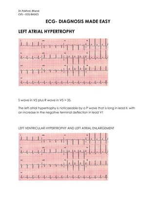
LEFT ATRIAL HYPERTROPHY
S wave in V2 plus R wave in V5 > 35.
The left atrial hypertrophy is noticaeable by a P wave that is long in lead II, with
an increase in the negative terminal deflection in lead V1
LEFT VENTRICULAR HYPERTROPHY AND LEFT ATRIAL ENLARGEMENT
Dr.Pokhrel, Bharat
CVS – ECG BASICS
Left ventricular hypertrophy (S wave V2 plus R wave of V5 greater than 35mm)
and left atrial enlargement (II and V1).
RIGHT VENTRICULAR HYPERTROPHY AND RIGHT ATRIAL ENLARGEMENT
right ventricular hypertrophy and right atrial enlargement
Dr.Pokhrel, Bharat
CVS – ECG BASICS
LEFT VENTRICULAR AND LEFT ATRIAL HYPERTROPHY
Left ventricular and left atrial hypertrophy.
The R wave in aVL is greater than 12mm.
The left atrial hypertrophy is barely noticeable by a P wave that is notched and
wide in lead II and with an increase in the negative terminal deflection in lead
V1.
Dr.Pokhrel, Bharat
CVS – ECG BASICS
RIGHT VENTRICULAR AND RIGHT ATRIAL HYPERTROPHY
Right ventricular and right atrial hypertrophy.
The R wave is greater than the S wave in V1 and the R wave gets progressively
smaller from V1 to V6. Normally, the R wave should increase from V1 to V6.
The right atrial hypertrophy is marked by peaked P waves in lead II and a large
intitial positive deflection of the P wave in lead V1.
BUNDLE BRANCH BLOCK
2ND DEGREE BUNDLE BRANCH BLOCK (MOBITZ TYPE II)
Dr.Pokhrel, Bharat
CVS – ECG BASICS
MOBITZ TYPE I
3RD DEGREE BUNDLE BRANCH BLOCK
complete heart block 3rd degree AV block
Dr.Pokhrel, Bharat
CVS – ECG BASICS
FASCICULAR BLOCK
ANTERIOR FASCICULAR BLOCK
Anterior fascicular block - the most common.
You will see left axis deviation (-30 to -90) and a small Q wave in lead I and an S
in lead III (Q1S3). The QRS will be slightly prolonged (0.1 - 0.12 sec).
Dr.Pokhrel, Bharat
CVS – ECG BASICS
POSTERIOR FASCICULAR BLOCK
Figure 40: Posterior fascicular block.
Posterior fascicular block - less common.
You will see right axis deviation, an S in lead I and an Q in lead III (S1Q3). The
QRS will be slightly prolonged (0.1 - 0.12 sec).
Dr.Pokhrel, Bharat
CVS – ECG BASICS
Figure 41: Right bundle branch block and left anterior fascicular block.
Bifascicular block.
This means two (2) of the three (3) fascicles (in diagram) are blocked. The most
important example is a right bundle branch block and a left anterior fascicular
block. Watch out for this. Only one fascicle is left for conduction, and if that
fasicle is intermittently blocked, the dangerous Mobitz 2 is set up!
"fascicular Blocks" may seem a bit complicated - simply remember that axis
deviation is the clue. In your differential, consider posterior fascicular blocks with
right axis deviation and consider anterior fascicular blocks with left axis
Dr.Pokhrel, Bharat
CVS – ECG BASICS
deviation. Fascicular blocks cause axis deviations, like infarcts and hypertrophy.
If you see a left or right axis deviation, first look for infarct or hypertrophy. If
neither are present, the remaining diagnosis of fascicular block is usually correct.
Review differential diagnosis of right and left axis deviation
Infarct
Accurate ECG interpretation in a patient with chest pain is critical. Basically,
there can be three types of problems - ischemia is a relative lack of blood
supply (not yet an infarct), injury is acute damage occurring right now, and
finally, infarct is an area of dead myocardium. It is important to realize that
certain leads represent certain areas of the left ventricle; by noting which leads
are involved, you can localize the process. The prognosis often varies
depending on which area of the left ventricle is involved (i.e. anterior wall
myocardial infarct generally has a worse prognosis than an inferior wall infarct).
V1-V2 anteroseptal wall
V3-V4 anterior wall
V5-V6 anterolateral wall
II, III,
aVF
inferior wall
I, aVL lateral wall
V1-V2
posterior wall
(reciprocal)
Infarct
1.
Ischemia
Represented by symmetrical T wave
inversion (upside down). The definitive leads for
ischemia are: I, II, V2 - V6.
Dr.Pokhrel, Bharat
CVS – ECG BASICS
2. Injury
Acute damage - look for elevated ST segments.
(Pericarditis and cardiac aneurysm can also
cause ST elevation; remember to correlate it
with the patient.
3. Infarct
Look for significant "patholgic" Q waves. To be
significant, a Q wave must be at least one small
box wide or one-third the entire QRS height.
Remember, to be a Q wave, the initial
deflection must be down; even a tiny initial
upward deflection makes the apparent Q wave
an R wave.
Figure 34: Ischemia: Note symmetric T wave inversions in leads I, V2-V5.
Dr.Pokhrel, Bharat
CVS – ECG BASICS
Figure 35: Injury: Note ST segment elevation in leads V2-V3 (anteroseptal/anterior
wall).
Figure 36: Infarct: Note Q waves in leads II, III, and aVF (inferior wall).
For the posterior wall, remember that vectors representing depolarization of the
anterior and posterior portion of the left ventricle are in opposite directions. So, a
posterior process shows up as opposite of an anterior process in V1. Instead of a
Q wave and ST elevation, you get an R wave and ST depression in V1.
Dr.Pokhrel, Bharat
CVS – ECG BASICS
Figure 37: Posterior wall infarct. Notice tall R wave in V1. Posterior wall infarcts are
often associated with inferior wall infarcts (Q waves in II, III and aVF).
Two other caveats: One is that normally the R wave gets larger as you go to V1
to V6. If there is no R wave "progression" from V1 to V6 this can also mean infarct.
The second caveat is that, with a left bundle branch block, you cannot
evaluate "infarct" on that ECG. In a patient with chest pain and left bundle
branch block, you must rely on cardiac enzymes (blood tests) and the history.
SYSTEMATIC INTERPRETATION GUIDELINES for Electrocardiograms
RATE
Rate calculation
Common method: 300-150-100-75-60-50
Mathematical method: 300/# large boxes between R waves
Six-second method: # R-R intervals x10
RHYTHM
Rhythm Guidelines:
1. Check the bottom rhythm strip for regularity, i.e. - regular, regularly irregular,
and irregularly irregular.
Dr.Pokhrel, Bharat
CVS – ECG BASICS
2. Check for a P wave before each QRS, QRS after each P.
3. Check PR interval (for AV blocks) and QRS (for bundle branch blocks). Check
for prolonged QT.
4. Recognize "patterns" such as atrial fibrillation, PVC's, PAC's, escape beats,
ventricular tachycardia, paroxysmal atrial tachycardia, AV blocks and bundle
branch blocks.
AXIS
Lead I
Lead
aVF
1. Normal axis (0 to +90 degrees) Positive Positive
2. Left axis deviation (-30 to -90) Also
check lead II. To be true left axis
deviation, it should also be down in
lead II.
Positive Negative
3. Right axis deviation (+90 to +180) Negative Positive
4. Indeterminate axis (-90 to -180) Negative Negative
Left axis deviation differential: LVH, left anterior fascicular block, inferior wall MI.
Right axis deviation differential: RVH, left posterior fascicular block, lateral wall
MI.
HYPERTROPHY
1. LVH -- left ventricular hypertrophy = S wave in V1 or V2 + R wave in V5 or V6 >
35mm or aVL R wave > 12mm.
2. RVH -- right ventricular hypertrophy = R wave > S wave in V1 and gets
progressively smaller to left V1-V6 (normally, R wave increases from V1-V6).
3. Atrial hypertrophy (leads II and V1)
Right atrial hypertrophy -- Peaked P wave in lead II > 2.5 mm in amplitude. V1
has increase in the initial positive direction.
Left atrial hypertrophy -- Notched wide (> 3mm) P wave in II. V1 has increase in
the terminal negative direction.
INFARCT
Ischemia
Represented by symmetrical T wave inversion
(upside down). Look in leads I, II, V2-V6.
Injury Acute damage -- look for elevated ST segments.
Infarct
"Pathologic" Q waves. To be significant, a Q
wave must be at least one small square wide or
one-third the entire QRS height.
Dr.Pokhrel, Bharat
CVS – ECG BASICS
Certain leads represent certain areas of the left ventricle:
V1-
V2
anteroseptal wall
II, III,
aVF
inferior wall
V3-
V4
anterior wall I, aVL lateral wall
V5-
V6
anterolateral
wall
V1-V2
posterior wall
(reciprocal)

Weitere ähnliche Inhalte

Was ist angesagt?

12 lead ecg
12 lead ecg12 lead ecg
12 lead ecg
whitmaha
 
Localization of MI on ECG
Localization of MI on ECGLocalization of MI on ECG
Localization of MI on ECG
Nooh Khushal
 
A Guide TO ECG Interpretation
A Guide TO ECG InterpretationA Guide TO ECG Interpretation
A Guide TO ECG Interpretation
meducationdotnet
 
ECG LOCALISATION OF CULPRIT ARTERY IN STEMI
ECG LOCALISATION OF CULPRIT ARTERY IN STEMIECG LOCALISATION OF CULPRIT ARTERY IN STEMI
ECG LOCALISATION OF CULPRIT ARTERY IN STEMI
Praveen Nagula
 
basic ecg diagnosis
basic ecg diagnosisbasic ecg diagnosis
basic ecg diagnosis
jaxboss
 
12 lead ecg
12 lead ecg12 lead ecg
12 lead ecg
jrwas
 

Was ist angesagt? (20)

12 lead ecg
12 lead ecg12 lead ecg
12 lead ecg
 
Localization of MI on ECG
Localization of MI on ECGLocalization of MI on ECG
Localization of MI on ECG
 
Myocardial infarction (MI) ecg localisation
Myocardial infarction (MI) ecg localisationMyocardial infarction (MI) ecg localisation
Myocardial infarction (MI) ecg localisation
 
Ecg changes in mi
Ecg changes in miEcg changes in mi
Ecg changes in mi
 
Tutorial in ecg
Tutorial in ecgTutorial in ecg
Tutorial in ecg
 
ECG Conduction Abnormalities
ECG Conduction AbnormalitiesECG Conduction Abnormalities
ECG Conduction Abnormalities
 
A Guide TO ECG Interpretation
A Guide TO ECG InterpretationA Guide TO ECG Interpretation
A Guide TO ECG Interpretation
 
ECG LOCALISATION OF CULPRIT ARTERY IN STEMI
ECG LOCALISATION OF CULPRIT ARTERY IN STEMIECG LOCALISATION OF CULPRIT ARTERY IN STEMI
ECG LOCALISATION OF CULPRIT ARTERY IN STEMI
 
basic ecg diagnosis
basic ecg diagnosisbasic ecg diagnosis
basic ecg diagnosis
 
Ecg basic guidelines
Ecg basic guidelinesEcg basic guidelines
Ecg basic guidelines
 
ECG Changes in Myocardial Infarction
ECG Changes in Myocardial InfarctionECG Changes in Myocardial Infarction
ECG Changes in Myocardial Infarction
 
ECG Learning
ECG Learning ECG Learning
ECG Learning
 
diagnosis of conduction disorders
diagnosis of conduction disordersdiagnosis of conduction disorders
diagnosis of conduction disorders
 
ECG In Ischemic Heart Disease - Dr Vivek Baliga Review
ECG In Ischemic Heart Disease - Dr Vivek Baliga ReviewECG In Ischemic Heart Disease - Dr Vivek Baliga Review
ECG In Ischemic Heart Disease - Dr Vivek Baliga Review
 
Ecg
EcgEcg
Ecg
 
Ecg final pp ts; 19 06-2012
Ecg final pp ts; 19 06-2012Ecg final pp ts; 19 06-2012
Ecg final pp ts; 19 06-2012
 
Role of ecg in pulmonology
Role of ecg in pulmonologyRole of ecg in pulmonology
Role of ecg in pulmonology
 
TCAD - Multi-lead ECG
TCAD - Multi-lead ECGTCAD - Multi-lead ECG
TCAD - Multi-lead ECG
 
12 lead ecg
12 lead ecg12 lead ecg
12 lead ecg
 
Part ecg stemi nstemi 2019
Part   ecg stemi nstemi  2019Part   ecg stemi nstemi  2019
Part ecg stemi nstemi 2019
 

Ähnlich wie Ecg made easy by pokhrel, bharat

E C G M O H A M E D R A M A D A N
E C G  M O H A M E D  R A M A D A NE C G  M O H A M E D  R A M A D A N
E C G M O H A M E D R A M A D A N
Mohamed Ramadan
 
ecg_systemic_approach_12-lead_compressed.pdf
ecg_systemic_approach_12-lead_compressed.pdfecg_systemic_approach_12-lead_compressed.pdf
ecg_systemic_approach_12-lead_compressed.pdf
jiregnaetichadako
 
Ekg Tutorial
Ekg TutorialEkg Tutorial
Ekg Tutorial
wfrumkin
 
Beginners Guide for ECG Interpretation
Beginners Guide for ECG InterpretationBeginners Guide for ECG Interpretation
Beginners Guide for ECG Interpretation
meducationdotnet
 
Basics of ECG.ppt dr.k.subramanyam
Basics of ECG.ppt dr.k.subramanyamBasics of ECG.ppt dr.k.subramanyam
Basics of ECG.ppt dr.k.subramanyam
Adarsh
 
Instrumental_and_Laboratory_Techniques_of_Examination_in_Pathology of CVS.ppt
Instrumental_and_Laboratory_Techniques_of_Examination_in_Pathology of CVS.pptInstrumental_and_Laboratory_Techniques_of_Examination_in_Pathology of CVS.ppt
Instrumental_and_Laboratory_Techniques_of_Examination_in_Pathology of CVS.ppt
HARSHIKARIZANI
 
Myocardial Ischemia and Infarction
Myocardial Ischemia and InfarctionMyocardial Ischemia and Infarction
Myocardial Ischemia and Infarction
mssa_500
 

Ähnlich wie Ecg made easy by pokhrel, bharat (20)

ECG BY Dr.MOHAMED RAMADAN
ECG BY Dr.MOHAMED RAMADANECG BY Dr.MOHAMED RAMADAN
ECG BY Dr.MOHAMED RAMADAN
 
E C G M O H A M E D R A M A D A N
E C G  M O H A M E D  R A M A D A NE C G  M O H A M E D  R A M A D A N
E C G M O H A M E D R A M A D A N
 
ecg_systemic_approach_12-lead_compressed.pdf
ecg_systemic_approach_12-lead_compressed.pdfecg_systemic_approach_12-lead_compressed.pdf
ecg_systemic_approach_12-lead_compressed.pdf
 
Ekg Tutorial
Ekg TutorialEkg Tutorial
Ekg Tutorial
 
Beginners Guide for ECG Interpretation
Beginners Guide for ECG InterpretationBeginners Guide for ECG Interpretation
Beginners Guide for ECG Interpretation
 
How to read ECG systematically with practice strips
How to read ECG systematically with practice strips How to read ECG systematically with practice strips
How to read ECG systematically with practice strips
 
Basics of ECG.ppt dr.k.subramanyam
Basics of ECG.ppt dr.k.subramanyamBasics of ECG.ppt dr.k.subramanyam
Basics of ECG.ppt dr.k.subramanyam
 
ECG: Indication and Interpretation
ECG: Indication and InterpretationECG: Indication and Interpretation
ECG: Indication and Interpretation
 
Basic of ECG and Easy Interpretation
Basic of ECG and Easy InterpretationBasic of ECG and Easy Interpretation
Basic of ECG and Easy Interpretation
 
PD. ECG 1.ppt
PD. ECG 1.pptPD. ECG 1.ppt
PD. ECG 1.ppt
 
Ecg ecg abnormalities
Ecg ecg abnormalitiesEcg ecg abnormalities
Ecg ecg abnormalities
 
Instrumental_and_Laboratory_Techniques_of_Examination_in_Pathology of CVS.ppt
Instrumental_and_Laboratory_Techniques_of_Examination_in_Pathology of CVS.pptInstrumental_and_Laboratory_Techniques_of_Examination_in_Pathology of CVS.ppt
Instrumental_and_Laboratory_Techniques_of_Examination_in_Pathology of CVS.ppt
 
Basics of ecg
Basics of ecgBasics of ecg
Basics of ecg
 
LOCALIZATION OF CULPRIT ARTERY IN MI.pptx
LOCALIZATION OF CULPRIT ARTERY IN MI.pptxLOCALIZATION OF CULPRIT ARTERY IN MI.pptx
LOCALIZATION OF CULPRIT ARTERY IN MI.pptx
 
ECG BASICS IN DETAIL
ECG BASICS IN DETAILECG BASICS IN DETAIL
ECG BASICS IN DETAIL
 
ECG Basics
ECG BasicsECG Basics
ECG Basics
 
Ecgs
EcgsEcgs
Ecgs
 
Myocardial Ischemia and Infarction
Myocardial Ischemia and InfarctionMyocardial Ischemia and Infarction
Myocardial Ischemia and Infarction
 
Characteristic of normal ECG
Characteristic of normal ECGCharacteristic of normal ECG
Characteristic of normal ECG
 
Ecg basic guidelines
Ecg basic guidelinesEcg basic guidelines
Ecg basic guidelines
 

Mehr von Bharat Pokhrel (14)

Covid 19 MindMap
Covid 19 MindMapCovid 19 MindMap
Covid 19 MindMap
 
Acute appendicitis
Acute appendicitisAcute appendicitis
Acute appendicitis
 
Prader willi syndrome
Prader willi syndromePrader willi syndrome
Prader willi syndrome
 
Complications of intubation
Complications of intubationComplications of intubation
Complications of intubation
 
Endotrachial intubation ppt
Endotrachial intubation pptEndotrachial intubation ppt
Endotrachial intubation ppt
 
Psoriatric arthritis
Psoriatric arthritisPsoriatric arthritis
Psoriatric arthritis
 
Congenital heart disease
Congenital heart diseaseCongenital heart disease
Congenital heart disease
 
Constipation
Constipation Constipation
Constipation
 
Prenatal care
Prenatal care Prenatal care
Prenatal care
 
Cleft lip and palate
Cleft lip and palateCleft lip and palate
Cleft lip and palate
 
Pyelonephritis pokhrel,bharat
Pyelonephritis  pokhrel,bharatPyelonephritis  pokhrel,bharat
Pyelonephritis pokhrel,bharat
 
Hcg and hpl
Hcg and hplHcg and hpl
Hcg and hpl
 
Cardiac diagnostic tests
Cardiac diagnostic testsCardiac diagnostic tests
Cardiac diagnostic tests
 
Upper limb arteries practice questions by pokhrel,bharat
Upper limb arteries practice questions by pokhrel,bharatUpper limb arteries practice questions by pokhrel,bharat
Upper limb arteries practice questions by pokhrel,bharat
 

Kürzlich hochgeladen

❤️Amritsar Escorts Service☎️9815674956☎️ Call Girl service in Amritsar☎️ Amri...
❤️Amritsar Escorts Service☎️9815674956☎️ Call Girl service in Amritsar☎️ Amri...❤️Amritsar Escorts Service☎️9815674956☎️ Call Girl service in Amritsar☎️ Amri...
❤️Amritsar Escorts Service☎️9815674956☎️ Call Girl service in Amritsar☎️ Amri...
Sheetaleventcompany
 
Goa Call Girl Service 📞9xx000xx09📞Just Call Divya📲 Call Girl In Goa No💰Advanc...
Goa Call Girl Service 📞9xx000xx09📞Just Call Divya📲 Call Girl In Goa No💰Advanc...Goa Call Girl Service 📞9xx000xx09📞Just Call Divya📲 Call Girl In Goa No💰Advanc...
Goa Call Girl Service 📞9xx000xx09📞Just Call Divya📲 Call Girl In Goa No💰Advanc...
Sheetaleventcompany
 
Premium Call Girls Nagpur {9xx000xx09} ❤️VVIP POOJA Call Girls in Nagpur Maha...
Premium Call Girls Nagpur {9xx000xx09} ❤️VVIP POOJA Call Girls in Nagpur Maha...Premium Call Girls Nagpur {9xx000xx09} ❤️VVIP POOJA Call Girls in Nagpur Maha...
Premium Call Girls Nagpur {9xx000xx09} ❤️VVIP POOJA Call Girls in Nagpur Maha...
Sheetaleventcompany
 
Dehradun Call Girls Service {8854095900} ❤️VVIP ROCKY Call Girl in Dehradun U...
Dehradun Call Girls Service {8854095900} ❤️VVIP ROCKY Call Girl in Dehradun U...Dehradun Call Girls Service {8854095900} ❤️VVIP ROCKY Call Girl in Dehradun U...
Dehradun Call Girls Service {8854095900} ❤️VVIP ROCKY Call Girl in Dehradun U...
Sheetaleventcompany
 
Kolkata Call Girls Service ❤️🍑 9xx000xx09 👄🫦 Independent Escort Service Kolka...
Kolkata Call Girls Service ❤️🍑 9xx000xx09 👄🫦 Independent Escort Service Kolka...Kolkata Call Girls Service ❤️🍑 9xx000xx09 👄🫦 Independent Escort Service Kolka...
Kolkata Call Girls Service ❤️🍑 9xx000xx09 👄🫦 Independent Escort Service Kolka...
Sheetaleventcompany
 
Call Girl in Chennai | Whatsapp No 📞 7427069034 📞 VIP Escorts Service Availab...
Call Girl in Chennai | Whatsapp No 📞 7427069034 📞 VIP Escorts Service Availab...Call Girl in Chennai | Whatsapp No 📞 7427069034 📞 VIP Escorts Service Availab...
Call Girl in Chennai | Whatsapp No 📞 7427069034 📞 VIP Escorts Service Availab...
amritaverma53
 
Premium Call Girls Dehradun {8854095900} ❤️VVIP ANJU Call Girls in Dehradun U...
Premium Call Girls Dehradun {8854095900} ❤️VVIP ANJU Call Girls in Dehradun U...Premium Call Girls Dehradun {8854095900} ❤️VVIP ANJU Call Girls in Dehradun U...
Premium Call Girls Dehradun {8854095900} ❤️VVIP ANJU Call Girls in Dehradun U...
Sheetaleventcompany
 
Call Girl In Indore 📞9235973566📞 Just📲 Call Inaaya Indore Call Girls Service ...
Call Girl In Indore 📞9235973566📞 Just📲 Call Inaaya Indore Call Girls Service ...Call Girl In Indore 📞9235973566📞 Just📲 Call Inaaya Indore Call Girls Service ...
Call Girl In Indore 📞9235973566📞 Just📲 Call Inaaya Indore Call Girls Service ...
Sheetaleventcompany
 

Kürzlich hochgeladen (20)

❤️Amritsar Escorts Service☎️9815674956☎️ Call Girl service in Amritsar☎️ Amri...
❤️Amritsar Escorts Service☎️9815674956☎️ Call Girl service in Amritsar☎️ Amri...❤️Amritsar Escorts Service☎️9815674956☎️ Call Girl service in Amritsar☎️ Amri...
❤️Amritsar Escorts Service☎️9815674956☎️ Call Girl service in Amritsar☎️ Amri...
 
Goa Call Girl Service 📞9xx000xx09📞Just Call Divya📲 Call Girl In Goa No💰Advanc...
Goa Call Girl Service 📞9xx000xx09📞Just Call Divya📲 Call Girl In Goa No💰Advanc...Goa Call Girl Service 📞9xx000xx09📞Just Call Divya📲 Call Girl In Goa No💰Advanc...
Goa Call Girl Service 📞9xx000xx09📞Just Call Divya📲 Call Girl In Goa No💰Advanc...
 
Ahmedabad Call Girls Book Now 9630942363 Top Class Ahmedabad Escort Service A...
Ahmedabad Call Girls Book Now 9630942363 Top Class Ahmedabad Escort Service A...Ahmedabad Call Girls Book Now 9630942363 Top Class Ahmedabad Escort Service A...
Ahmedabad Call Girls Book Now 9630942363 Top Class Ahmedabad Escort Service A...
 
Premium Call Girls Nagpur {9xx000xx09} ❤️VVIP POOJA Call Girls in Nagpur Maha...
Premium Call Girls Nagpur {9xx000xx09} ❤️VVIP POOJA Call Girls in Nagpur Maha...Premium Call Girls Nagpur {9xx000xx09} ❤️VVIP POOJA Call Girls in Nagpur Maha...
Premium Call Girls Nagpur {9xx000xx09} ❤️VVIP POOJA Call Girls in Nagpur Maha...
 
Race Course Road } Book Call Girls in Bangalore | Whatsapp No 6378878445 VIP ...
Race Course Road } Book Call Girls in Bangalore | Whatsapp No 6378878445 VIP ...Race Course Road } Book Call Girls in Bangalore | Whatsapp No 6378878445 VIP ...
Race Course Road } Book Call Girls in Bangalore | Whatsapp No 6378878445 VIP ...
 
Dehradun Call Girls Service {8854095900} ❤️VVIP ROCKY Call Girl in Dehradun U...
Dehradun Call Girls Service {8854095900} ❤️VVIP ROCKY Call Girl in Dehradun U...Dehradun Call Girls Service {8854095900} ❤️VVIP ROCKY Call Girl in Dehradun U...
Dehradun Call Girls Service {8854095900} ❤️VVIP ROCKY Call Girl in Dehradun U...
 
Kolkata Call Girls Service ❤️🍑 9xx000xx09 👄🫦 Independent Escort Service Kolka...
Kolkata Call Girls Service ❤️🍑 9xx000xx09 👄🫦 Independent Escort Service Kolka...Kolkata Call Girls Service ❤️🍑 9xx000xx09 👄🫦 Independent Escort Service Kolka...
Kolkata Call Girls Service ❤️🍑 9xx000xx09 👄🫦 Independent Escort Service Kolka...
 
Chandigarh Call Girls Service ❤️🍑 9809698092 👄🫦Independent Escort Service Cha...
Chandigarh Call Girls Service ❤️🍑 9809698092 👄🫦Independent Escort Service Cha...Chandigarh Call Girls Service ❤️🍑 9809698092 👄🫦Independent Escort Service Cha...
Chandigarh Call Girls Service ❤️🍑 9809698092 👄🫦Independent Escort Service Cha...
 
❤️Call Girl Service In Chandigarh☎️9814379184☎️ Call Girl in Chandigarh☎️ Cha...
❤️Call Girl Service In Chandigarh☎️9814379184☎️ Call Girl in Chandigarh☎️ Cha...❤️Call Girl Service In Chandigarh☎️9814379184☎️ Call Girl in Chandigarh☎️ Cha...
❤️Call Girl Service In Chandigarh☎️9814379184☎️ Call Girl in Chandigarh☎️ Cha...
 
Cardiac Output, Venous Return, and Their Regulation
Cardiac Output, Venous Return, and Their RegulationCardiac Output, Venous Return, and Their Regulation
Cardiac Output, Venous Return, and Their Regulation
 
💰Call Girl In Bangalore☎️63788-78445💰 Call Girl service in Bangalore☎️Bangalo...
💰Call Girl In Bangalore☎️63788-78445💰 Call Girl service in Bangalore☎️Bangalo...💰Call Girl In Bangalore☎️63788-78445💰 Call Girl service in Bangalore☎️Bangalo...
💰Call Girl In Bangalore☎️63788-78445💰 Call Girl service in Bangalore☎️Bangalo...
 
Call Girl in Chennai | Whatsapp No 📞 7427069034 📞 VIP Escorts Service Availab...
Call Girl in Chennai | Whatsapp No 📞 7427069034 📞 VIP Escorts Service Availab...Call Girl in Chennai | Whatsapp No 📞 7427069034 📞 VIP Escorts Service Availab...
Call Girl in Chennai | Whatsapp No 📞 7427069034 📞 VIP Escorts Service Availab...
 
Premium Call Girls Dehradun {8854095900} ❤️VVIP ANJU Call Girls in Dehradun U...
Premium Call Girls Dehradun {8854095900} ❤️VVIP ANJU Call Girls in Dehradun U...Premium Call Girls Dehradun {8854095900} ❤️VVIP ANJU Call Girls in Dehradun U...
Premium Call Girls Dehradun {8854095900} ❤️VVIP ANJU Call Girls in Dehradun U...
 
Kolkata Call Girls Shobhabazar 💯Call Us 🔝 8005736733 🔝 💃 Top Class Call Gir...
Kolkata Call Girls Shobhabazar  💯Call Us 🔝 8005736733 🔝 💃  Top Class Call Gir...Kolkata Call Girls Shobhabazar  💯Call Us 🔝 8005736733 🔝 💃  Top Class Call Gir...
Kolkata Call Girls Shobhabazar 💯Call Us 🔝 8005736733 🔝 💃 Top Class Call Gir...
 
Call Girl In Indore 📞9235973566📞 Just📲 Call Inaaya Indore Call Girls Service ...
Call Girl In Indore 📞9235973566📞 Just📲 Call Inaaya Indore Call Girls Service ...Call Girl In Indore 📞9235973566📞 Just📲 Call Inaaya Indore Call Girls Service ...
Call Girl In Indore 📞9235973566📞 Just📲 Call Inaaya Indore Call Girls Service ...
 
tongue disease lecture Dr Assadawy legacy
tongue disease lecture Dr Assadawy legacytongue disease lecture Dr Assadawy legacy
tongue disease lecture Dr Assadawy legacy
 
ANATOMY AND PHYSIOLOGY OF REPRODUCTIVE SYSTEM.pptx
ANATOMY AND PHYSIOLOGY OF REPRODUCTIVE SYSTEM.pptxANATOMY AND PHYSIOLOGY OF REPRODUCTIVE SYSTEM.pptx
ANATOMY AND PHYSIOLOGY OF REPRODUCTIVE SYSTEM.pptx
 
Independent Bangalore Call Girls (Adult Only) 💯Call Us 🔝 7304373326 🔝 💃 Escor...
Independent Bangalore Call Girls (Adult Only) 💯Call Us 🔝 7304373326 🔝 💃 Escor...Independent Bangalore Call Girls (Adult Only) 💯Call Us 🔝 7304373326 🔝 💃 Escor...
Independent Bangalore Call Girls (Adult Only) 💯Call Us 🔝 7304373326 🔝 💃 Escor...
 
Exclusive Call Girls Bangalore {7304373326} ❤️VVIP POOJA Call Girls in Bangal...
Exclusive Call Girls Bangalore {7304373326} ❤️VVIP POOJA Call Girls in Bangal...Exclusive Call Girls Bangalore {7304373326} ❤️VVIP POOJA Call Girls in Bangal...
Exclusive Call Girls Bangalore {7304373326} ❤️VVIP POOJA Call Girls in Bangal...
 
Kolkata Call Girls Naktala 💯Call Us 🔝 8005736733 🔝 💃 Top Class Call Girl Se...
Kolkata Call Girls Naktala  💯Call Us 🔝 8005736733 🔝 💃  Top Class Call Girl Se...Kolkata Call Girls Naktala  💯Call Us 🔝 8005736733 🔝 💃  Top Class Call Girl Se...
Kolkata Call Girls Naktala 💯Call Us 🔝 8005736733 🔝 💃 Top Class Call Girl Se...
 

Ecg made easy by pokhrel, bharat

  • 1. Dr.Pokhrel, Bharat CVS – ECG BASICS ECG- DIAGNOSIS MADE EASY LEFT ATRIAL HYPERTROPHY S wave in V2 plus R wave in V5 > 35. The left atrial hypertrophy is noticaeable by a P wave that is long in lead II, with an increase in the negative terminal deflection in lead V1 LEFT VENTRICULAR HYPERTROPHY AND LEFT ATRIAL ENLARGEMENT
  • 2. Dr.Pokhrel, Bharat CVS – ECG BASICS Left ventricular hypertrophy (S wave V2 plus R wave of V5 greater than 35mm) and left atrial enlargement (II and V1). RIGHT VENTRICULAR HYPERTROPHY AND RIGHT ATRIAL ENLARGEMENT right ventricular hypertrophy and right atrial enlargement
  • 3. Dr.Pokhrel, Bharat CVS – ECG BASICS LEFT VENTRICULAR AND LEFT ATRIAL HYPERTROPHY Left ventricular and left atrial hypertrophy. The R wave in aVL is greater than 12mm. The left atrial hypertrophy is barely noticeable by a P wave that is notched and wide in lead II and with an increase in the negative terminal deflection in lead V1.
  • 4. Dr.Pokhrel, Bharat CVS – ECG BASICS RIGHT VENTRICULAR AND RIGHT ATRIAL HYPERTROPHY Right ventricular and right atrial hypertrophy. The R wave is greater than the S wave in V1 and the R wave gets progressively smaller from V1 to V6. Normally, the R wave should increase from V1 to V6. The right atrial hypertrophy is marked by peaked P waves in lead II and a large intitial positive deflection of the P wave in lead V1. BUNDLE BRANCH BLOCK 2ND DEGREE BUNDLE BRANCH BLOCK (MOBITZ TYPE II)
  • 5. Dr.Pokhrel, Bharat CVS – ECG BASICS MOBITZ TYPE I 3RD DEGREE BUNDLE BRANCH BLOCK complete heart block 3rd degree AV block
  • 6. Dr.Pokhrel, Bharat CVS – ECG BASICS FASCICULAR BLOCK ANTERIOR FASCICULAR BLOCK Anterior fascicular block - the most common. You will see left axis deviation (-30 to -90) and a small Q wave in lead I and an S in lead III (Q1S3). The QRS will be slightly prolonged (0.1 - 0.12 sec).
  • 7. Dr.Pokhrel, Bharat CVS – ECG BASICS POSTERIOR FASCICULAR BLOCK Figure 40: Posterior fascicular block. Posterior fascicular block - less common. You will see right axis deviation, an S in lead I and an Q in lead III (S1Q3). The QRS will be slightly prolonged (0.1 - 0.12 sec).
  • 8. Dr.Pokhrel, Bharat CVS – ECG BASICS Figure 41: Right bundle branch block and left anterior fascicular block. Bifascicular block. This means two (2) of the three (3) fascicles (in diagram) are blocked. The most important example is a right bundle branch block and a left anterior fascicular block. Watch out for this. Only one fascicle is left for conduction, and if that fasicle is intermittently blocked, the dangerous Mobitz 2 is set up! "fascicular Blocks" may seem a bit complicated - simply remember that axis deviation is the clue. In your differential, consider posterior fascicular blocks with right axis deviation and consider anterior fascicular blocks with left axis
  • 9. Dr.Pokhrel, Bharat CVS – ECG BASICS deviation. Fascicular blocks cause axis deviations, like infarcts and hypertrophy. If you see a left or right axis deviation, first look for infarct or hypertrophy. If neither are present, the remaining diagnosis of fascicular block is usually correct. Review differential diagnosis of right and left axis deviation Infarct Accurate ECG interpretation in a patient with chest pain is critical. Basically, there can be three types of problems - ischemia is a relative lack of blood supply (not yet an infarct), injury is acute damage occurring right now, and finally, infarct is an area of dead myocardium. It is important to realize that certain leads represent certain areas of the left ventricle; by noting which leads are involved, you can localize the process. The prognosis often varies depending on which area of the left ventricle is involved (i.e. anterior wall myocardial infarct generally has a worse prognosis than an inferior wall infarct). V1-V2 anteroseptal wall V3-V4 anterior wall V5-V6 anterolateral wall II, III, aVF inferior wall I, aVL lateral wall V1-V2 posterior wall (reciprocal) Infarct 1. Ischemia Represented by symmetrical T wave inversion (upside down). The definitive leads for ischemia are: I, II, V2 - V6.
  • 10. Dr.Pokhrel, Bharat CVS – ECG BASICS 2. Injury Acute damage - look for elevated ST segments. (Pericarditis and cardiac aneurysm can also cause ST elevation; remember to correlate it with the patient. 3. Infarct Look for significant "patholgic" Q waves. To be significant, a Q wave must be at least one small box wide or one-third the entire QRS height. Remember, to be a Q wave, the initial deflection must be down; even a tiny initial upward deflection makes the apparent Q wave an R wave. Figure 34: Ischemia: Note symmetric T wave inversions in leads I, V2-V5.
  • 11. Dr.Pokhrel, Bharat CVS – ECG BASICS Figure 35: Injury: Note ST segment elevation in leads V2-V3 (anteroseptal/anterior wall). Figure 36: Infarct: Note Q waves in leads II, III, and aVF (inferior wall). For the posterior wall, remember that vectors representing depolarization of the anterior and posterior portion of the left ventricle are in opposite directions. So, a posterior process shows up as opposite of an anterior process in V1. Instead of a Q wave and ST elevation, you get an R wave and ST depression in V1.
  • 12. Dr.Pokhrel, Bharat CVS – ECG BASICS Figure 37: Posterior wall infarct. Notice tall R wave in V1. Posterior wall infarcts are often associated with inferior wall infarcts (Q waves in II, III and aVF). Two other caveats: One is that normally the R wave gets larger as you go to V1 to V6. If there is no R wave "progression" from V1 to V6 this can also mean infarct. The second caveat is that, with a left bundle branch block, you cannot evaluate "infarct" on that ECG. In a patient with chest pain and left bundle branch block, you must rely on cardiac enzymes (blood tests) and the history. SYSTEMATIC INTERPRETATION GUIDELINES for Electrocardiograms RATE Rate calculation Common method: 300-150-100-75-60-50 Mathematical method: 300/# large boxes between R waves Six-second method: # R-R intervals x10 RHYTHM Rhythm Guidelines: 1. Check the bottom rhythm strip for regularity, i.e. - regular, regularly irregular, and irregularly irregular.
  • 13. Dr.Pokhrel, Bharat CVS – ECG BASICS 2. Check for a P wave before each QRS, QRS after each P. 3. Check PR interval (for AV blocks) and QRS (for bundle branch blocks). Check for prolonged QT. 4. Recognize "patterns" such as atrial fibrillation, PVC's, PAC's, escape beats, ventricular tachycardia, paroxysmal atrial tachycardia, AV blocks and bundle branch blocks. AXIS Lead I Lead aVF 1. Normal axis (0 to +90 degrees) Positive Positive 2. Left axis deviation (-30 to -90) Also check lead II. To be true left axis deviation, it should also be down in lead II. Positive Negative 3. Right axis deviation (+90 to +180) Negative Positive 4. Indeterminate axis (-90 to -180) Negative Negative Left axis deviation differential: LVH, left anterior fascicular block, inferior wall MI. Right axis deviation differential: RVH, left posterior fascicular block, lateral wall MI. HYPERTROPHY 1. LVH -- left ventricular hypertrophy = S wave in V1 or V2 + R wave in V5 or V6 > 35mm or aVL R wave > 12mm. 2. RVH -- right ventricular hypertrophy = R wave > S wave in V1 and gets progressively smaller to left V1-V6 (normally, R wave increases from V1-V6). 3. Atrial hypertrophy (leads II and V1) Right atrial hypertrophy -- Peaked P wave in lead II > 2.5 mm in amplitude. V1 has increase in the initial positive direction. Left atrial hypertrophy -- Notched wide (> 3mm) P wave in II. V1 has increase in the terminal negative direction. INFARCT Ischemia Represented by symmetrical T wave inversion (upside down). Look in leads I, II, V2-V6. Injury Acute damage -- look for elevated ST segments. Infarct "Pathologic" Q waves. To be significant, a Q wave must be at least one small square wide or one-third the entire QRS height.
  • 14. Dr.Pokhrel, Bharat CVS – ECG BASICS Certain leads represent certain areas of the left ventricle: V1- V2 anteroseptal wall II, III, aVF inferior wall V3- V4 anterior wall I, aVL lateral wall V5- V6 anterolateral wall V1-V2 posterior wall (reciprocal)