DIGESTIVE SYSTEM
•
0 gefällt mir•82 views

it is useful for the Diploma in pharmacy and Bachelor of Pharmacy students and Doctor of Pharmacy Students and MBBS and BDS

Empfohlen
Weitere ähnliche Inhalte
Ähnlich wie DIGESTIVE SYSTEM
Ähnlich wie DIGESTIVE SYSTEM (20)
Mehr von SURESH BABU EMANDI DEPARTMENT OF PHARMACOGNOSY Vikas Institute of Pharmaceutical scienes
Mehr von SURESH BABU EMANDI DEPARTMENT OF PHARMACOGNOSY Vikas Institute of Pharmaceutical scienes (20)
Kürzlich hochgeladen
Kürzlich hochgeladen (20)
DIGESTIVE SYSTEM
- 1. DIGIVE SYSTEMEST SURESH BABU EMANDI M.Pharm Vikas Institute of Pharmaceutical Sciences NearAir Port, Rajahmundry-533103.
- 2. Introduction • Digestion is defined as the process by which food is broken down into simple chemical substances that can be absorbed and used as nutrients by the body. • Most of the substances in the diet cannot be utilized as such. These substances must be broken into smaller particles, so that they can be absorbed into blood and distributed to various parts of the body for utilization. Digestive system is responsible for these functions.
- 3. Introduction • Digestive process is accomplished by mechanical and enzymatic breakdown of food into simpler chemical compounds. A normal young healthy adult consumes about 1 kg of solid diet and about 1 to 2 liter of liquid diet every day. All these food materials are subjected to digestive process, before being absorbed into blood and distributed to the tissues of the body. Digestive system plays the major role in the digestion and absorption of food substances.
- 4. Functions of digestive system • 1. Ingestion or consumption of food substances. • 2. Breaking them into small particles. • 3. Transport of small particles to different areas of the digestive tract. • 4. Secretion of necessary enzymes and other substances for digestion. • 5. Digestion of the food particles. • 6. Absorption of the digestive products (nutrients) • 7. Removal of unwanted substances from the body.
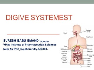
- 6. FUNCTIONALANATOMYOF DIGESTIVE SYSTEM • Digestive system is made up of gastrointestinal tract (GI tract) or alimentary canal and accessory organs, which help in the process of digestion and absorption. • GI tract is a tubular structure extending from the mouth up to anus, with a length of about 30 feet. • It opens to the external environment on both ends. • GI tract is formed by two types of organs: • 1. Primary digestive organs. • 2. Accessory digestive organs.
- 7. 1. Primary Digestive Organs • Primary digestive organs are the organs where actual digestion takes place. • Primary digestive organs are: • Mouth • Pharynx • Esophagus • Stomach • Duodenum • Small intestine • Large intestine. • Rectum and Anus.
- 8. 2. AccessoryDigestive Organs • Accessory digestive organs are those which help primary digestive organs in the process of digestion. • Accessory digestive organs are: • i. Teeth • ii. Tongue • iii. Salivary glands • iv. Exocrine part of pancreas • v. Liver • vi. Gallbladder.
- 9. Mouth and Salivary Glands
- 10. FUNCTIONALANATOMY OF MOUTH • Mouth is otherwise known as oral cavity or buccal cavity. It is formed by cheeks, lips and palate. • It encloses the teeth, tongue and salivary glands. • Mouth opens anteriorly to the exterior through lips and posteriorly through faces into the pharynx. • Digestive juice present in the mouth is saliva, which is secreted by the salivary glands.
- 11. FUNCTIONS OF MOUTH • Primary function of mouth is eating and it has few other • important functions also. • Functions of mouth include: • 1. Ingestion of food materials • 2. Chewing the food and mixing it with saliva • 3. Appreciation of taste of the food • 4. Transfer of food (bolus) to the esophagus by • swallowing • 5. Role in speech • 6. Social functions such as smiling and other • expressions.
- 12. SALIVARY GLANDS • In humans, the saliva is secreted by three pairs of • Major (larger) salivary glands • Minor (small) salivary glands.
- 13. MAJORSALIVARYGLANDS • Major glands are: • 1. Parotid glands • 2. Submaxillary or submandibular glands • 3. Sublingual glands.
- 14. 1. Parotid Glands • Parotid glands are the largest of all salivary glands, situated at the side of the face just below and in front of the ear. Each gland weighs about 20 to 30 g in adults. Secretions from these glands are emptied into the oral cavity by Stensen duct. This duct is about 35 mm to 40 mm long and opens inside the cheek against the upper second molar tooth.
- 15. 2. Submaxillary Glands • Submaxillary glands or submandibular glands are located in submaxillary triangle, medial to mandible. • Each gland weighs about 8 to 10 g. • Saliva from these glands is emptied into the oral cavity by Wharton duct, which is about 40 mm long. • The duct opens at the side of frenulum of tongue, by means of a small opening on the summit of papilla called caruncula sublingualis.
- 16. 3. Sublingual Glands • Sublingual glands are the smallest salivary glands situated in the mucosa at the floor of the mouth. • Each gland weighs about 2 to 3 g. • Saliva from these glands is poured into 5 to 15 small ducts called ducts of Rivinus. • These ducts open on small papillae beneath the tongue. One of the ducts is larger and it is called Bartholin duct. • It drains the anterior part of the gland and opens on caruncula sublingualis near the opening of submaxillary duct.
- 17. MINORSALIVARYGLANDS • 1. Lingual Mucus Glands Lingual mucus glands are situated in posterior one third of the tongue, behind circumvallate papillae and at the tip and margins of tongue. • 2. Lingual Serous Glands Lingual serous glands are located near circumvallate papillae and filiform papillae. • 3. Buccal Glands Buccal glands or molar glands are present between the mucus membrane and buccinator muscle. Four to five of these are larger and situated outside buccinator, around the terminal part of parotid duct. • 4. Labial Glands Labial glands are situated beneath the mucus membrane around the orifice of mouth • 5. Palatal Glands Palatal glands are found beneath the mucus membrane of the soft palate.
- 18. STRUCTURE AND DUCT SYSTEM OF SALIVARYGLANDS • Salivary glands are formed by acini or alveoli. • Each acinus is formed by a small group of cells which surround a central globular cavity. Central cavity of each acinus is continuous with the lumen of the duct. The fine duct draining each acinus is called intercalated duct. Many intercalated ducts join together to form intralobular duct. • Few intralobular ducts join to form interlobular ducts,which unite to form the main duct of the gland. • A gland with this type of structure and duct system is called racemose type (racemose = bunch of grapes).
- 20. Contributionby each major salivary gland is • i. Parotid glands : 25% • ii. Submaxillary glands : 70% • iii. Sublingual glands : 5%.
- 21. PROPERTIESOF SALIVA 1. Volume: 1000 mL to 1500 mL of saliva is secreted per day and it is approximately about 1 mL/minute. 2. Reaction: Mixed saliva from all the glands is slightly acidic with pH of 6.35 to 6.85. 3. Specific gravity: It ranges between 1.002 and 1.012 4. 4. Tonicity: Saliva is hypotonic to plasma.
- 22. COMPOSITION OF SALIVA • Mixed saliva contains 99.5% water and 0.5% solids.
- 23. FUNCTIONS OF SALIVA • Saliva is a very essential digestive juice. Since it has many functions, its absence leads to many inconveniences. ➢Preparation of food for swallowing. ➢Appreciation of taste. ➢Digestive function ➢Role in speech ➢Excretory function. ➢Regulation of body temperature. ➢Regulation of water balance
- 24. PREPARATIONOF FOODFOR SWALLOWING ➢When food is taken into the mouth, it is moistened and dissolved by saliva. ➢The mucus membrane of mouth is also moistened by saliva. ➢It facilitates chewing. ➢By the movement of tongue, the moistened and masticated ➢Food is rolled into a bolus. ➢Mucin of saliva lubricates the bolus and facilitates swallowing.
- 25. Digestivefunction • Saliva has three digestive enzymes, • Salivary amylase, • Maltase • Lingual lipase
- 26. APPRECIATIONOF TASTE • Taste is a chemical sensation. By its solvent action, • saliva dissolves the solid food substances, so that the dissolved substances can stimulate the taste buds. The stimulated taste buds recognize the taste.
- 27. Role in speech • By moistening and lubricating soft parts of mouth and lips, saliva helps in speech. • If the mouth becomes dry, articulation and pronunciation becomes difficult.
- 28. CLEANSING ANDPROTECTIVE FUNCTIONS • i. Due to the constant secretion of saliva, the mouth and teeth are rinsed and kept free off food debris, shed epithelial cells and foreign particles. In this way, saliva prevents bacterial growth by removing materials, which may serve as culture media for the bacterial growth. • ii. Enzyme lysozyme of saliva kills some bacteria such as staphylococcus, streptococcus and brucella.
- 29. EXCRETORYFUNCTION • Many substances, both organic and inorganic, are • excreted in saliva. • It excretes substances like mercury, potassium iodide, lead, and thiocyanate. • Saliva also excretes some viruses such as those causing rabies and mumps.
- 30. REGULATIONOF BODYTEMPERATURE • In dogs and cattle, excessive dripping of saliva during panting helps in the loss of heat and regulation of body temperature. However, in human beings, sweat glands play a major role in temperature regulation and saliva does not play any role in this function.
- 31. REGULATION OF WATER BALANCE • When the body water content decreases, salivary secretion also decreases. • This causes dryness of the mouth and induces thirst. When water is taken, it quenches the thirst and restores the body water content.
- 32. Stomach
- 33. Structureof stomach • The stomach is a J-shaped dilated portion of the alimentary tract situated in the epigastric, umbilical and left hypochondriac regions of the abdominal cavity. • Stomach is a hollow organ situated just below the diaphragm on the left side in the abdominal cavity. • Volume of empty stomach is 50 mL. • Under normal conditions, it can expand to accommodate 1 L to 1.5 L of solids and liquids. However, it is capable of expanding still further up to 4 L.
- 34. PARTS OF STOMACH stomach has four parts: • 1. Cardiac region • 2. Fundus • 3. Body or corpus • 4. Pyloric region. • 1. Cardiac Region • Cardiac region is the upper part of the stomach where esophagus opens. The opening is guarded by a sphincter called cardiac sphincter, which opens only towards stomach. This portion is also known as cardiac end.
- 35. 2. Fundus • Fundus is a small dome shaped structure. It is elevated above the level of esophageal opening 3. Body or Corpus Body is the largest part of stomach forming about 75% to 80% of the whole stomach. It extends from just below the fundus up to the pyloric region. 4. Pyloric Region Pyloric region has two parts, antrum and pyloric canal. The body of stomach ends in antrum.
- 36. • Junction between body and antrum is marked by an angular notch called incisura angularis. • Antrum is continued as the narrow canal, which is called pyloric canal or pyloric end. • Pyloric canal opens into first part of small intestine called duodenum. • The opening of pyloric canal is guarded by a sphincter called pyloric sphincter. It opens towards duodenum. Stomach has two curvatures. • One on the right side is lesser curvature and the other on left side is greater curvature.
- 37. STRUCTURE OF STOMACH WALL Stomach wall is formed by four layers of structures: 1. Outer serous layer: Formed by peritoneum. 2. Muscular layer: Made up of three layers of smooth muscle fibers, namely inner oblique, middle circular and outer longitudinal layers. 3. Submucus layer: Formed by areolar tissue, blood vessels, lymph vessels and Meissner nerve plexus. 4. Inner mucus layer: Lined by mucussecreting columnar epithelial cells. The gastric glands are situated in this layer. Under resting conditions, the mucosa of the stomach is thrown into many folds. These folds are called rugae. The rugae disappear when the stomach is distended after meals. Throughout the inner mucus layer, small depressions called gastric pits are present. Glands of the stomach open into these pits. Inner surface of mucus layer is covered by 2 mm thick mucus.
- 38. GLANDS OF STOMACH – GASTRIC GLANDS • Glands of the stomach or gastric glands are tubular structures made up of different types of cells. These glands open into the stomach cavity via gastric pits.
- 39. CLASSIFICATION OF GLANDS OF THE STOMACH • Gastric glands are classified into three types, on the basis of their location in the stomach: • 1. Fundic glands or main gastric glands or oxyntic glands: Situated in body and fundus of stomach • 2. Pyloric glands: Present in the pyloric part of the stomach • 3. Cardiac glands: Located in the cardiac region of the stomach
- 40. Parietal cells are different from other cells of the gland because of the presence of canaliculi (singular = canaliculus). Parietal cells empty their secretions into the lumen of the gland through the canaliculi. But, other cells empty their secretions directly into lumen of the gland.
- 41. STRUCTURE OF GASTRIC GLANDS 1. Fundic Glands Fundic glands are considered as the typical gastric glands. • These glands are long and tubular. • Each gland has three parts, viz. body, neck and isthmus. Cells of fundic glands • 1. Chief cells or pepsinogen cells • 2. Parietal cells or oxyntic cells • 3. Mucus neck cells • 4. Enterochromaffin (EC) cells or Kulchitsky cells • 5. Enterochromaffinlike (ECL) cells.
- 42. Pyloric Glands • Pyloric glands are short and tortuous in nature. • These glands are formed by • G cells, • Mucus cells, • EC cells and • ECL cells.
- 43. FUNCTIONS OF GASTRIC GLANDS • Function of the gastric gland is to secrete gastric juice. • Secretory activities of different cells of gastric glands and enteroendocrine cells.
- 44. Cardiac Glands Cardiac glands are also short and tortuous in structure, with many mucus cells. EC cells, ECL cells Chief cells are also present in the cardiac glands
- 45. 1. MECHANICAL FUNCTION i. Storage Function • Food is stored in the stomach for a long period, i.e. for 3 to 4 hours and emptied into the intestine slowly. • The maximum capacity of stomach is up to 1.5 L. Slow emptying of stomach provides enough time for proper digestion and absorption of food substances in the small intestine. ii. Formation of Chyme • Peristaltic movements of stomach mix the bolus with gastric juice and convert it into the semisolid material known as chyme
- 46. DIGESTIVEFUNCTION • „ DIGESTIVE FUNCTION Gastric juice acts mainly on proteins. Proteolytic enzymes of the gastric juice are pepsin and rennin.
- 47. • Gastric juice also contains some other enzymes like gastric lipase, gelatinase, urase and gastric amylase. Pepsin Pepsin is secreted as inactive pepsinogen. • Pepsinogen is converted into pepsin by hydrochloric acid. Optimum pH for activation of pepsinogen is below 6. • Action of pepsin Pepsin converts proteins into proteoses, peptones and polypeptides. • Pepsin also causes curdling and digestion of milk (casein). • Gastric Lipase Gastric lipase is a weak lipolytic enzyme when compared to pancreatic lipase. • It is active only when the pH is between 4 and 5 and becomes inactive at a pH below
- 48. PROTECTIVEFUNCTION • FUNCTION OF MUCUS • Mucus is a mucoprotein, secreted by mucus neck cells of the gastric glands and surface mucus cells in fundus, body and other parts of stomach. It protects the gastric wall by the following ways: Mucus: i. Protects the stomach wall from irritation or mechanical injury, by virtue of its high viscosity
- 49. HEMOPOIETICFUNCTION • Intrinsic factor of Castle, secreted by parietal cells of gastric glands plays an important role in erythropoiesis. • It is necessary for the absorption of vitamin B12 (which is called extrinsic factor) from GI tract into the blood. • Vitamin B12 is an important maturation factor during erythropoiesis. • Absence of intrinsic factor in gastric juice causes deficiency of vitamin B12, leading to pernicious anemia
- 50. EXCRETORYFUNCTION • Many substances like toxins, alkaloids and metals are excreted through gastric juice
- 51. GASTRICOMPOSITIONOF C JUICEPROPERTIES Gastric juice is a mixture of secretions from different gastric glands. PROPERTIES OF GASTRIC JUICE • Volume : 1200 mL/day to 1500 mL/day. • Reaction : Gastric juice is highly acidic with a pH of 0.9 to 1.2. Acidity of gastric juice is due to the presence of hydrochloric acid. • Specific gravity : 1.002 to 1.004
- 52. COMPOSITION OF GASTRIC JUICE • Gastric juice contains 99.5% of water and 0.5% solids. Solids are organic and inorganic substances.for composition of gastric juice.
- 53. COMPOSITION OF GASTRIC JUICE
- 54. FUNCTIONS OF GASTRIC JUICE • FUNCTIONS OF HYDROCHLORIC ACID • Hydrochloric acid is present in the gastric juice: • i. Activates pepsinogen into pepsin • ii. Kills some of the bacteria entering the stomach along with food substances. This action is called bacteriolytic action • iii. Provides acid medium, which is necessary for the action of hormones.