Anatomy of Blood vessels of abdomen pelvic cavities. Portacaval & Cavacaval Anastomoses. Fetal Circulation
•Als PPT, PDF herunterladen•
23 gefällt mir•4,527 views

1. The abdominal aorta a. the parietal branches b. the visceral branches 2. The common iliac arteries and veins 3. The external iliac artery and veins 4. The internal iliac artery and veins 5. The inferior vena cava 6. The portal vein 7. The cavacacal Anastomoses 8. The portacaval Anastomoses 9. The Fetal Circulation

Empfohlen
Weitere ähnliche Inhalte
Was ist angesagt?
Was ist angesagt? (20)
Ähnlich wie Anatomy of Blood vessels of abdomen pelvic cavities. Portacaval & Cavacaval Anastomoses. Fetal Circulation
Ähnlich wie Anatomy of Blood vessels of abdomen pelvic cavities. Portacaval & Cavacaval Anastomoses. Fetal Circulation (20)
Mehr von Eneutron
Mehr von Eneutron (20)
Kürzlich hochgeladen
Kürzlich hochgeladen (20)
Anatomy of Blood vessels of abdomen pelvic cavities. Portacaval & Cavacaval Anastomoses. Fetal Circulation
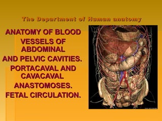
- 1. The Department of Human anatomyThe Department of Human anatomy ANATOMY OF BLOODANATOMY OF BLOOD VESSELS OFVESSELS OF ABDOMINALABDOMINAL AND PELVIC CAVITIES.AND PELVIC CAVITIES. PORTACAVALPORTACAVAL ANDAND CAVACAVACAVALCAVAL AANASTOMOSESNASTOMOSES.. FETAL CIRCULATION.FETAL CIRCULATION.
- 2. PLANPLAN THE ABDOMINAL AORTATHE ABDOMINAL AORTA a) the parietal branchesa) the parietal branches b) the visceral branchesb) the visceral branches THE COMMON ILIAC ARTERIES AND VEINS THE EXTERNAL ILIAC ARTERY AND VEINS THE INTERNAL ILIAC ARTERY AND VEINS THE INFERIOR VENA CAVATHE INFERIOR VENA CAVA THE PORTAL VEINTHE PORTAL VEIN THE CAVATHE CAVACAVALCAVAL AANASTOMOSESNASTOMOSES THETHE PORTACAVALPORTACAVAL AANASTOMOSESNASTOMOSES THE FETAL CIRCULATION.THE FETAL CIRCULATION.
- 3. THE ABDOMINAL AORTATHE ABDOMINAL AORTA Relations of the abdominal aorta: The abdominal aorta begins its course from the aortic hiatus of the diaphragm and terminates with the aortic bifurcation at L4. The abdominal aorta descends along the left side of vertebral column. The inferior vena cava resides on the right. The abdominal aorta gives rise to parietal and visceral (paired and unpaired) branches
- 4. TheThe abdominalabdominal aortaaorta bifurcates anteriorbifurcates anterior to the left side of L4 intoto the left side of L4 into thethe common iliaccommon iliac arteriesarteries. These two. These two arteries diverge andarteries diverge and further divide into thefurther divide into the externalexternal andand internalinternal iliac arteriesiliac arteries on eachon each side at the level of theside at the level of the lumbosacral intervertebrallumbosacral intervertebral disc.disc.
- 5. SponsoredSponsored Medical Lecture Notes –Medical Lecture Notes – All SubjectsAll Subjects USMLE Exam (America) –USMLE Exam (America) – PracticePractice
- 6. The parietal branchesThe parietal branches ofof abdominal aortaabdominal aorta are likeare like the followingthe following:: thethe inferior phrenic arteryinferior phrenic artery,, arteriaarteria phrenica inferior,phrenica inferior, paired, it arisespaired, it arises from the uppermost segment of thefrom the uppermost segment of the abdominal aorta. It supplies theabdominal aorta. It supplies the diaphragm and the suprarenal glanddiaphragm and the suprarenal gland thethe lumbar arterieslumbar arteries,, arteriaearteriae lumbaleslumbales (4 pairs) run laterally(4 pairs) run laterally crossing the bodies of lumbarcrossing the bodies of lumbar vertebrae. They supply the respectivevertebrae. They supply the respective vertebrae, the muscles of back andvertebrae, the muscles of back and abdominal wall.abdominal wall. thethe median sacral arterymedian sacral artery,, arteriaarteria sacralis medianasacralis mediana is the unpairedis the unpaired branch that arises from the aorticbranch that arises from the aortic bifurcation. It descends along thebifurcation. It descends along the pelvic surface of sacrum to the lesserpelvic surface of sacrum to the lesser pelvispelvis
- 7. THE VISCERAL BRANCHES of abdominal aorta The paired branches: -Suprarenal artery -Renal artery -Ovarian artery/Testicular The unpaired branches: •Celiac artery: supplies the abdominal viscera •Left gastric artery: supplies the stomach and esophagus •Splenic artery: supplies the spleen, stomach and pancreas •Common hepatic artery: supplies the liver •Superior mesenteric artery: supplies the pancreas, small intestine and colon •Inferior mesenteric artery: supplies the descending colon and rectum
- 9. TheThe external iliacexternal iliac arteryartery gives off thegives off the inferior epigastricinferior epigastric andand deep circumflexdeep circumflex iliac arteriesiliac arteries andand continues as thecontinues as the femoral artery in thefemoral artery in the lower limb.lower limb. TheThe internal iliacinternal iliac arteryartery gives manygives many branches and thesebranches and these are divided intoare divided into anterior and posterioranterior and posterior trunk divisions.trunk divisions.
- 10. Branches of the Posterior Trunk of the Internal Iliac Artery - The iliolumbar artery ascends to the medial border of the psoas major muscle. -The lateral sacral arteries. -The superior gluteal artery. -The inferior gluteal artery.
- 11. Branches from the Anterior Trunk of Internal Iliac Artery 1.The superior vesical artery. 2.The inferior vesical artery (in males). 3.The middle rectal artery. 4.The uterine artery (in females). 5.The vaginal artery (corresponds to the inferior vesical artery of males). 6.The obturator artery. 7.The internal pudendal artery: The artery gives the branches as follows: - the inferior rectal artery. - the artery of bulb of penis, - the dorsal artery of penis (clitoris), - the deep artery of penis (clitoris), - the perineal artery, - the posterior labial branches (the posterior scrotal branches), - the inferior gluteal artery.
- 12. Figure 21.31 The Venous Drainage of theThe Venous Drainage of the Abdomen and ChestAbdomen and Chest
- 13. TheThe inferior venainferior vena cavacava ((v. cava Inferiorv. cava Inferior),), the thickestthe thickest venous trunk in the body,venous trunk in the body, lies in the abdominal cavitylies in the abdominal cavity close to the right side of theclose to the right side of the aorta.aorta. Lying in theLying in the sulcus venaesulcus venae cavaecavae on the posterioron the posterior surface of the liver, thesurface of the liver, the inferior vena cava theninferior vena cava then passes through the foramenpasses through the foramen venae cavaevenae cavae of theof the diaphragm into the thoracicdiaphragm into the thoracic cavity and immediatelycavity and immediately drains into the right atrium.drains into the right atrium.
- 14. TheThe parietalparietal veins:veins: (1)(1) the right and leftthe right and left lumbar veinslumbar veins (vv. lumbales dextrae(vv. lumbales dextrae andand sinistraesinistrae vv. lumbalesvv. lumbales ascendensascendens));; (2(2) the phrenic veins) the phrenic veins (vv. phrenicae(vv. phrenicae inferiores).inferiores). Tributaries of the inferior vena cava.Tributaries of the inferior vena cava. Venous drainage of abdominal organs notVenous drainage of abdominal organs not drained by the hepatic portal vein.drained by the hepatic portal vein. They are divided into parietal and visceral veins.
- 15. 1)1) the testicularthe testicular veinsveins (vv.(vv. testiculares) intesticulares) in males (ovarianmales (ovarian veins in females)veins in females) 2)2) the renal veinsthe renal veins (vv. renales);(vv. renales); 3)3) the rightthe right suprarenal veinsuprarenal vein (v.(v. suprarenalissuprarenalis dextra);dextra); 4)4) the hepatic veinsthe hepatic veins (vv. hepaticae)(vv. hepaticae) The visceral veins:
- 16. Veins of Hepatic PortalVeins of Hepatic Portal SystemSystem Drains blood fromDrains blood from viscera (stomach,viscera (stomach, spleen and intestines)spleen and intestines) to liver so thatto liver so that nutrients are absorbednutrients are absorbed It is formed by the 1. Superior mesenteric vein 2. Inferior mesenteric vein 3.Splenic vein. The portal vein and its tributaries
- 17. The cavaThe cava--cavalcaval aanastomosesnastomosesThe anterior abdominal wall: - the superior epigastric veins(SVC system) and the inferior epigastric veins(IVC system).
- 18. The posterior abdominal wall: -the vertebral venous plexuses, -the sacral and lumbar veins (IVC system) and the vertebral, azygos and hemiazygos veins(SVC system), -the azygos and hemiazygos veins(SVC system) and ascending lumbar veins (IVC system) The cavacaval anastomoses
- 19. The porto-caval anastomoses: - The oesophageal (SVC system) and left gastric veins (HPV system). - The paraumbilical veins (HPV system) and the veins of anterior abdominal wall (SVC and IVC systems). - The superior rectal veins (HPV system) and middle, inferior rectal veins (IVC system).
- 20. - Pair of umbilical arteries carry deoxygenated blood & wastes to placenta. - Umbilical vein carries oxygenated blood and nutrients from the placenta. Fetal Circulation
- 21. Facilitates gasFacilitates gas and nutrientand nutrient exchangeexchange betweenbetween maternal andmaternal and fetal blood.fetal blood. The blood itselfThe blood itself does not mix.does not mix. The Placenta
- 22. Some blood from the umbilical vein enters the portal circulation allowing the liver to process nutrients. The majority of the blood enters the ductus venosus, a shunt which bypasses the liver and puts blood into the hepatic veins. Umbilical vein to portal circulation
- 23. Foramen ovale Blood is shunted from right atrium to left atrium, skipping the lungs. More than one-third of blood takes this route. Is a valve with two flaps that prevent back-flow. The blood pumped from the right ventricle enters the pulmonary trunk. Most of this blood is shunted into the aortic arch through the ductus arteriosus. Ductus arteriousus
- 24. - The change from fetal to postnatal circulation happens very quickly. - Changes are initiated by baby’s first breath. What happens at birth?
- 25. Foramen ovaleForamen ovale Closes soon after birth,Closes soon after birth, fuses completely in firstfuses completely in first year.year. Ductus arterioususDuctus arteriousus Closes soon after birth,Closes soon after birth, becomes ligamentumbecomes ligamentum arteriousum in about 3arteriousum in about 3 months.months. Ductus venosusDuctus venosus Ligamentum venosumLigamentum venosum Umbilical arteriesUmbilical arteries Medial umbilical ligamentsMedial umbilical ligaments Umbilical veinUmbilical vein Ligamentum teresLigamentum teres Аfter birth
- 26. Problem with persistence of fetal circulation Patent (open) ductus arteriosus and patent foramen ovale each characterize about 8% of congenital heart defects. Both cause a mixing of oxygen-rich and oxygen-poor blood; blood reaching tissues not fully oxygenated. Can cause cyanosis. Surgical correction now available, ideally completed around age two. Many of these defects go undetected until child is at least school age.
- 27. Thank you for attention!Thank you for attention!