SlideShare ist ein Scribd-Unternehmen logo
1 von 77
- Dr. Dona Bhattacharya
1.   Introduction
2.   Objectives
3.   Alveolar atrophy
4.   Diagnosis & treatment planning
5.   Ridge correction procedures
       a)   Alveoloplasty
       b)   Mylohyoid reduction
       c)   Tuberosity reduction
       d)   Genial tubercles reduction
       e)   Removal of tori
       f)   Removal of exostoses
       g)   Removal of undercuts
6.   Ridge augmentation
7.   Conclusion
8.   References
∆ Preprosthetic surgery refers to the surgical procedures that can modify the
oral anatomy to facilitate the retention of conventional dentures.

∆ According to the Glossary of Prosthodontic Terms (7), preprosthetic
surgery is defined as surgical procedures designed to facilitate fabrication
or to improve the prognosis of prosthodontic care.

∆ According to Bruce Donoff, preprosthetic surgery is that part of the
oral and maxillofacial surgery designed to establish the best hard and
soft tissue bases for prosthetic appliances.
∆   Elimination of disease

∆ Conservation of oral structures


∆ Provide residual tissue to withstand masticatory forces


∆ Maintain function


∆ Esthetics
∆   The term alveolar atrophy refers to the regression of the
teeth-supporting, crescent-shaped osseous part of the upper and
lower jaw.
Causes:

∆ Periodontal diseases
∆ Trauma
∆ Pt factors (age, gender, skeletal morphology)
∆ Endocrine & metabolic disorders (hyperparathyrodism,Ca defeciency)
∆ Dietary considerations
∆ Mechanical factors (extractions,removable denture wearers,
  combination syndrome)
Patterns of bone loss

       ∆ The results of Talgren’ s studies indicate that changes under the
         denture base more often occur in the mandible.(4:1)

       ∆ The difference in resorption of the jaws increases within the first year
         of denture wearing, which proves that the mandible cannot resist the
         strong bite forces under the denture base.

       ∆ According to Klemetti initially resorption starts on the alveolar part of
         the mandible, and the rest of the mandible remains unchanged.

       ∆ Resorption is faster in the labial and buccal parts of the alveolar ridge.


(Resorptive Changes of Maxillary and Mandibular Bone Structures; Dubravka Knezovi et al, Acta
Stomat Croat 2002; 261-265)
Class                  Characteristics                           Treatment

  I     Alveolar ridge (AR) adequate in height but Hydroxyapatite (HA) alone
        inadequate in width, usually with lateral
        deficiency or undercut areas

 II     AR deficient in both height & width and has a HA alone
        knife edge appearance
 III    AR resorbed to level of the basilar bone, HA alone or mixed with
        producing concave form on posterior areas of the autogenous cancellous bone
        mandible and sharp bony ridge form with mobile
        soft tissue in the maxilla

 IV     Resorption of the basilar bone, producing pencil- HA mixed with autogenous
        thin, flat mandible or flat maxilla               cancellous bone


                           (Mercier,1995)
Modifications:

                                                                           Class II-no wall
                                                                           defect/buccal
                                                                           wall/multiwall
                                                                           defect

                                                                           Class VI-
                                                                           marginal
                                                                           resection
                                                                           /continuity
                                                                           defect


Atrophy of the Residual Alveolar Ridge Following Tooth Loss in a Historical Population; Reich,
                           Karoline et al;"Oral Diseases 17, 1 (2010)
Functional effects of edentulism:

∆ The maxillomandibular relationship is altered in all spatial dimensions.

∆ Progression toward decreased overall lower facial height, leading to the
typical overclosed appearance.

∆ Progressive instability of conventional soft tissue
Ideal denture base has following characteristics:

a)   Adequate bony support
b)   Soft tissue coverage
c)   No undercuts or protuberances
d)   No sharp ridges
e)   Adequate sulci
f)   Absence of peripheral scar bands
g)   no muscle fibres to mobilize prosthesis
h)   No soft tissue folds/hypertrophies
i)   No neoplastic lesions
j)   Proper maxillomandibular arch relationships
k)   Adequate palatal vault/tuberosity notching
1.   History
        ∆      Chief complaint
        ∆      Medical history

2.   Physical examination
            Soft tissues
                  a)   Presence of mass
                  b)   Tenderness
                  c)   Frena
                  d)   Mucous membrane
                  e)   Muscle movements
                  f)   Relation of oral mucosa to gingiva
   Hard tissues
               a)   Undercuts
               b)   Bony prominences
               c)   Sharp ridges
               d)   Ridge form
               e)   Ridge parallelism
               f)   Tuberosity notching

           Maxillo-mandibular relation
           Dentition

3.   Investigations
            Radiographic
               a)   Gen condition of dentition
               b)   Bone resorption
               c)   Proximity to imp structures
               d)   Maxillo-mandibular relation

           Lab investigations
Patient selection:

∆ General physical status
∆ Age
∆ Anatomic factors
Preprosthetic procedures

Ridge correction
•   Alveoloplasty
•   Mylohyoid reduction
•   Tuberosity reduction
•   Genial tubercles reduction
•   Removal of tori
•   Removal of exostoses
•   Removal of undercuts


Ridge extension
• vestibuloplasty


Ridge augmentation
• Maxillary
• Mandibular
Defined as surgical recontouring of alveolar process

History:

    ∆   Willard(1853) –removal of interdental papilla ,permitting edge to edge closure

    ∆ Beers(1876): radical alveolectomy

    ∆ De van(1930): trend towards conservatism had begun

    ∆ Molt(1923):use of study casts in planning alveolectomy

    ∆ Dean(1936):interseptal alveoloplasty

    ∆ Obwegesser(1966):modification of dean’s technique

    ∆ Michael & Barsoum(1976): study on post operative resorption
Principles:

     1.   Optimal ridge contour
     2.   Permit early construction of dentures
     3.   Preservation of alveolar bone
     4.   Broad alveolar ridges
     5.   Reduction of irregularities
     6.   Rounding off sharp ridges
     7.   Preserve cortical bone as much as possible
     8.   Defer surgery 4-6 weeks in case of severe periodontitis
Types


  Alveolar     Simple         Labial &          Dean’s      Obwegesser’s
compression alveoloplasty   buccal cortical   intraseptal    technique
∆ Easiest & quickest method
∆ Involves compression of cortical plates with fingers
∆ Reduction in socket width
Indications:
   ∆   Reduction of buccal/labial plate
   ∆   Extraction of single/multiple teeth


Technique:
   ∆   Single tooth extraction
   ∆   Multiple teeth extraction
   ∆   Over erupted teeth
Principles:

a) Reduction of labial/alveolar prominences
b) Muscle attachments are undisturbed
c) Intact periosteum
d) Preserve cortical bone
e) Less post-op resorption
Indications:

∆ immediate dentures
∆ quadrant extraction


Technique:

Mac Kay’s modification(1964)
1966

Indication

-premaxillary protrusion

Technique

Advantages
Extreme resorption results
in sharp, pointed ridge that
cuts into mucoperiosteum
on pressure application.

Pain occurs on wearing
dentures.
Technique
Gillies(1956): Mylohyoid ridge should be
reduced if found at same or higher level
than alveolar process

Roberts(1977): Reduction of mylohyoid
ridge & extension of posterior lingual
denture flange into retromylohyoid fossa

Howe(1964): Mylohyoid ridge reduction
is the most useful single operation
Technique (Trauner)
Obwegesser modification
Excess tissue in the region of the
maxillary tuberosity may become so
large that it:

∆ Impinge upon the mandible during
mastication.
∆ Interfere with denture construction,
insertion and seating


Complication of tuberosity reduction
-expanded tuberosity in proximity to
sinus
3 techniques:

 Removal of tubercle followed by
allowing genial muscle to reattach
on its own.

 Removal of tubercle followed by
repositioning of muscle with
sutures fastened to chin.

 Removal of tubercle followed by
transposition of muscle to inferior
border.
∆ Torus mandibular is an exostosis
  found on the lingual surface of the
  mandible opposite the canine and
  premolars region.

∆ Present in 8% of the population,
  with equal frequency in males and
  females


∆ Usually bilateral, (80% of affected
  patients), may be single, multiple
  or lobulated.


∆ Etiology:   unknown, functional
  reaction to masticatory forces.
Indications for removal:

∆   Tori causing lingual undercuts and interfering with lingual flange
       extension of the planned prosthesis.
∆   When the mucosal covering is ulcerated.
∆   Large tori interfering with speech and deglutition

Technique

Complications
∆ Torus     palatinus present itself as an
    outgrowth in the midline of the palate.
∆   Shapes (single dome shaped, spindle
    shaped, nodular, lobular or multiple).
∆   Present in approximately 25% of all
    females
∆   Etiology unknown
∆   Composed of cortical bone; may have a
    cancellous component
Indications for removal:

   ∆ An extremely large torus filling the palatal
       vault.
   ∆   A torus that extend beyond the posterior dam
       area.
   ∆   Traumatized mucosa over the torus.
   ∆   Deep bony undercuts interfering with
       denture insertion and stability.
   ∆   Interference     with    function   (speech,
       deglutition).
   ∆   Psychological considerations (malignancy
       phobia).
Technique

Position: head tilted
backward
Complications:

   ∆ Haemorrhage
   ∆ Hematoma formation.
   ∆ Nasal or antral perforation.
   ∆ Sloughing and necrosis of palatal tissues.
   ∆ Fracture of palatine bone.
Found in maxillary molar
region.

Preservation of vascular
supply: main concern during
surgery
Caused by resorption in
apical areas.

Treatment:
     ∆ Excision
     ∆ Filling of undercut
Technique
Indications for Ridge Augmentation

Progressive     loss    of    denture     stability    and   retention.

Loss of alveolar ridge height, width and decreased vestibular depth
and denture bearing area.

Considerable basal bone resorption in the mandible, resulting in
neurosensory disturbances.

Increased susceptibility to fracture of the atrophic jaws.

Replacement of necessary supportive bone.

Altered interarch relationship
Ridge Augmentation


         Maxillary augmentation                                           Mandibular augmentation


                                                             Superior
                                                              border      Inferior border                                   Onlay
Onlay bone
                              Interpositio                  augmentati     augmentation                                   grafting:
 grafting –     Alloplastic                      Sinus                                       Interpositional    Visor
                                  nal /                      on (Iliac    (Autogenous or                                 Autogenou
Autogenou          onlay                     lift procedu                                      / sandwich      osteoto
                 grafting      sandwich                      crest, rib   allogenic freeze                               s, allograft
s / allogenic                                      re                                          bone grafts       my
                                 grafts                        graft,     dried cadaveric                                    and
    grafts
                                                            hydroxyapa       mandible)                                   alloplastic
                                                               tite)
Graft:
portion of a tissue or organ that after removal from its origin or donor site is
positioned or inserted at a different place with the objective of reinforcing the
existing tissues &/or correcting a structural defect.
Classification


                                   According to
According to     According to
                                   embryologic
 structure          source
                                      origin


      Cortical         Autograft      Membranous


    Cancellous         Allograft      Endochondral


     Cortico-
                       Xenograft
    cancellous


                       Alloplast
Autogenous Grafts


       Distant sites                          Local sites
•Rib                                 •Chin
•Iliac crest                         •Body and ramus
•Calvarium                           •ZM buttress
•Fibula                              •Coronoid
•Tibia
AUGMENTATION OF SUPERIOR
BORDER OF MANDIBLE (Davis,
1970)

Indications:
Remaining bone < 10 mm
Ability of patient to tolerate procedure

Donor considerations

Recipient site
Kerfing of rib graft
Augmentation of inferior border of
mandible

Indications:
   ∆ Remaining bone < 10 mm
   ∆ Risk of pathologic #
   ∆ Management of malunion or non
      union of #

Donor considerations

Recipient site
ADVANTAGES
                          No vestibule obliteration
                          No dehiscence
                          Less pain
                          Better # stabilization
                          2o sulculoplasty not required
                          Less bone resorption
                          Indicated for pencil thin ridges
                          Easier to perform skin graft vestibuloplasty


              DISADVANTAGES
Scarring
Presence of loose submandibular tissue
Does not correct superior irregularities
AUGMENTATION OF MANDIBLE BY PEDICLED FLAPS


   Horizontal                  Vertical
   osteotomy/sandwich          osteotomy/visor
   technique                   technique
Horizontal osteotomy (Danielson and
Nemarich)/sandwich technique

Indication
   ∆ reasonable amt of bone above
     mandibular canal
   ∆ b/l dimension<12-15mm
Technique

Donor site
Recipient site

Lekkas modification
Vertical osteotomy (Harle,1975)/visor
osteotomy

Indications
   ∆ little bone above mandibular canal


Technique
Combined vertical and horizontal osteotomy (Koomen et al)

Advantages:

   ∆ Less risk of #
   ∆ Better sup & post repositioning of segment
   ∆ Correction of mild-moderate AP discrepancies
   ∆ Increase in amt of augmentation


Technique

Stoelinga modification
Bell & mc bride(1977)
Augmentation with synthetic graft materials:

Hydroxyapatite is the prototype of the nonresorbable ceramic bone
substitutes. It is a calcium phosphate material having physical and chemical
characteristic nearly identical to dental enamel and cortical bone.
Technique
Advantages:

∆ Simple surgical technique suitable as an office procedure.
∆ No donor site is required to obtain autogenous bone graft
    material unless a composite graft is being accomplished.
∆   HA is totally biocompatible and nonresorbable
∆   Composite grafting can easily be accomplished as in severe class
    III and IV cases.
∆   Vestibular extension after alveolar augmentation is possible after
    3 months of primary healing.
∆   Local augmentation is possible such as in bridge pontic areas.
∆   Metallic implant systems through HA augmented ridges are
    possible.
Complications:

   ∆ Dehiscence with extrusion of particles
   ∆ Abrasion through the mucosa with extrusion of the HA implant
   ∆ Infection
   ∆ Abnormal color is noted under the mucosa
   ∆ Mental nerve neuropathy
The use of particulate bone with membrane
coverage allows for both horizontal and vertical
augmentation of the mandible. The membrane
is designed to prevent infiltration of the
particulate graft with connective tissue and
allow bone to infiltrate into the particulate
graft mass rather than connective tissue, with
the formation of sufficient bone.


Disadvantage:

∆ premature exposure      of   the membrane
through the mucosa.
∆ infection


Used for ant maxillary combination syndrome
Grafting bone on the superior surface of the residual alveolar cortical bone
is accomplished by first gaining access to the cortical bone, placing and
securing a bone graft to the region to be augmented, and closing the soft
tissue.

Indication: class V

Advantage:

1.   avoidance of direct damage to the IAN
2.   ease of placement of the graft
3.    immediate postoperative vertical augmentation.

Disadvantage:
 incision breakdown over the graft can result in a reduction of the long-
term augmentation
Mandibular Tori as a Source for Onlay Bone Graft Augmentation: A Surgical
                    Procedure; Scott D. Ganz JPPA;2007
After alveolar bone osteotomy,distractor device is placed in
                transport segment, which remains vascularized via periosteum

Latency
period(5-
7 days)
                            Bony segment subjected to traction
Distraction
period(0.5-
1mm/day
 1-4 times              Activation of tissue growth & regeneration

Consolidation
period(8-12
weeks)
                    Formation of distraction callus, matures into bone
Indications:


  ∆ Moderate-severe alveolar bone defects


  ∆ Segmental deficiencies


  ∆ Adjuvant to other grafts


  ∆ Less b/l width of ridges
Simple, less resorption, include teeth,
implants in transport segment, less time
Accurate diagnosis of the problem areas during denture
construction and determination of the necessity of surgery is
accomplished by careful evaluation of the information
systematically obtained from the patient.
As conservation is the philosophy of surgical patient
management. therefore every attempt should be made to preserve
as much as oral structures as possible.
Proper knowledge of the available surgical procedures helps in
achieving the best results.
1.   Preprosthetic oral & maxillofacial surgery-Starshak &
     Sanders
2.   Textbook of oral & maxillofacial surgery- Laskin vol II
3.   Principles of oral & maxillofacial surgery-Peterson
4.   Textbook of oral & maxillofacial surgery- Fonseca vol 7
5.   Textbook of oral & maxillofacial surgery- Kruger
6.   Textbook of oral & maxillofacial Surgery – Archer
7.   Textbook of oral & maxillofacial surgery- Killey And Kay
8.   Bone grafting in oral implantology: Alfaro
9.    Alveolar bone grafting techniques for dental implant preparation-
      OMFS,Aug 2010
10.   Sugar,Hopkins et al:A sandwich mandibular osteotomy, BJOMS, 1982,
      20:168
11.   Interpositional Osteotomy for Posterior Mandible Ridge
      Augmentation Michael S. Block, DMD,* Christopher J. Haggerty.JOMS
      67:31-39, 2009, Suppl 3
12.   Distraction implants: a new operative technique for alveolar ridge
      augmentation      Alexander Gaggl,        Gfinter Schultes,         Hans
      K~ircherJournal of Cranio-Maxilloj'acial Surge , (1999) 27, 214-221
13.   Reconstruction of the severely atrophic mandible with iliac crest
      grafts and endosteal implants: a report of two cases; O’Connell J.E.
      ,Galvin M, Journal of the Irish Dental Association 2009; 55 (5): 237-241.
14.   Mandibular Tori as a Source for Onlay Bone Graft Augmentation:A
      Surgical Procedure Scott D. Ganz,JPPAD
Pre prosthetic surgery (2)

Weitere ähnliche Inhalte

Was ist angesagt?

Inferior Alveolar Nerve Block
Inferior Alveolar Nerve BlockInferior Alveolar Nerve Block
Inferior Alveolar Nerve Block
shabeel pn
 

Was ist angesagt? (20)

5. Vestibuloplasty.pptx
5. Vestibuloplasty.pptx5. Vestibuloplasty.pptx
5. Vestibuloplasty.pptx
 
Ridge augmentation seminar
Ridge augmentation seminarRidge augmentation seminar
Ridge augmentation seminar
 
Vestibuloplasty
VestibuloplastyVestibuloplasty
Vestibuloplasty
 
Resective osseous surgery
Resective osseous surgeryResective osseous surgery
Resective osseous surgery
 
Kaban protocol tmj ankylosis treatment orignal 1990
Kaban protocol tmj ankylosis treatment orignal  1990Kaban protocol tmj ankylosis treatment orignal  1990
Kaban protocol tmj ankylosis treatment orignal 1990
 
Genioplasty
GenioplastyGenioplasty
Genioplasty
 
Bsso
BssoBsso
Bsso
 
Management of Mandibular Fractures
Management of Mandibular FracturesManagement of Mandibular Fractures
Management of Mandibular Fractures
 
Periodontal flap
Periodontal flapPeriodontal flap
Periodontal flap
 
Apicoectomy
ApicoectomyApicoectomy
Apicoectomy
 
Mucogingival surgery in periodontics
Mucogingival surgery in periodonticsMucogingival surgery in periodontics
Mucogingival surgery in periodontics
 
Pre-Prosthetic Surgery
Pre-Prosthetic SurgeryPre-Prosthetic Surgery
Pre-Prosthetic Surgery
 
Condylar fractures
Condylar fracturesCondylar fractures
Condylar fractures
 
Management of impacted3rd molar
Management of impacted3rd molarManagement of impacted3rd molar
Management of impacted3rd molar
 
Principles of suture and flap design
Principles of suture and flap designPrinciples of suture and flap design
Principles of suture and flap design
 
apicoectomy
apicoectomyapicoectomy
apicoectomy
 
Impaction
Impaction Impaction
Impaction
 
Inferior Alveolar Nerve Block
Inferior Alveolar Nerve BlockInferior Alveolar Nerve Block
Inferior Alveolar Nerve Block
 
Gingival recession
Gingival recession Gingival recession
Gingival recession
 
Soft tissue changes in preprosthetic surgery
Soft tissue changes in preprosthetic surgerySoft tissue changes in preprosthetic surgery
Soft tissue changes in preprosthetic surgery
 

Andere mochten auch

pre-prosthetic surgery
 pre-prosthetic surgery pre-prosthetic surgery
pre-prosthetic surgery
vasanramkumar
 

Andere mochten auch (20)

Pre prosthetic surgery
Pre prosthetic surgeryPre prosthetic surgery
Pre prosthetic surgery
 
Pre prosthetic surgery
Pre prosthetic surgeryPre prosthetic surgery
Pre prosthetic surgery
 
Vestibuloplasty /certified fixed orthodontic courses by Indian dental academy
Vestibuloplasty /certified fixed orthodontic courses by Indian dental academy Vestibuloplasty /certified fixed orthodontic courses by Indian dental academy
Vestibuloplasty /certified fixed orthodontic courses by Indian dental academy
 
Ridge Augmentation Procedures
Ridge Augmentation Procedures Ridge Augmentation Procedures
Ridge Augmentation Procedures
 
Pre prosthetic surgeries
Pre prosthetic surgeriesPre prosthetic surgeries
Pre prosthetic surgeries
 
pre-prosthetic surgery
 pre-prosthetic surgery pre-prosthetic surgery
pre-prosthetic surgery
 
SPACES OF HEAD AND NECK
SPACES OF HEAD AND NECKSPACES OF HEAD AND NECK
SPACES OF HEAD AND NECK
 
The Controlled Assisted Ridge Expansion Technique for Implant placement in An...
The Controlled Assisted Ridge Expansion Technique for Implant placement in An...The Controlled Assisted Ridge Expansion Technique for Implant placement in An...
The Controlled Assisted Ridge Expansion Technique for Implant placement in An...
 
Soft tissue ridge augmentation
Soft tissue ridge augmentationSoft tissue ridge augmentation
Soft tissue ridge augmentation
 
H ridge augmentation with a collagen membrane and combination of particulated...
H ridge augmentation with a collagen membrane and combination of particulated...H ridge augmentation with a collagen membrane and combination of particulated...
H ridge augmentation with a collagen membrane and combination of particulated...
 
Vertical Ridge Augmentation and Dental Implant Restoration
Vertical Ridge Augmentation and Dental Implant RestorationVertical Ridge Augmentation and Dental Implant Restoration
Vertical Ridge Augmentation and Dental Implant Restoration
 
Ridge preservation & augmentation /cosmetic dentistry course
Ridge preservation & augmentation /cosmetic dentistry courseRidge preservation & augmentation /cosmetic dentistry course
Ridge preservation & augmentation /cosmetic dentistry course
 
Vertical ridge augmentation
Vertical ridge augmentationVertical ridge augmentation
Vertical ridge augmentation
 
Bone grafting
Bone graftingBone grafting
Bone grafting
 
Bone grafting
Bone graftingBone grafting
Bone grafting
 
Bone grafts in oral surgery
Bone grafts in oral surgeryBone grafts in oral surgery
Bone grafts in oral surgery
 
Bone grafts
Bone graftsBone grafts
Bone grafts
 
Bone grafts
Bone grafts Bone grafts
Bone grafts
 
surgical interventions in orthodontics
surgical interventions in orthodonticssurgical interventions in orthodontics
surgical interventions in orthodontics
 
Blunt trauma &amp; blow out fracture
Blunt trauma  &amp; blow out fractureBlunt trauma  &amp; blow out fracture
Blunt trauma &amp; blow out fracture
 

Ähnlich wie Pre prosthetic surgery (2)

Periodontal Flap Surgery
Periodontal Flap SurgeryPeriodontal Flap Surgery
Periodontal Flap Surgery
Wendy Jeng
 

Ähnlich wie Pre prosthetic surgery (2) (20)

Furcation involvement and management
Furcation involvement and managementFurcation involvement and management
Furcation involvement and management
 
Denuded root coverage
Denuded root coverageDenuded root coverage
Denuded root coverage
 
Mandibular defects maxillofacial prosthesis
Mandibular defects maxillofacial prosthesis Mandibular defects maxillofacial prosthesis
Mandibular defects maxillofacial prosthesis
 
mandibulardefectsfinal-181206222021.pdf
mandibulardefectsfinal-181206222021.pdfmandibulardefectsfinal-181206222021.pdf
mandibulardefectsfinal-181206222021.pdf
 
Obturators
ObturatorsObturators
Obturators
 
32(new).preprosthetic surgical procedures (n)
32(new).preprosthetic surgical procedures (n)32(new).preprosthetic surgical procedures (n)
32(new).preprosthetic surgical procedures (n)
 
Sequelae caused by wearing dentures
Sequelae caused by wearing denturesSequelae caused by wearing dentures
Sequelae caused by wearing dentures
 
Furcation involvement
Furcation involvementFurcation involvement
Furcation involvement
 
Periodontal Flap Surgery
Periodontal Flap SurgeryPeriodontal Flap Surgery
Periodontal Flap Surgery
 
Resective osseous surgery
Resective osseous surgeryResective osseous surgery
Resective osseous surgery
 
Endodontic surgeries /orthodontics courses
Endodontic surgeries /orthodontics coursesEndodontic surgeries /orthodontics courses
Endodontic surgeries /orthodontics courses
 
Maxillary Orthognathic surgery
Maxillary Orthognathic surgeryMaxillary Orthognathic surgery
Maxillary Orthognathic surgery
 
TMJ Ankylosis.pptx
TMJ Ankylosis.pptxTMJ Ankylosis.pptx
TMJ Ankylosis.pptx
 
PREPROSTHETIC SURGERY 2.pptx
PREPROSTHETIC SURGERY 2.pptxPREPROSTHETIC SURGERY 2.pptx
PREPROSTHETIC SURGERY 2.pptx
 
Lefort 1 osteotomy
Lefort 1 osteotomyLefort 1 osteotomy
Lefort 1 osteotomy
 
Biomechanics and treatment of skeletal deformities part 2
Biomechanics and treatment of skeletal deformities part 2Biomechanics and treatment of skeletal deformities part 2
Biomechanics and treatment of skeletal deformities part 2
 
PREPROSTHETIC SURGERY.pdf
PREPROSTHETIC SURGERY.pdfPREPROSTHETIC SURGERY.pdf
PREPROSTHETIC SURGERY.pdf
 
BONE-DESTRUCTION-PATTERNS-20208171318390.pptx
BONE-DESTRUCTION-PATTERNS-20208171318390.pptxBONE-DESTRUCTION-PATTERNS-20208171318390.pptx
BONE-DESTRUCTION-PATTERNS-20208171318390.pptx
 
SURGICAL MANAGEMENT OF COMPLETE DENTURE PATIENT
SURGICAL MANAGEMENT OF COMPLETE DENTURE PATIENTSURGICAL MANAGEMENT OF COMPLETE DENTURE PATIENT
SURGICAL MANAGEMENT OF COMPLETE DENTURE PATIENT
 
Restoration of midfacial defects
Restoration of midfacial defectsRestoration of midfacial defects
Restoration of midfacial defects
 

Pre prosthetic surgery (2)

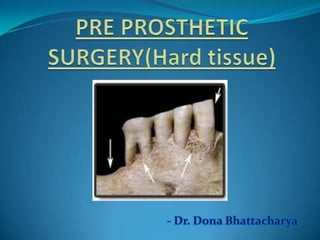
  • 1. - Dr. Dona Bhattacharya
  • 2. 1. Introduction 2. Objectives 3. Alveolar atrophy 4. Diagnosis & treatment planning 5. Ridge correction procedures a) Alveoloplasty b) Mylohyoid reduction c) Tuberosity reduction d) Genial tubercles reduction e) Removal of tori f) Removal of exostoses g) Removal of undercuts 6. Ridge augmentation 7. Conclusion 8. References
  • 3. ∆ Preprosthetic surgery refers to the surgical procedures that can modify the oral anatomy to facilitate the retention of conventional dentures. ∆ According to the Glossary of Prosthodontic Terms (7), preprosthetic surgery is defined as surgical procedures designed to facilitate fabrication or to improve the prognosis of prosthodontic care. ∆ According to Bruce Donoff, preprosthetic surgery is that part of the oral and maxillofacial surgery designed to establish the best hard and soft tissue bases for prosthetic appliances.
  • 4. Elimination of disease ∆ Conservation of oral structures ∆ Provide residual tissue to withstand masticatory forces ∆ Maintain function ∆ Esthetics
  • 5. The term alveolar atrophy refers to the regression of the teeth-supporting, crescent-shaped osseous part of the upper and lower jaw.
  • 6. Causes: ∆ Periodontal diseases ∆ Trauma ∆ Pt factors (age, gender, skeletal morphology) ∆ Endocrine & metabolic disorders (hyperparathyrodism,Ca defeciency) ∆ Dietary considerations ∆ Mechanical factors (extractions,removable denture wearers, combination syndrome)
  • 7. Patterns of bone loss ∆ The results of Talgren’ s studies indicate that changes under the denture base more often occur in the mandible.(4:1) ∆ The difference in resorption of the jaws increases within the first year of denture wearing, which proves that the mandible cannot resist the strong bite forces under the denture base. ∆ According to Klemetti initially resorption starts on the alveolar part of the mandible, and the rest of the mandible remains unchanged. ∆ Resorption is faster in the labial and buccal parts of the alveolar ridge. (Resorptive Changes of Maxillary and Mandibular Bone Structures; Dubravka Knezovi et al, Acta Stomat Croat 2002; 261-265)
  • 8. Class Characteristics Treatment I Alveolar ridge (AR) adequate in height but Hydroxyapatite (HA) alone inadequate in width, usually with lateral deficiency or undercut areas II AR deficient in both height & width and has a HA alone knife edge appearance III AR resorbed to level of the basilar bone, HA alone or mixed with producing concave form on posterior areas of the autogenous cancellous bone mandible and sharp bony ridge form with mobile soft tissue in the maxilla IV Resorption of the basilar bone, producing pencil- HA mixed with autogenous thin, flat mandible or flat maxilla cancellous bone (Mercier,1995)
  • 9. Modifications: Class II-no wall defect/buccal wall/multiwall defect Class VI- marginal resection /continuity defect Atrophy of the Residual Alveolar Ridge Following Tooth Loss in a Historical Population; Reich, Karoline et al;"Oral Diseases 17, 1 (2010)
  • 10. Functional effects of edentulism: ∆ The maxillomandibular relationship is altered in all spatial dimensions. ∆ Progression toward decreased overall lower facial height, leading to the typical overclosed appearance. ∆ Progressive instability of conventional soft tissue
  • 11. Ideal denture base has following characteristics: a) Adequate bony support b) Soft tissue coverage c) No undercuts or protuberances d) No sharp ridges e) Adequate sulci f) Absence of peripheral scar bands g) no muscle fibres to mobilize prosthesis h) No soft tissue folds/hypertrophies i) No neoplastic lesions j) Proper maxillomandibular arch relationships k) Adequate palatal vault/tuberosity notching
  • 12. 1. History ∆ Chief complaint ∆ Medical history 2. Physical examination  Soft tissues a) Presence of mass b) Tenderness c) Frena d) Mucous membrane e) Muscle movements f) Relation of oral mucosa to gingiva
  • 13. Hard tissues a) Undercuts b) Bony prominences c) Sharp ridges d) Ridge form e) Ridge parallelism f) Tuberosity notching  Maxillo-mandibular relation  Dentition 3. Investigations  Radiographic a) Gen condition of dentition b) Bone resorption c) Proximity to imp structures d) Maxillo-mandibular relation  Lab investigations
  • 14. Patient selection: ∆ General physical status ∆ Age ∆ Anatomic factors
  • 15. Preprosthetic procedures Ridge correction • Alveoloplasty • Mylohyoid reduction • Tuberosity reduction • Genial tubercles reduction • Removal of tori • Removal of exostoses • Removal of undercuts Ridge extension • vestibuloplasty Ridge augmentation • Maxillary • Mandibular
  • 16. Defined as surgical recontouring of alveolar process History: ∆ Willard(1853) –removal of interdental papilla ,permitting edge to edge closure ∆ Beers(1876): radical alveolectomy ∆ De van(1930): trend towards conservatism had begun ∆ Molt(1923):use of study casts in planning alveolectomy ∆ Dean(1936):interseptal alveoloplasty ∆ Obwegesser(1966):modification of dean’s technique ∆ Michael & Barsoum(1976): study on post operative resorption
  • 17. Principles: 1. Optimal ridge contour 2. Permit early construction of dentures 3. Preservation of alveolar bone 4. Broad alveolar ridges 5. Reduction of irregularities 6. Rounding off sharp ridges 7. Preserve cortical bone as much as possible 8. Defer surgery 4-6 weeks in case of severe periodontitis
  • 18. Types Alveolar Simple Labial & Dean’s Obwegesser’s compression alveoloplasty buccal cortical intraseptal technique
  • 19. ∆ Easiest & quickest method ∆ Involves compression of cortical plates with fingers ∆ Reduction in socket width
  • 20. Indications: ∆ Reduction of buccal/labial plate ∆ Extraction of single/multiple teeth Technique: ∆ Single tooth extraction ∆ Multiple teeth extraction ∆ Over erupted teeth
  • 21.
  • 22. Principles: a) Reduction of labial/alveolar prominences b) Muscle attachments are undisturbed c) Intact periosteum d) Preserve cortical bone e) Less post-op resorption
  • 23. Indications: ∆ immediate dentures ∆ quadrant extraction Technique: Mac Kay’s modification(1964)
  • 25. Extreme resorption results in sharp, pointed ridge that cuts into mucoperiosteum on pressure application. Pain occurs on wearing dentures.
  • 27. Gillies(1956): Mylohyoid ridge should be reduced if found at same or higher level than alveolar process Roberts(1977): Reduction of mylohyoid ridge & extension of posterior lingual denture flange into retromylohyoid fossa Howe(1964): Mylohyoid ridge reduction is the most useful single operation
  • 30. Excess tissue in the region of the maxillary tuberosity may become so large that it: ∆ Impinge upon the mandible during mastication. ∆ Interfere with denture construction, insertion and seating Complication of tuberosity reduction -expanded tuberosity in proximity to sinus
  • 31. 3 techniques: Removal of tubercle followed by allowing genial muscle to reattach on its own. Removal of tubercle followed by repositioning of muscle with sutures fastened to chin. Removal of tubercle followed by transposition of muscle to inferior border.
  • 32.
  • 33. ∆ Torus mandibular is an exostosis found on the lingual surface of the mandible opposite the canine and premolars region. ∆ Present in 8% of the population, with equal frequency in males and females ∆ Usually bilateral, (80% of affected patients), may be single, multiple or lobulated. ∆ Etiology: unknown, functional reaction to masticatory forces.
  • 34. Indications for removal: ∆ Tori causing lingual undercuts and interfering with lingual flange extension of the planned prosthesis. ∆ When the mucosal covering is ulcerated. ∆ Large tori interfering with speech and deglutition Technique Complications
  • 35.
  • 36. ∆ Torus palatinus present itself as an outgrowth in the midline of the palate. ∆ Shapes (single dome shaped, spindle shaped, nodular, lobular or multiple). ∆ Present in approximately 25% of all females ∆ Etiology unknown ∆ Composed of cortical bone; may have a cancellous component
  • 37. Indications for removal: ∆ An extremely large torus filling the palatal vault. ∆ A torus that extend beyond the posterior dam area. ∆ Traumatized mucosa over the torus. ∆ Deep bony undercuts interfering with denture insertion and stability. ∆ Interference with function (speech, deglutition). ∆ Psychological considerations (malignancy phobia).
  • 39. Complications: ∆ Haemorrhage ∆ Hematoma formation. ∆ Nasal or antral perforation. ∆ Sloughing and necrosis of palatal tissues. ∆ Fracture of palatine bone.
  • 40. Found in maxillary molar region. Preservation of vascular supply: main concern during surgery
  • 41.
  • 42. Caused by resorption in apical areas. Treatment: ∆ Excision ∆ Filling of undercut
  • 44.
  • 45. Indications for Ridge Augmentation Progressive loss of denture stability and retention. Loss of alveolar ridge height, width and decreased vestibular depth and denture bearing area. Considerable basal bone resorption in the mandible, resulting in neurosensory disturbances. Increased susceptibility to fracture of the atrophic jaws. Replacement of necessary supportive bone. Altered interarch relationship
  • 46. Ridge Augmentation Maxillary augmentation Mandibular augmentation Superior border Inferior border Onlay Onlay bone Interpositio augmentati augmentation grafting: grafting – Alloplastic Sinus Interpositional Visor nal / on (Iliac (Autogenous or Autogenou Autogenou onlay lift procedu / sandwich osteoto grafting sandwich crest, rib allogenic freeze s, allograft s / allogenic re bone grafts my grafts graft, dried cadaveric and grafts hydroxyapa mandible) alloplastic tite)
  • 47. Graft: portion of a tissue or organ that after removal from its origin or donor site is positioned or inserted at a different place with the objective of reinforcing the existing tissues &/or correcting a structural defect.
  • 48. Classification According to According to According to embryologic structure source origin Cortical Autograft Membranous Cancellous Allograft Endochondral Cortico- Xenograft cancellous Alloplast
  • 49. Autogenous Grafts Distant sites Local sites •Rib •Chin •Iliac crest •Body and ramus •Calvarium •ZM buttress •Fibula •Coronoid •Tibia
  • 50.
  • 51. AUGMENTATION OF SUPERIOR BORDER OF MANDIBLE (Davis, 1970) Indications: Remaining bone < 10 mm Ability of patient to tolerate procedure Donor considerations Recipient site
  • 52. Kerfing of rib graft
  • 53.
  • 54. Augmentation of inferior border of mandible Indications: ∆ Remaining bone < 10 mm ∆ Risk of pathologic # ∆ Management of malunion or non union of # Donor considerations Recipient site
  • 55. ADVANTAGES No vestibule obliteration No dehiscence Less pain Better # stabilization 2o sulculoplasty not required Less bone resorption Indicated for pencil thin ridges Easier to perform skin graft vestibuloplasty DISADVANTAGES Scarring Presence of loose submandibular tissue Does not correct superior irregularities
  • 56. AUGMENTATION OF MANDIBLE BY PEDICLED FLAPS Horizontal Vertical osteotomy/sandwich osteotomy/visor technique technique
  • 57. Horizontal osteotomy (Danielson and Nemarich)/sandwich technique Indication ∆ reasonable amt of bone above mandibular canal ∆ b/l dimension<12-15mm
  • 59. Vertical osteotomy (Harle,1975)/visor osteotomy Indications ∆ little bone above mandibular canal Technique
  • 60.
  • 61. Combined vertical and horizontal osteotomy (Koomen et al) Advantages: ∆ Less risk of # ∆ Better sup & post repositioning of segment ∆ Correction of mild-moderate AP discrepancies ∆ Increase in amt of augmentation Technique Stoelinga modification
  • 62.
  • 63. Bell & mc bride(1977)
  • 64. Augmentation with synthetic graft materials: Hydroxyapatite is the prototype of the nonresorbable ceramic bone substitutes. It is a calcium phosphate material having physical and chemical characteristic nearly identical to dental enamel and cortical bone.
  • 66. Advantages: ∆ Simple surgical technique suitable as an office procedure. ∆ No donor site is required to obtain autogenous bone graft material unless a composite graft is being accomplished. ∆ HA is totally biocompatible and nonresorbable ∆ Composite grafting can easily be accomplished as in severe class III and IV cases. ∆ Vestibular extension after alveolar augmentation is possible after 3 months of primary healing. ∆ Local augmentation is possible such as in bridge pontic areas. ∆ Metallic implant systems through HA augmented ridges are possible.
  • 67. Complications: ∆ Dehiscence with extrusion of particles ∆ Abrasion through the mucosa with extrusion of the HA implant ∆ Infection ∆ Abnormal color is noted under the mucosa ∆ Mental nerve neuropathy
  • 68. The use of particulate bone with membrane coverage allows for both horizontal and vertical augmentation of the mandible. The membrane is designed to prevent infiltration of the particulate graft with connective tissue and allow bone to infiltrate into the particulate graft mass rather than connective tissue, with the formation of sufficient bone. Disadvantage: ∆ premature exposure of the membrane through the mucosa. ∆ infection Used for ant maxillary combination syndrome
  • 69. Grafting bone on the superior surface of the residual alveolar cortical bone is accomplished by first gaining access to the cortical bone, placing and securing a bone graft to the region to be augmented, and closing the soft tissue. Indication: class V Advantage: 1. avoidance of direct damage to the IAN 2. ease of placement of the graft 3. immediate postoperative vertical augmentation. Disadvantage: incision breakdown over the graft can result in a reduction of the long- term augmentation
  • 70. Mandibular Tori as a Source for Onlay Bone Graft Augmentation: A Surgical Procedure; Scott D. Ganz JPPA;2007
  • 71. After alveolar bone osteotomy,distractor device is placed in transport segment, which remains vascularized via periosteum Latency period(5- 7 days) Bony segment subjected to traction Distraction period(0.5- 1mm/day 1-4 times Activation of tissue growth & regeneration Consolidation period(8-12 weeks) Formation of distraction callus, matures into bone
  • 72. Indications: ∆ Moderate-severe alveolar bone defects ∆ Segmental deficiencies ∆ Adjuvant to other grafts ∆ Less b/l width of ridges
  • 73. Simple, less resorption, include teeth, implants in transport segment, less time
  • 74. Accurate diagnosis of the problem areas during denture construction and determination of the necessity of surgery is accomplished by careful evaluation of the information systematically obtained from the patient. As conservation is the philosophy of surgical patient management. therefore every attempt should be made to preserve as much as oral structures as possible. Proper knowledge of the available surgical procedures helps in achieving the best results.
  • 75. 1. Preprosthetic oral & maxillofacial surgery-Starshak & Sanders 2. Textbook of oral & maxillofacial surgery- Laskin vol II 3. Principles of oral & maxillofacial surgery-Peterson 4. Textbook of oral & maxillofacial surgery- Fonseca vol 7 5. Textbook of oral & maxillofacial surgery- Kruger 6. Textbook of oral & maxillofacial Surgery – Archer 7. Textbook of oral & maxillofacial surgery- Killey And Kay 8. Bone grafting in oral implantology: Alfaro
  • 76. 9. Alveolar bone grafting techniques for dental implant preparation- OMFS,Aug 2010 10. Sugar,Hopkins et al:A sandwich mandibular osteotomy, BJOMS, 1982, 20:168 11. Interpositional Osteotomy for Posterior Mandible Ridge Augmentation Michael S. Block, DMD,* Christopher J. Haggerty.JOMS 67:31-39, 2009, Suppl 3 12. Distraction implants: a new operative technique for alveolar ridge augmentation Alexander Gaggl, Gfinter Schultes, Hans K~ircherJournal of Cranio-Maxilloj'acial Surge , (1999) 27, 214-221 13. Reconstruction of the severely atrophic mandible with iliac crest grafts and endosteal implants: a report of two cases; O’Connell J.E. ,Galvin M, Journal of the Irish Dental Association 2009; 55 (5): 237-241. 14. Mandibular Tori as a Source for Onlay Bone Graft Augmentation:A Surgical Procedure Scott D. Ganz,JPPAD

Hinweis der Redaktion

  1. Tuberoplasty,sinus lift