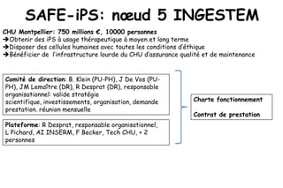
1. SAFE-iPS: nœud 5 INGESTEM
CHU Montpellier: 750 millions €, 10000 personnes
Obtenir des iPS à usage thérapeutique à moyen et long terme
Disposer des cellules humaines avec toutes les conditions d’éthique
Bénéficier de l’infrastructure lourde du CHU d’assurance qualité et de maintenance
Comité de direction: B. Klein (PU-PH), J De Vos (PU-
PH), JM Lemaître (DR), R Desprat (DR), responsable
organisationnel: valide stratégie
scientifique, investissements, organisation, demande Charte fonctionnement
prestation. réunion mensuelle
Contrat de prestation
Plateforme: R Desprat, responsable organisationnel,
L Pichard, AI INSERM, F Becker, Tech CHU, + 2
personnes
2. Institute of Research in Biotherapy
Director: Pr B. Klein
Beta Innov Horiba ABX
Immune molecules Rare cells
INSERM-UM1 , 870 m2 Common facilities, 658 m2 University hospital R&D, 1000 m2
Multiple Myeloma Cell Administrative & technical staff.
Plasticity, Stem Cells and Niche. BK G Berthaud & M Frei Monitoring of Novel Therapeutics
Pr Klein, CHRU
Early Embryonic Development. SH Common platforms: TL3,
Region Clinical proteomic
cryogeny, molecular
equipments, laundery, meeting
platform. Pr Lehmann, CHRU
Hepatic differentiation of stem rooms
cells and biotherapy of liver
diseases. MD Identification of rare cells
High density DNA Affymetrix Dr Vendrell, CHRU
microarray platform. CHRU-
Immune system control of INSERM
hematological neoplasias. MV , V Pantesco, T Commes Diabetes Cellular Therapy
Laboratory : Pancreatic Beta Cell
Survival and Regeneration. Dr
Montpellier Rio Imaging
Pathophysiology, diagnosis and cell Dalle , Pr Renard, dr Wojtusciszyn
Cytometry Platform
therapy of neurodegenerative disorders. Dr Duperray, INSERM CHRU
SL
Safe Induced pluripotent stem cells
Biomathematics and
Genomic instability of pluripotent stem bioinformatics service B. Klein, J De Vos, JM lemaitre, CHRU
cells. JDV Dr Reme, Pr Commes,
Ecell France
Bioinformatics & high speed sequencing. C. Jorgensen
T Commes
Cell Therapy Unit In vitro fertilization and Department of Clinical Hematology Hepatogastroenterology Endocrinology
Dr De Vos, CHRU PGD. Pr and Oncology. Pr Rossi, CHRU Pr Navarro and Pr Blanc, Pr Bringer and Pr Renard,
Hammamah, CHRU CHRU CHRU
3. Node 5: Institute of research in Biotherapy, CHU Montpellier
Platform to produce iPS.
Romain Desprat, Objective. Produce iPS for academic and private SAFE-IPS platform
laboratories.
Direction committee: B. Klein
Quality control tests for safe iPSCs (WP4). John De Vos, J De Vos, JM Lemaître,
1. Optimize and develop quality controls for safe iPSC. 2. Quantify recurrent stresses and
consecutive genetic abnormalities during iPSC production to improve production of safe
iPSCs. 3. Provide tools to dissect the nature and cause of genetic abnormalities in iPSCs R Desprat,
related to early replicative stress. L Pichard, INSERM assistant
engineer
Non-integrative methodologies for reprogramming. F Becker, CHU technician
Jean Marc Lemaître, Objective. Development of non-integrative
reprogramming methodologies for SAFE-IPS obtaining . Temporary staffs
2 people
Projects promoting SAFE-IPS development
Projects using SAFE IPS platform
JM Lemaître, INSERM B. Klein, CHU-INSERM-UM1 U1040
Pluripotency to study and revert Pluripotency to revert oncogene C Hamel, INSERM-UM1 U1051
senescence involved in aging and induced senescence and study cancer Obtaining pluripotent stem cell from
genetic diseases cell plasticity patients with genetic blindness as tools to
monitor gene therapy methodologies
J De Vos, CHU-INSERM-UM1 U1040 M Daujat, CHU-INSERM-UM1 U1040
Identification and mastering the Pluripotency as a tool to get human
S Lehmann, INSERM-UM1 U1040
critical steps creating genomic hepatocyte cells for cell therapy use
Obtaining pluripotent stem cell from
instability throughout pluripotency patients with Alzheimer’s disease
induction and maintenance S Hamamah, CHU-INSERM-UM1 U1040
Immortalized human cumulus cells as a C Jorgensen, INSERM-UM1 U844
tool to induce and maintain full and safe Obtaining pluripotent stem cell from
pluripotency patients with osteoarthritis
4. Activité de la platforme
Octobre 2012-Décembre 2013 :
Journée porte ouverte à l’IRB afin de présenter le projet INGESTEM et les services offerts par la plateforme :
Plus de 80 personnes présentes
Mise en place Administrative de la plateforme :
-Ecriture d’une chartre de fonctionnement et de contrats de reprogrammation, (définie le cadre légal de la
prestation apportée)
-Mise à disposition de la totalité du budget INGESTEM dédié à l’IRB-Montpellier via le CHU,
-Signature d’un d’accord de transfert de technologie entre DNAvec (Invitrogen) pour produire le virus Sendai
(OKSM facteurs) sur Montpellier (IRB) grâce aux infrastructures spécifiques de l’IRB (P2/P3).
-Mise en place d’une collaboration technique avec Dr.Roux (Genève) spécialisés sur le virus sendai afin
d’exprimer de nouveaux facteurs (Lin28, …) et de conseils pour résoudre les potentiels problèmes de production
et d’expression
Mise en place Technique : Ecriture des protocoles de routine et de reprogrammation à partir de Fibroblastes :
Feeders SNL, production virus (Lentivirus, rétrovirus non intégratifs, virus sendai…)
Mise en place Pratique : Constitution de stock de feeders (SNL), plasmides, achats milieux, primers, etc …
Mise en place Stratégique : Point de départ Lentivirus/Rétrovirus afin de qualifier le personnel et les protocoles
et les control qualités pour évoluer ensuite sur une stratégie non intégratives.
Mise en Place Organisationnelle : Réunion Comité de direction tous les semaines
5. Objectifs à court term (1-2 mois) : Objectifs
-Qualification de la platforme
-Détermination des meilleures conditions de culture pour reprogrammer
-Création de « custom chips » afin d’analyser l’expression et les changements de CNV/SNP :Sur les
genes définissant l’état fibroblastique et iPS.
-Demande de financement via le CHU
Objectifs à moyen term (1 an) : Mise en place des méthodes de reprogrammation non intégratives
avec une maitrise totale des différents aspects de la production permettant une transition
potentielle clinique plus probable :
-Production du Virus Sendai (DNAvec) sur Montpellier et Production de beta-FGF sur Montpellier:
Permettant une diminution des coûts associes aux milieux -> plus d’utilisateurs
-Production de proteines (oct4,KLF, Sox…) pour faire des iPS
-Optimisation des conditions de culture pour produires des iPS.
-Création d’un blog/site web (plus de flexibilité) et/ou en utilisant un system déjà existant
(INGESTEM), permettant un partage plus fluide des protocols et publications, photo des cellules,
questions associées à la culture des iPS….
-Collaboration avec ReproCell afin de créer des ateliers ou période d’apprentissage sponsorise par
ReproCell et Ozyme pour les milieux .
Objectifs à long term (3 ans):
- indépendance financière de la plateforme et identifications des facteurs
6. Contrôle de la qualitée des iPS :Test de pluripotence
Qualité du contrôle
Test de pluripotence But pour démontrer la
pluripotence
Morphologie des colonies Vérifier que les iPS ont une morphologie similaire au hES Faible
Marquage de la pluripotence:Oct4, Tra-1-60,Sox2, Tra-1-80,
Immunostaining Moyen
Nanog et SSEA
Détecte le niveau et la qualité d'expression des genes :Oct4,
RT-PCR Moyen-Bon
Tra-1-60,Sox2, Tra-1-80, Nanog et SSEA
Capacité à se différencier dans les trois feuillets
Génération d'EB embryonnaires (ecto,endo, mésoderme et disparition des Moyen-Bon
marqueurs de la pluripotence)
Microarray ou RNA-seq Comparaison avec hES Moyen-Bon
Capacité à se différencier dans les trois feuillets
Teratoma formation Bon
embryonnaires (ecto,endo, mésoderme ) IN VIVO
7. Contrôle des Conditions de culture
Xvivo configuration :
-Incubateurs ne peuvent pas être
contaminés par les manipulateurs ou l’air
de la pièce.
-Les cellules ne subissent AUCUN
changements dans les paramètres de
culture contrôlé par le Xvivo (atmosphère
composition hypoxie, température,…) qui
s’ils ne sont pas maitrise participent à
l’apparition d’aberration chromosomique.
8. Pluripotent genes in Multiple Myeloma
malignant plasma cells
Cancer that develops in the bone marrow
Invasion by a clone of malignant plasma cells
Destruction of the bone tissue
4000 newly-diagnosed patients/year in France
25000 in Europe and 25000 in USA
Still fatal disease in 2012
Median overall survival: 5 years depending on age and treatment
9. Fixed Somatic mutations
Germinal Recombination linked
with isotype switching
Center Primary
reaction genetic events
Cell cycle
KLF4 expression
IgH@
translocations
Hyperdiploidy
Normal plasma Quiescent cells? Deletion.
G0 Long lived: several yearsCyclin D1-3
cells
Loss of Rb
Ras mutations
Block in G1.
GO G1 Oncogene induced
MGUS senescence
Diffferent Clones
Block in G1.
Smouldering G Oncogene induced
G1
Multiple 0 senescence
Myeloma Diffferent Clones
T(4;14)
Myc rarrangement and
Heterogeneous disease
Clonal dominance
Copy number
variations
upregulation
G
Multiple 0
G1 From 0% to 4% MMCs in S Mutations
Myeloma phase. Median 0.4%. Strong P53 deletions
Sox 2 expression
Clonal dominance
prognosis factor NF-Kappa B
Myc
overexpressi
S/ Highly proliferating on
Extramedullary G1 G2
proliferation /M
10. Role of myc, sox2, KLF4, Oct4 to
drive self-renewal in a putative
myeloma stem cell
Hinweis der Redaktion
The ability to differentiate into almost all tissue types is the hallmark of human pluripotent stem cells (hPSCs). However, as we study the biology of hPSCs for future clinical applications – whether they be under the influence of growth factors (1–4), pro-survival cocktails (5,6), genetic modification (7–10), or other manipulations (1) – pluripotency testing remains a fundamental component of every research design. Assays generally used to test such “stemness” include genomic profiling for relative quantification of pluripotency genes, immunocytochemistry to detect pluripotency markers, embryoid body formation to test 3-germ-layer differentiation capability in vitro or in vivo, and teratoma formation to test 3-germ-layer differentiation capability in vivo (Table 1). Notwithstanding the in vitro assays, teratoma formation in vivo is considered the most stringent of pluripotency assays because it provides more reliable and comprehensive confirmation than does testing cells on a simplified, artificial petri dish. This in vivo assay, coupled with noninvasive, longitudinal imaging, have proven to be invaluable not only in the visualization of stem cell survival and migration post-delivery, but also have been crucial in studying the safety and viability of future stem cell applications (11–13).