SlideShare ist ein Scribd-Unternehmen logo
1 von 91
ZYGOMATICO MAXILLARY
FRACTURE
PRESENTED BY :
PROF.DR.SHIVARAJ.S.WAGDARGI
DEPT OF ORAL AND MAXILLOFACIAL SURGERY
NAVODAYA DENTAL COLLEGE RAICHUR
CONTENTS
• INTRODUCTION
• CLASSIFICATION.
• EXAMINATION.
• SIGNS AND SYMPTOMS.
• MANAGEMENT.
• SURGICAL APPROACHES.
• COMPLICATIONS.
• REFERENCES
INTRODUCTION
 The zygoma or malar complex forms the central support of the cheek and is a strong
buttress of the lateral and middle third of the facial skeleton
• Zygomatic or malar fracture are the terms commonly used to described fractures that
involve the lateral one third of the middle face. Other names for this fracture are:
 Zygomaticomaxillary complex
 Zygomaticomaxillary compound
 zygomatico orbital
 Zygomatic complex
 Malar
 Trimalar
 Tripod
CLASSIFIACTION
Knight and North (1961)
Rowe and Killey (1968)
Yanagisawa ( 1973)
Larsen and Thomson (1978)
Rowe and Williams (1985)
Spiessl & Schroll (1972)
Poswillo’s classification
Markus zing classification
Manson and colleages
Henderssons classification
Zingg classification
Ozyagzan classification
According to Knight and North (1961)
Group I – No significant displacement.
Group II – Arch fracture.
Group III – Unrotated body fracture
(downward and backward).
Group IV – Medially rotated body fracture
(inward and backward).
Group V – Lateral rotated body fracture.
Group VI – Complex body fractures.
According to Larsen and Thomsen (1968)
 Stable fractures that shows - minimal or no displacement ( no
intervention)
Stable fracture – with displacement ( no fixation)
 Unstable fracture with great displacement and disruption at
frontozygomatic suture and comminuted fractures.
Fractures of zygomatic arch are classified again into:
Minimum or no displacement.
V type in fracture.
Comminuted fracture.
ROWE’S AND KILLEY (1968)
 Type I : No significant displacement
 Type II : Fracture of the zygomatic arch
 Type III : Rotation around horizontal axis
- Inward displacement of orbital rim
- Outward displacement of orbital rim
 Type IV : Rotation around longitudinal axis
- Medial displacement of frontal process
- Lateral displacement of frontal process
 Type V : Displacement of the complex
enbloc - Medial
-Inferior
- lateral (Rare)
 Type VI : Displacement of orbitoantral
partition - Inferiorly
-Superiorly
 Type VII : Displacement of orbital rim
segments
 Type VIII : Complex comminuted fractures
ROWE & WILLIAMS CLASSIFICATION
Fractures unstable after
elevation
a)Arch only (inferiorly displaced)
b)Rotation around horizontal axis
i)medially
ii)Laterally
c)Dislocations en bloc
i) inferiorly
ii)medially
iii)Postero-laterally
Fractures stable after elevation
a)Arch only (medially displaced)
b)Rotation around the vertical axis
i)medially
ii)Laterally
SPIESSLAND SCHROLL (1972)
• TYPE 1- zygomatic arch fracture.
• TYPE 2- zygomatic - complex fracture with no significant displacement.
• TYPE 3- zygomatic complex fracture - partial medial displacement. (Kinking
at F-Z suture)
• TYPE 4 - Z.C with total medial displacement(complex fracture of F-Z suture).
• TYPE 5- Z.C dorsal displacement (2 fracture sites in zygomatic arch).
• TYPE 6- Z.C, inferior displacement.
• TYPE 7- Z.C, comminuted fracture.
YANAGISAWA(1973)
• GROUP 1- non displaced fracture- no treatment.
• GROUP 2- arch fracture(pure)
• GROUP 3- medial/lateral rotation around vertical axis.
• GROUP 4- medial/ lateral rotation around longitudinal axis.
• GROUP 5-medial/ lateral displacement without rotation.
• GROUP 6- isolated rim fracture.
• GROUP 7- all complex fracture.
EBERHARD KRUGER(1986)
FRACTURE OF ZYGOMA
• No displacement.
• Partial medial
• Total medial
• Dorsal displacement.
• Inferior displacement.
• Comminuted fracture.
MANSON& COLLEAGUES(1990)
Based on
- Amount of energy dissipated by the facial bones.
- Secondary to the traumatic force.
- Findings in CT scans
High energy fractures Lower energy fractures
• Extreme displacement. Displacement
• Comminution of articulations. NIL
• Segmentation of bones.
Treatment- Treatment-
• Require extensive exposure Less agressive
and Aggressive fixation.
ANATOMY
PRINCIPAL STRUCTURE OF MIDFACE
MAJOR ROLE IN FACIAL CONTOUR
4 PROCESSES -ORBITAL
-FRONTAL
-TEMPORAL
-MAXILLARY
4 ARTICULATIONS -FRONTAL
-TEMPORAL
-MAXILLARY
-SPHENOID
MUSCLE ATTACHMENTS - MASSETER
-TEMPORAL FASCIA
ANATOMY - ZYGOMA
ARTICULATIONS
MUSCLE FORCES ON ZYGOMA
FRACTURE PATTERNS
• Depends on- DIRECTION & MAGNITUDE OF FORCE
• Fracture lines pass through lines of greatest weakness
• Malar bone represent strong bone on fragile suppport
• Force appiled on zygoma - distributed through four processes
to adjacent articulating bone –weaker than zygoma
• Three lines of fracture extend from IOF
Anteromedial – orbital floor
Superolateral -
Inferior direction
Clinical examination
1.NEUROLOGICAL STATUS.
• First step is to assess neurological
status…….
• Associated neurologic injury was
encountered in 57% of patients.
2.VISUAL STATUS
3.EXAMINATION OF ZYGOMA
INSPECTION
Symmetry of arches.
Pupillary level.
Presence of orbital oedema.
Sub-conjunctival ecchymosis.
Ant & lat projection of zygoma.
Intra-oral.
• PALPATION
-Systematic & thorough.
-Compare one side with another.
SIGNS AND SYMPTOMS
• Periorbital ecchymosis and edema.
• Flattening of the malar prominence.
• Flattening of the zygomatic arch.
• Pain.
• Ecchymosis of the maxillary buccal sulcus.
• Deformity of the zygomatic buttress of the maxilla.
• Deformity of the infra-orbital margin.
• Trismus.
• Abnormal nerve sensibility.
• Epistasis.
• Subconjunctival ecchymosis.
• Crepitation from air emphysema.
• Displacement of palpebral fissure.
• Diplopia.
• Enopthalmos.
Orbital signs and symptoms
Periorbital
Oedema
Circumorbital
ecchymosis
Subconjunctival
haemorrhage
Orbital
Emphysema Diplopia
Unequal
pupillary
levels
Flattening of malar prominence
• Characteristic sign
• Presence of edema flattening may be difficult to discern
Flattening of zygomatic arch
• Loss of normal convex curvature
Displacement of
palpebral fissure Deformity of orbital margin
ORBITAL EXAMINATION
FORCED DUCTION TEST
• To determine presence of
mechanical obstruction to
globe movement.
• Test done to assess the cause
of reduced movement of
extraocular muscles
following trauma to mid-face
region.
• It helps to determine if the
cause is mechanical/
neurological.
DIPLOPIA
RADIOGRAPHIC EXAMINATION
 Postero-anterior oblique view (OM/PNS view): excellent assessment of
sinuses and their walls, zygoma and its processes and rims of orbit
 Submentovertex view is specific for zygomatic arch fractures
CT SCANS
TREATMENT
STEPS IN TREATING ZMC FRACTURE
1. Prophylactic antibiotics.
2. Anesthesia.
3. Clinical examination and forced duction test.
4. Protection of the globe.
5. Antiseptic preparation.
6. Reduction of the fracture.
7. Assessment of reduction.
8. Determination of necessity for fixation.
9. Application of fixation device.
10. Assessment of ocular motility.
11. Bone graft for extra orbital osseous defects.
12. Post surgical ocular examinations.
13. Post surgical images.
NEED FOR FIXATION
Indications for fixation
1. Comminuted fracture fragments.
2. Doubt regarding the stability
Role of MASSETER in displacement:
• Albright and McFarland recommended IMF following fracture reduction
helps to reduce the pull of the masseter muscle on the repositioned ZMC.
• Ellis et al reviewed series of isolated ZMC fractures treated by different
approaches and fixation schemes and found no evidence of post reduction
instability
TREATMENT MODALITIES
NO TREATMENT
INDIRECT REDUCTION-
• Gillies approach.
• Keens approach.
• Lateral coronoid approach.
• Eyebrow approach.
• Percutaneous approach.
• Transnasal approach.
DIRECT REDUCTION
FIXATION.
Coronal approach/bicoronal.
Upper eyelid/ blepharoplasty.
Supraorbital eyebrow approach.
Transconjunctival .
Subcilliary.
Infraorbital.
Maxillary vestibular approach
TEMPORARY SUPPORT
INDIRECT FIXATION-
• Internal pin fixation.
• Transfixation with kirshner wire.
DIRECT FIXATION-
• Transosseous wiring.
• Bony platting.
NO TREATMENT
NON-DISPLACED FRACTURES-9-50 % OF ZMC #
MEDICAL CONTRAINDICATIONS
MINIMAL DEGREE OF DISPLACEMENT.
UNLIKELY TO RESULT IN
COSMETIC DEFORMITY
DISTURBANCE OF VISION
PARESTHESIA
IMPAIRED MANDIBULAR MOVEMENTS.
DECISION TO INTERVENE
DEPENDS ON
SIGNS & SYMPTOMS.
FUNCTIONAL IMPAIRMENT.
PRESENCE OF OPHTHALMIC INJURIES.
PROGRESSIVE PROPTOSIS.
VISUAL ACUITY DETERIORATION.
VISUAL INTEGRITY ON UNAFFECTED SITE.
OTHER RELATED FACIAL INJURIES.
MEDICAL CONDITION OF PATIENT.
DELAYED TREATMENT
1. MANIPULATION OF ORBITAL BONES
2. RISK OF EYEBALL INJURY.
3. EXISTING HAEMORRGHAGE.
4. PROGRESSIVE PROPTOSIS.
5. PRE-EXISTING BLINDNESS IN OTHER EYE.
6. GENERAL MEDICAL CONDITION.
7. NEUROLOGICAL STATUS QUESTIONABLE.
8. GROSS OEDEMA.
9. CRITICAL PERIOD - 5-10th DAY
DELAYED TREATMENT
ADVANTAGES
OEDEMA RESOLVES- DETAILED EXAMINATION OF
EYE.
ANTRUM CLEARS - BETTER RADIOGRAPH.
HAEMATOMA STILL NOT ORGANISED –DISSECTION
EASIER.
>5-10 DAYS
DIFFICULTY IN DISIMPACTING & REDUCING.
PHYSIOLOGICAL RESORPTION OF # MARGINS.
INTERDIGITATION LESS ACCURATE.
INELASTICITY OF FIBROUS TISSUE AT SITE OF
MALUNION.
IMMEDIATE TREATMENT
• IN THE ABSENCE OF COMPLICATIONS.
• NO OEDEMA.
ADVANTAGES
EXCELLENT REDUCTION -NOT COMPROMISED
NO SOFT TISSUE SCARRING
NO CHANGE IN MORPHOLOGY
SUPERIOR FACIAL SOFT TISSUE CONTOUR.
APPROACHES:
 Temporal approach –
 Buccal sulcus approach –
Gillies (1927)
Keen (1909),
Balasubramanium (1967)
 Lateral coronoid approach – Quinn (1977)
 Eyebrow approach -
 Percutaneous approach -
Dingman & Natvig (1964)
Stroymeyer (1844)
INDIRECT REDUCTION
TEMPORAL APPROACH
Popular approach
First described by Gillies & coworkers in 1927 – arch fractures
ADVANTAGES :
 Allows application of greater amount of controlled force to
disimpact even the most difficult zygomatic fracture.
 For treatment of fractures which are consolidated already
 Quick and simple method
DISADVANTAGE:
 Encountered temporal vessels- hemorrhage
Incision – 2.5cm, through skin & subcutaneous tissue
White glistening surface Temporalis muscle bulge
PLACEMENT OF ROWES
ZYGOMATIC ELEV
ATOR
AND ELEVATION.
BUCCALSULCUS APPROACH
Keen’s Technique (1909)
ADVANTAGES:
Avoidance of any external scar.
Used for both arch and ZMC fracture
DISADVANTAGES:
Unstable fractures – external incision for fixation
TECHNIQUE:
•A small incision (approximately 1 cm) is made in the mucobuccal fold, beneath the
zygomatic buttress of the maxilla.
•Sharp end of periosteal elevator – inserted & swept in a sweeping motion to
contact infratemporal surface of maxilla & zygoma.
•A heavier instrument inserted behind the infratemporal surface of the zygoma,
using superior, lateral & anterior force to reduce the bone.
LATERAL CORONOID APPROACH
Quinnin1977–described
Usedonlyforarchfractures
TECHNIQUE :
•3 to 4 cm incision -anterior border of the ramus.
•Insision made - to the depth of the temporal muscle
insertion (not to the bone)
•Instrument between the temporal muscle and the
zygomatic arch - readily palpable.
•A flat-bladed instrument- inserted into the pocket&
arch is elevated while palpating the arch extraorally.
ELEVATION FROM EYEBROWAPPROACH
•Used for both ZMC & arch fracture
ADVANTAGE:
•Fracture at the orbital rim is visualized
directly
•Fixation can be done with same incision.
DISADVANTAGE:
•Difficult to generate a large amount of force,
especially in the superior direction.
DINGMAN ZYGOMATIC
ELEVATOR
PERCUTANEOUS APPROACH
•Most simple of all techniques as no soft tissue dissection is necessary
•Direct route to elevate the depressed zygoma is through the skin
surface of the face overlying the zygoma.
ADVANTAGE:
Produces forces anteriorly, laterally, and superiorly in a very direct
manner
DISADVANTAGE:
Scar on the face in a very noticeable location.
ELEVATION OF THE ZYGOMA WITH A
BONE HOOK
•Strohmeyer in 1844
•Poswillo`s
intersecting lines.
•Stab incision made
and hook inserted.
•Apply strong traction.
Stacey bone hook
Carrol-Girard bone screw
Elongated corkscrew with a T- bar handle.
Contains threads on its working end.
Screw is threaded into body of zygoma
following a hole placement & is used as a
handle to reduce fracture
Can control ZMC position in all 3 planes of
space.
LARGE BONE SCREW
SURGICALAPPROACHES TO ZMC
 Maxillary vestibular approach
 Supraorbital eyebrow approach
 Upper eyelid approach
 Lower eyelid approach
 Transconjunctival approach
 Coronal approach
DIRECT REDUCTION
MAXILLARY VESTIBULAR APPROACH
• Most useful when performing any of
wide variety of procedures in the
midface.
• Safe access to the entire facial surface
of the midfacial skeleton- from the
zygomatic arch to the infraorbital rim
to the frontal process of the maxilla.
ADVANTAGE:
• Hidden intraoral scar.
• Rapid and simple approach
• Complications are few.
TECHNIQUE
Subperiosteal Dissection
V –Y Closure
Incision through the mucosa, submucosa, facial
musculature,and periosteum (3-5mm superior to
mucogingival junction)
Infraorbital rim, anterior
maxilla & ZMC Buttress -
treated
LATERAL BROW APPROACH
Popular approach - Access to the lateral
orbital rim and the frontozygomatic suture
ADVANTAGE:
Simple, safe and rapid approach
Scar is usually hidden within the confines of
the eyebrow
No important neurovascular structures are
involved in this approach.
DISADVANTAGE :
 In individual who has no eyebrows extending laterally and
inferiorly along the orbital margin, this approach is undesirable.
 Incisions made along the lateral orbital rim outside of the
eyebrow are very conspicuous in such individuals, and another
type of incision may be indicated.
 Extremely limited access.
TECHNIQUE
•Incision – parallel to the eyebrow hair.
•The incision is made through skin and subcutaneous tissue
to the level of the periosteum in one stroke. (2cm)
•Incision through periosteum along lateral orbital rim and
subperiosteal dissection into lacrimal fossa.
•Because of the concavity just behind the orbital rim in this area,
the periosteal elevator is oriented laterally as dissection proceeds
posteriorly.
Closure:
The incision is closed in two layers, the periosteum and the skin.
UPPER EYELID APPROACH
 The upper eyelid approach to the superolateral orbital rim is also called UPPER
BLEPHAROPLASTY, UPPER EYELID CREASE, & SUPRATARSAL FOLD
APPROACH.
 In this approach, a natural skin crease in the upper eyelid is used to make the
incision.
Advantage: ADVANTAGE:
Inconspicuous scar it creates - best
approaches to the region.
TECHNIQUE
Closure:The wound is closed in two layers, periosteum and skin/muscle.
To facilitate retraction of the skin/muscle flap, it can be widely undermined
laterally and retracted with small retractors.
Periosteum is divided 2-3mm posterior to the orbital rim with a scalpel.
Sagittal section through orbit and globe showing dissection
between orbicularis oculi muscle and the orbital septum
Incision – begins 10mm superior to upper lid margin
& lies 6mm above lateral canthus as it extends laterally.
The initial incision is made through skin and muscle.
LOWER EYELID APPROACH
SUBTARSAL approach:
• Frequently employed – access to infraorbital rim & orbital floor
• Along natural skin crease at/below the level of tarsus, approx half
distance between lash margin and orbital rim
ADVANTAGE:
• Relatively easy
• Scar is imperceptible
• Minimal complication
SUBCILIARY approach:
• Also called INFRACILIARY APPROACH/ BLEPHAROPLASTY
• Incision is - approx 2 mm below the eyelashes and can be extended laterally as necessary
(top dashed line) along the natural crease inferior to lateral canthal ligament.
ADVANTAGE:
• Imperceptible scar
• Simultaneously exposes infraorbital
& F-Z areas.
DISADVANTAGE:
• Technically difficult
• Risk of postoperative ectropion
TECHNIQUE
•Skin incision
•Dissect b/w skin & muscle
until orbital rim is reached
•2nd incision – through muscle
& periosteum to bone
•Incision – skin & muscle at
same level
•Dissect down anterior to
orbital septum to orbital rim
•Combination of these – STEPPED
incision
•Subcutaneous dissection toward
rim – few mm
•Followed by muscle incision at
lower level & then following orbital
septum to rim.
•Fine scissors are used during dissection –spreading motion.
•Incision through periosteum – 3-4mm below rim
TRANSCONJUNCTIVALAPPROACH
 Originally described by Bourguet in 1928.
 Also called INFERIOR FORNIX APPROACH.
 2 types:
 Preseptal (Tessier)
 Retroseptal (Tenzel&Miller) approaches.
 Converse & colleagues added a lateral canthotomy to
transconjunctival retroseptal incision for improved lateral
exposure.
RETROSEPTAL PRESEPTAL
TECHNIQUE
Sagital section through orbit showing preseptal and
retroseptal placement of incision.
Initial incision for lateral canthotomy
Initial canthopexy incision to dissect in the
subconjunctival plane. The dissection should be just
below the tarsal plate and extend no farther medially
than the lacrimal punctum.
Closure of transconjunctival incision and inferior
canthopexy
ADVANTAGE :
 Produce excellent cosmetic results because the scar is
hidden in the conjunctiva.
 If a canthotomy is performed in conjunction with the
approach, the only visible scar is the lateral extension,
which heals with an inconspicuous scar.
 Rapid and no skin or muscle dissection is necessary.
DISADVANTAGE :
• medial extent of the incision is limited by the lacrimal
drainage system.
CORONALAPPROACH
 The coronal or bi-frontal incision is a
versatile surgical approach to the upper
and middle regions of the facial skeleton,
including the zygomatic arch
ADVANTAGE:
•The surgical scar is hidden within the hairline.
•Excellent acess to orbits, zygoma & arch.
TECHNIQUE
Incision placement:
•Hairline of patient
•Amount of inferior access
•Incision – skin, subcutaneous tissue & galea
•Lateral aspect of skull- superficial layer of temporal fascia incised 2cm supr
to zygomatic arch
•Dissect inferiorly - zygomatic arch
•Periosteal incision – along the superior aspect of arch
•Pericranium is incised along lateral orbital rim – FZ suture
•Wound is closed in layers – fascia , galea, skin.
HEMICORONAL APPROACH
MODIFICATION OF HEMICORONAL
APPROACH
•The anterior arm of the incision is
curved downward toward the superior
wall of the orbit before it reaches the
vertex of the skull within the hairline.
•The ‘backcut’ provides excellent
exposure of the entire zygomatic
complex and the arch.
• Aesthetic and is less invasive thereby
being quite acceptable by patients.
Journal of Maxillofacial and Oral Surgery 2010
Volume 9, Number 3, 270-272
Common methods include
wire osteosynthesis and
rigid fixation by plates
Less common methods include
external pin fixation and
maxillary antral support
IMMOBILIZATION
PIN FIXATION
 External pin fixation
Can be used for fractures that demonstrate
an intact body of the zygoma but severe
communition at the junction with the
surrounding bones
 Internal pin fixation
Was introduced by Fryer and results in
stable entity and relatively free of
complications
Techniques make use of K-wire placement
SINUS PACKING SUPPORT
 Gauze or balloon can be used to
provide inferior support to the
zygoma
 Lateral wall is approached
through a Caldwell-Luc
 ½ inch gauze dipped in antibiotic
of choice is placed along the
floor anteroposteriorly
 Antral balloon can be used by it is
relatively imprecise and cannot
adapt to the topography
FIXATION TECHNIQUES
INDIRECT FIXATION
• Zygomatic bone - rigidly secured to facial skeleton.
INDICATION
• Gross loss of bone in FZ region and infra orbital rim.
 ZYGOMATICO-ZYGOMATIC
 NASO-ZYGOMATIC
 ZYGOMATICO-PALATAL
 MAXILLO-ZYGOMATIC
 FRONTO-ZYGOMATIC
 CRANIO-ZYGOMATIC
INDIRECT
FIXATION
TRANSFACIAL WIRE
TRANSNASALWIRE
ZYGOMATICOMAXILLARY ZYGOMATICOPALATAL
EXTERNAL FIXATION
Accomplished with wires suspended from plaster head caps,
head frames and by pins connected to one another with
universal joints and cold cure acrylic.
ADVANTAGES-
1. Three dimensional stability.
2. Minimal scarring.
3. Adjustability of the reduction.
DISADVANTAGES-
1. Patient comfort is compromised.
2. Need for specific hardware.
3. Lack of usefulness in comminuted fractures.
DIRECT FIXATION
• TRANS-OSSEOUS WIRING/ OSTEOSYNTHESIS
FZ SUTURE
INFRA ORBITAL MARGIN
• BONE PLATES
FIXATION TECHNIQUES - PRINCIPLES
1. Use self-threading bone screws.
2. Use hardware that will not scatter postoperative CT scans.
3. Place at least two screws through the plate on each side of the fracture.
4. Avoid important anatomic structures.
•Use Y,L,T shaped plates where fracture line
in the zmc buttress region is low.
•Prevents damage to the roots and nerve
bundle.
5. Use a thin plate as possible in the periorbital areas.
6. Place as many bone plates in as many locations as
necessary for ensuring stability.
7. If concomitant fractures of other midfacial bones exist,
it will be necessary to apply fixation devices more
liberally.
8. In areas of comminution or bone loss, span the gap
with the bone plate.
BONE PLATES
FOUR POINT FIXATION-
 COMMINUTED ZMC FRACTURES
SITES OF FIXATION-
1. F-Z SUTURE.
2. INFRAORBITAL RIM.
3. ZYGOMATIC ARCH.
4. MAXILLARY BUTTRESS.
THREE POINT FIXATION-
 NON-COMMINUTED ZMC FRACTURES
 SITES OF FIXATION-
•F-Z SUTURE
• INFRAORBITAL RIM
• ZYGOMATIC ARCH
(OR)
MAXILLARY BUTTRESS.
TWO POINT FIXATION-
 SIMPLE NON-COMMINUTED ZMC
FRACTURES
 SITES OF FIXATION-
F-Z SUTURE
 INFRAORBITAL RIM
OR
MAXILLARY BUTTRESS
PLACEMENT OF FIRST PLATE FIXATION OF FIRST PLATE
PLACEMENT OF SECOND PLATE PLACEMENT OF THIRD PLATE
COMPLICATIONS
Complications of periorbital incision
1. Minor - dehiscence
hematoma /seroma
lymphedema
2. Vertical shortening of lower lid
prevention - superior support of lower lid for several days( best
achieved with frost sutures).
3. Ectropion – associated with subciliary incision and
trans conjunctival incision(mild /moderate/severe)
4. Entropion – inward curl of lower eyelid
-occurs less commonly but more
distressing (subciliary incision )
5.Infraorbital nerve injury
 Either direct injury to nerve due to trauma or
iatrogenic
 Mostly these injuries are temporary(neuropraxia) due to
stretching or compression of infraorbital nerve.
 Markedly displaced fractures - neurotmesis can occur
 Patient may complain of numbness , different
sensation and pain on heat /cold / light touch .
 ZMC fracture which are treated with rigid fixation
– early recovery of neurosensory deficit .
6.Persistent Diplopia
 Binocular diplopia initially present with ZMC fracture should
resolve within 5-7 days after fracture treatment
 Result of
 edema or hematoma of one or more
extraocular muscles or their nerves
 Introrbital edema
 Ocassionally muscle entrapment
 If persists, it may be due to scar contracture and adhesions either
within the ocular muscles or between them.
7.Enopthalmos
 Most commonly caused by increased volume of orbit
 Difficult to correct secondarily, however
improvement is possible.
 Surgery can be done to reduce orbital volume by
– reconstructing the internal orbit
- by placing a space occupying material behind
the globe
( glass beads , silicon sheets , sponges , teflon beads ,
cartilage graft, hydroxyapatite, metallic mesh or plate)
8.Blindness
Occasionally reported after ZMC fracture
 Causes- direct damage to optic nerve
- hemorrhage into optic sheath
- intraocular edema
- retrobulbar hemorrhage
9.Maxillary sinusitis
 Caused by inflammation of sinus membrane and occlusion of
ostium.
 Usually respond to antibiotic and decongestant therapy.
10.Ankylosis of zygoma to coronoid process
- very rare
- when noted usually fibrous.
Causes-
- improper reduction of zygoma leaving arch in close
proximity to coronoid process.
- untreated zygomatic fracture
- post-operative infection
11. Malunion of the zygoma
Signs and symptoms –
•Flattening of malar prominence
• Enopthalmos
•Altered pupillary level
• Limitation of mandibular movements
Treatment:
- camouflaging the defect with implant or transplant
- repositioning of malpositioned bone
CONCLUSION
 Surgical intervention is an effective treatment modality
of depressed ZMC fracture, whereas a nonsurgical
approach is often used for nondisplaced fractures.
 Most of them can be treated solely by an intraoral
approach & rigid fixation at ZM buttress.
 Further exposure of FZ junction or infra orbital rim is
necessary for severely displaced fractures, requiring
additional fixation.
REFERENCES
• ORAL AND MAXILLOFACIAL TRAUMA-FONSECA
• PETERSON’S PRINCIPLES OF ORAL AND MAXILLOFACIAL SURGERY
• ROWE AND WILLIAMS MAXILLOFACIAL TRAUMATOLOGY
• TEXTBOOK OF ORAL AND MAXILLOFACIAL SURGERY

Weitere ähnliche Inhalte

Was ist angesagt?

Zmc fracture..by Dr.GPK/ Dr.G.P.Kumar/Dr.G.Padmanabha Kumar
Zmc fracture..by Dr.GPK/ Dr.G.P.Kumar/Dr.G.Padmanabha KumarZmc fracture..by Dr.GPK/ Dr.G.P.Kumar/Dr.G.Padmanabha Kumar
Zmc fracture..by Dr.GPK/ Dr.G.P.Kumar/Dr.G.Padmanabha KumarPadmanabha Kumar G.P.
 
Surgical approaches to tmj
Surgical approaches to tmjSurgical approaches to tmj
Surgical approaches to tmjAditi Rajvanshi
 
Mandibular osteotomies in orthognathic surgery of Face
Mandibular osteotomies in orthognathic surgery of FaceMandibular osteotomies in orthognathic surgery of Face
Mandibular osteotomies in orthognathic surgery of FaceSapna Vadera
 
Condylar fractures
Condylar fracturesCondylar fractures
Condylar fracturesZeeshan Arif
 
Temporomandibular joint ankylosis and its management
Temporomandibular joint ankylosis and its managementTemporomandibular joint ankylosis and its management
Temporomandibular joint ankylosis and its managementDibya Falgoon Sarkar
 
Zmc fracture
Zmc fractureZmc fracture
Zmc fractureRam Yadav
 
Zygomatic complex fractures
Zygomatic complex fracturesZygomatic complex fractures
Zygomatic complex fracturesMahak Ralli
 
NOE fractures
NOE fractures NOE fractures
NOE fractures anchalag8
 
Zygomatic maxillary complex fracture
Zygomatic maxillary complex fractureZygomatic maxillary complex fracture
Zygomatic maxillary complex fracturejosna thankachan
 
Zygomatic arch fracture
Zygomatic arch fractureZygomatic arch fracture
Zygomatic arch fracturemostafa heeba
 
Journal club on Mandibular fracture after third molar
Journal club on Mandibular fracture after third molarJournal club on Mandibular fracture after third molar
Journal club on Mandibular fracture after third molarDr Bhavik Miyani
 

Was ist angesagt? (20)

Zmc fracture..by Dr.GPK/ Dr.G.P.Kumar/Dr.G.Padmanabha Kumar
Zmc fracture..by Dr.GPK/ Dr.G.P.Kumar/Dr.G.Padmanabha KumarZmc fracture..by Dr.GPK/ Dr.G.P.Kumar/Dr.G.Padmanabha Kumar
Zmc fracture..by Dr.GPK/ Dr.G.P.Kumar/Dr.G.Padmanabha Kumar
 
Surgical approaches to tmj
Surgical approaches to tmjSurgical approaches to tmj
Surgical approaches to tmj
 
Mandibular osteotomies in orthognathic surgery of Face
Mandibular osteotomies in orthognathic surgery of FaceMandibular osteotomies in orthognathic surgery of Face
Mandibular osteotomies in orthognathic surgery of Face
 
Le fort i maxillary osteotomy
Le fort i maxillary osteotomyLe fort i maxillary osteotomy
Le fort i maxillary osteotomy
 
Condylar fractures
Condylar fracturesCondylar fractures
Condylar fractures
 
Temporomandibular joint ankylosis and its management
Temporomandibular joint ankylosis and its managementTemporomandibular joint ankylosis and its management
Temporomandibular joint ankylosis and its management
 
Zmc fracture
Zmc fractureZmc fracture
Zmc fracture
 
Pediatric facial injuries
Pediatric facial injuriesPediatric facial injuries
Pediatric facial injuries
 
Zygomatic complex fractures
Zygomatic complex fracturesZygomatic complex fractures
Zygomatic complex fractures
 
Nasal and noe fractures
Nasal and noe fracturesNasal and noe fractures
Nasal and noe fractures
 
Lefort 1 osteotomy
Lefort 1 osteotomyLefort 1 osteotomy
Lefort 1 osteotomy
 
Bad splits in bsso
Bad splits in bssoBad splits in bsso
Bad splits in bsso
 
3 approaches to the tmj
3 approaches to the tmj3 approaches to the tmj
3 approaches to the tmj
 
NOE fractures
NOE fractures NOE fractures
NOE fractures
 
Orthognathic surgery
Orthognathic surgeryOrthognathic surgery
Orthognathic surgery
 
Zygomatic maxillary complex fracture
Zygomatic maxillary complex fractureZygomatic maxillary complex fracture
Zygomatic maxillary complex fracture
 
Condylar fractures
Condylar fracturesCondylar fractures
Condylar fractures
 
Zygomatic fractures
Zygomatic fracturesZygomatic fractures
Zygomatic fractures
 
Zygomatic arch fracture
Zygomatic arch fractureZygomatic arch fracture
Zygomatic arch fracture
 
Journal club on Mandibular fracture after third molar
Journal club on Mandibular fracture after third molarJournal club on Mandibular fracture after third molar
Journal club on Mandibular fracture after third molar
 

Ähnlich wie ZMC Fracture.pptx

Zygomatico Maxillary Complex Fractures.pptx
Zygomatico Maxillary Complex Fractures.pptxZygomatico Maxillary Complex Fractures.pptx
Zygomatico Maxillary Complex Fractures.pptxNeha Chodankar
 
Dentist in pune.(BDS. MDS) - Dr. Amit T. Suryawanshi..Zygomaticomaxillary com...
Dentist in pune.(BDS. MDS) - Dr. Amit T. Suryawanshi..Zygomaticomaxillary com...Dentist in pune.(BDS. MDS) - Dr. Amit T. Suryawanshi..Zygomaticomaxillary com...
Dentist in pune.(BDS. MDS) - Dr. Amit T. Suryawanshi..Zygomaticomaxillary com...All Good Things
 
Zmc fractures and management
Zmc fractures and managementZmc fractures and management
Zmc fractures and managementBharath omfs
 
Management of zygomaticomaxillary complex fractures ih
Management of zygomaticomaxillary complex fractures   ihManagement of zygomaticomaxillary complex fractures   ih
Management of zygomaticomaxillary complex fractures ihitrat hussain
 
Condylar fractures
Condylar fractures Condylar fractures
Condylar fractures MalikAshim
 
Facial Fractures I
Facial Fractures IFacial Fractures I
Facial Fractures IHadi Munib
 
Fractures of middle third of face
Fractures of middle third of faceFractures of middle third of face
Fractures of middle third of faceMohammed Shalik
 
Facio maxillary injuries
Facio maxillary injuriesFacio maxillary injuries
Facio maxillary injuriesrks sivasankar
 
MANDIBULAR CONDYLAR FRACTURES 12345.pptx
MANDIBULAR CONDYLAR FRACTURES 12345.pptxMANDIBULAR CONDYLAR FRACTURES 12345.pptx
MANDIBULAR CONDYLAR FRACTURES 12345.pptxambikaluthra3
 
Radius and Ulna Shaft Fracture
Radius and Ulna Shaft  FractureRadius and Ulna Shaft  Fracture
Radius and Ulna Shaft FractureDr Sandip Biswas
 
MANDIBULAR FRACTURES MANAGEMENT PROTOCO
MANDIBULAR  FRACTURES MANAGEMENT PROTOCOMANDIBULAR  FRACTURES MANAGEMENT PROTOCO
MANDIBULAR FRACTURES MANAGEMENT PROTOCOEUROUNDISA
 
Mandible fracture by Dr. Shivani Taank
Mandible fracture by Dr. Shivani TaankMandible fracture by Dr. Shivani Taank
Mandible fracture by Dr. Shivani TaankShivaniTaank
 
Zygomatic complex fractures
Zygomatic complex fracturesZygomatic complex fractures
Zygomatic complex fracturesAhmed Adawy
 
upper limb trauma.pptx
upper limb trauma.pptxupper limb trauma.pptx
upper limb trauma.pptxrohanjohnjacob
 

Ähnlich wie ZMC Fracture.pptx (20)

Zygomatico Maxillary Complex Fractures.pptx
Zygomatico Maxillary Complex Fractures.pptxZygomatico Maxillary Complex Fractures.pptx
Zygomatico Maxillary Complex Fractures.pptx
 
Dentist in pune.(BDS. MDS) - Dr. Amit T. Suryawanshi..Zygomaticomaxillary com...
Dentist in pune.(BDS. MDS) - Dr. Amit T. Suryawanshi..Zygomaticomaxillary com...Dentist in pune.(BDS. MDS) - Dr. Amit T. Suryawanshi..Zygomaticomaxillary com...
Dentist in pune.(BDS. MDS) - Dr. Amit T. Suryawanshi..Zygomaticomaxillary com...
 
ZYGOMATIC COMPLEX FRACTURE DAVISpptx
ZYGOMATIC COMPLEX FRACTURE DAVISpptxZYGOMATIC COMPLEX FRACTURE DAVISpptx
ZYGOMATIC COMPLEX FRACTURE DAVISpptx
 
Zmc fractures and management
Zmc fractures and managementZmc fractures and management
Zmc fractures and management
 
ZMC Fracture
ZMC FractureZMC Fracture
ZMC Fracture
 
Management of zygomaticomaxillary complex fractures ih
Management of zygomaticomaxillary complex fractures   ihManagement of zygomaticomaxillary complex fractures   ih
Management of zygomaticomaxillary complex fractures ih
 
ZYGOMATIC COMPLEX FRACTURE
ZYGOMATIC COMPLEX FRACTUREZYGOMATIC COMPLEX FRACTURE
ZYGOMATIC COMPLEX FRACTURE
 
Condylar fractures
Condylar fractures Condylar fractures
Condylar fractures
 
Facial Fractures I
Facial Fractures IFacial Fractures I
Facial Fractures I
 
Fractures of middle third of face
Fractures of middle third of faceFractures of middle third of face
Fractures of middle third of face
 
Facio maxillary injuries
Facio maxillary injuriesFacio maxillary injuries
Facio maxillary injuries
 
MANDIBULAR CONDYLAR FRACTURES 12345.pptx
MANDIBULAR CONDYLAR FRACTURES 12345.pptxMANDIBULAR CONDYLAR FRACTURES 12345.pptx
MANDIBULAR CONDYLAR FRACTURES 12345.pptx
 
Trauma 3
Trauma 3 Trauma 3
Trauma 3
 
Condylar fracture
Condylar fractureCondylar fracture
Condylar fracture
 
Radius and Ulna Shaft Fracture
Radius and Ulna Shaft  FractureRadius and Ulna Shaft  Fracture
Radius and Ulna Shaft Fracture
 
MANDIBULAR FRACTURES MANAGEMENT PROTOCO
MANDIBULAR  FRACTURES MANAGEMENT PROTOCOMANDIBULAR  FRACTURES MANAGEMENT PROTOCO
MANDIBULAR FRACTURES MANAGEMENT PROTOCO
 
Mandible fracture by Dr. Shivani Taank
Mandible fracture by Dr. Shivani TaankMandible fracture by Dr. Shivani Taank
Mandible fracture by Dr. Shivani Taank
 
Zygomatic complex fractures
Zygomatic complex fracturesZygomatic complex fractures
Zygomatic complex fractures
 
upper limb trauma.pptx
upper limb trauma.pptxupper limb trauma.pptx
upper limb trauma.pptx
 
maxillofacial trauma
maxillofacial traumamaxillofacial trauma
maxillofacial trauma
 

Mehr von DentalYoutube

Introduction to Dental Anatomy.ppt
Introduction to Dental Anatomy.pptIntroduction to Dental Anatomy.ppt
Introduction to Dental Anatomy.pptDentalYoutube
 
rationale of endodontic treatment.pptx
rationale of endodontic treatment.pptxrationale of endodontic treatment.pptx
rationale of endodontic treatment.pptxDentalYoutube
 
diseasesofpulpandperiapicaltissues.pptx
diseasesofpulpandperiapicaltissues.pptxdiseasesofpulpandperiapicaltissues.pptx
diseasesofpulpandperiapicaltissues.pptxDentalYoutube
 
cleaningandshaping.pptx
cleaningandshaping.pptxcleaningandshaping.pptx
cleaningandshaping.pptxDentalYoutube
 
castrestorations-170210145741-converted.pptx
castrestorations-170210145741-converted.pptxcastrestorations-170210145741-converted.pptx
castrestorations-170210145741-converted.pptxDentalYoutube
 
caseselectionfinal-170514163212.pptx
caseselectionfinal-170514163212.pptxcaseselectionfinal-170514163212.pptx
caseselectionfinal-170514163212.pptxDentalYoutube
 
MERCURY TOXICITY.pptx
MERCURY TOXICITY.pptxMERCURY TOXICITY.pptx
MERCURY TOXICITY.pptxDentalYoutube
 
contactsandcontours.pptx
contactsandcontours.pptxcontactsandcontours.pptx
contactsandcontours.pptxDentalYoutube
 
resorption-160714175024.pptx
resorption-160714175024.pptxresorption-160714175024.pptx
resorption-160714175024.pptxDentalYoutube
 
NON CARIOUS LESIONS AND MANAGEMENT.pptx
NON CARIOUS LESIONS AND MANAGEMENT.pptxNON CARIOUS LESIONS AND MANAGEMENT.pptx
NON CARIOUS LESIONS AND MANAGEMENT.pptxDentalYoutube
 
introduction-ENDODONTICS.pptx
introduction-ENDODONTICS.pptxintroduction-ENDODONTICS.pptx
introduction-ENDODONTICS.pptxDentalYoutube
 
Intracanal Medicaments.pptx
Intracanal Medicaments.pptxIntracanal Medicaments.pptx
Intracanal Medicaments.pptxDentalYoutube
 
PAIN CONTROL in operative dentistry.pptx
PAIN CONTROL in operative dentistry.pptxPAIN CONTROL in operative dentistry.pptx
PAIN CONTROL in operative dentistry.pptxDentalYoutube
 

Mehr von DentalYoutube (20)

Maxillary sinus.ppt
Maxillary sinus.pptMaxillary sinus.ppt
Maxillary sinus.ppt
 
Introduction to Dental Anatomy.ppt
Introduction to Dental Anatomy.pptIntroduction to Dental Anatomy.ppt
Introduction to Dental Anatomy.ppt
 
rationale of endodontic treatment.pptx
rationale of endodontic treatment.pptxrationale of endodontic treatment.pptx
rationale of endodontic treatment.pptx
 
diseasesofpulpandperiapicaltissues.pptx
diseasesofpulpandperiapicaltissues.pptxdiseasesofpulpandperiapicaltissues.pptx
diseasesofpulpandperiapicaltissues.pptx
 
cleaningandshaping.pptx
cleaningandshaping.pptxcleaningandshaping.pptx
cleaningandshaping.pptx
 
castrestorations-170210145741-converted.pptx
castrestorations-170210145741-converted.pptxcastrestorations-170210145741-converted.pptx
castrestorations-170210145741-converted.pptx
 
caseselectionfinal-170514163212.pptx
caseselectionfinal-170514163212.pptxcaseselectionfinal-170514163212.pptx
caseselectionfinal-170514163212.pptx
 
sealers.pptx
sealers.pptxsealers.pptx
sealers.pptx
 
MERCURY TOXICITY.pptx
MERCURY TOXICITY.pptxMERCURY TOXICITY.pptx
MERCURY TOXICITY.pptx
 
DFG.pptx
DFG.pptxDFG.pptx
DFG.pptx
 
contactsandcontours.pptx
contactsandcontours.pptxcontactsandcontours.pptx
contactsandcontours.pptx
 
resorption-160714175024.pptx
resorption-160714175024.pptxresorption-160714175024.pptx
resorption-160714175024.pptx
 
pulpirritants.pptx
pulpirritants.pptxpulpirritants.pptx
pulpirritants.pptx
 
pulp responses.pptx
pulp responses.pptxpulp responses.pptx
pulp responses.pptx
 
NON CARIOUS LESIONS AND MANAGEMENT.pptx
NON CARIOUS LESIONS AND MANAGEMENT.pptxNON CARIOUS LESIONS AND MANAGEMENT.pptx
NON CARIOUS LESIONS AND MANAGEMENT.pptx
 
introduction-ENDODONTICS.pptx
introduction-ENDODONTICS.pptxintroduction-ENDODONTICS.pptx
introduction-ENDODONTICS.pptx
 
Intracanal Medicaments.pptx
Intracanal Medicaments.pptxIntracanal Medicaments.pptx
Intracanal Medicaments.pptx
 
cavity designs.pptx
cavity designs.pptxcavity designs.pptx
cavity designs.pptx
 
airabrasion.pptx
airabrasion.pptxairabrasion.pptx
airabrasion.pptx
 
PAIN CONTROL in operative dentistry.pptx
PAIN CONTROL in operative dentistry.pptxPAIN CONTROL in operative dentistry.pptx
PAIN CONTROL in operative dentistry.pptx
 

Kürzlich hochgeladen

Asthma Review - GINA guidelines summary 2024
Asthma Review - GINA guidelines summary 2024Asthma Review - GINA guidelines summary 2024
Asthma Review - GINA guidelines summary 2024Gabriel Guevara MD
 
Call Girls Thane Just Call 9910780858 Get High Class Call Girls Service
Call Girls Thane Just Call 9910780858 Get High Class Call Girls ServiceCall Girls Thane Just Call 9910780858 Get High Class Call Girls Service
Call Girls Thane Just Call 9910780858 Get High Class Call Girls Servicesonalikaur4
 
call girls in green park DELHI 🔝 >༒9540349809 🔝 genuine Escort Service 🔝✔️✔️
call girls in green park  DELHI 🔝 >༒9540349809 🔝 genuine Escort Service 🔝✔️✔️call girls in green park  DELHI 🔝 >༒9540349809 🔝 genuine Escort Service 🔝✔️✔️
call girls in green park DELHI 🔝 >༒9540349809 🔝 genuine Escort Service 🔝✔️✔️saminamagar
 
Call Girls Electronic City Just Call 7001305949 Top Class Call Girl Service A...
Call Girls Electronic City Just Call 7001305949 Top Class Call Girl Service A...Call Girls Electronic City Just Call 7001305949 Top Class Call Girl Service A...
Call Girls Electronic City Just Call 7001305949 Top Class Call Girl Service A...narwatsonia7
 
Housewife Call Girls Bangalore - Call 7001305949 Rs-3500 with A/C Room Cash o...
Housewife Call Girls Bangalore - Call 7001305949 Rs-3500 with A/C Room Cash o...Housewife Call Girls Bangalore - Call 7001305949 Rs-3500 with A/C Room Cash o...
Housewife Call Girls Bangalore - Call 7001305949 Rs-3500 with A/C Room Cash o...narwatsonia7
 
Call Girl Bangalore Nandini 7001305949 Independent Escort Service Bangalore
Call Girl Bangalore Nandini 7001305949 Independent Escort Service BangaloreCall Girl Bangalore Nandini 7001305949 Independent Escort Service Bangalore
Call Girl Bangalore Nandini 7001305949 Independent Escort Service Bangalorenarwatsonia7
 
Mumbai Call Girls Service 9910780858 Real Russian Girls Looking Models
Mumbai Call Girls Service 9910780858 Real Russian Girls Looking ModelsMumbai Call Girls Service 9910780858 Real Russian Girls Looking Models
Mumbai Call Girls Service 9910780858 Real Russian Girls Looking Modelssonalikaur4
 
Call Girl Service Bidadi - For 7001305949 Cheap & Best with original Photos
Call Girl Service Bidadi - For 7001305949 Cheap & Best with original PhotosCall Girl Service Bidadi - For 7001305949 Cheap & Best with original Photos
Call Girl Service Bidadi - For 7001305949 Cheap & Best with original Photosnarwatsonia7
 
Glomerular Filtration and determinants of glomerular filtration .pptx
Glomerular Filtration and  determinants of glomerular filtration .pptxGlomerular Filtration and  determinants of glomerular filtration .pptx
Glomerular Filtration and determinants of glomerular filtration .pptxDr.Nusrat Tariq
 
Call Girls Jp Nagar Just Call 7001305949 Top Class Call Girl Service Available
Call Girls Jp Nagar Just Call 7001305949 Top Class Call Girl Service AvailableCall Girls Jp Nagar Just Call 7001305949 Top Class Call Girl Service Available
Call Girls Jp Nagar Just Call 7001305949 Top Class Call Girl Service Availablenarwatsonia7
 
Book Call Girls in Kasavanahalli - 7001305949 with real photos and phone numbers
Book Call Girls in Kasavanahalli - 7001305949 with real photos and phone numbersBook Call Girls in Kasavanahalli - 7001305949 with real photos and phone numbers
Book Call Girls in Kasavanahalli - 7001305949 with real photos and phone numbersnarwatsonia7
 
Call Girls Service In Shyam Nagar Whatsapp 8445551418 Independent Escort Service
Call Girls Service In Shyam Nagar Whatsapp 8445551418 Independent Escort ServiceCall Girls Service In Shyam Nagar Whatsapp 8445551418 Independent Escort Service
Call Girls Service In Shyam Nagar Whatsapp 8445551418 Independent Escort Serviceparulsinha
 
Call Girls Service Nandiambakkam | 7001305949 At Low Cost Cash Payment Booking
Call Girls Service Nandiambakkam | 7001305949 At Low Cost Cash Payment BookingCall Girls Service Nandiambakkam | 7001305949 At Low Cost Cash Payment Booking
Call Girls Service Nandiambakkam | 7001305949 At Low Cost Cash Payment BookingNehru place Escorts
 
Aspirin presentation slides by Dr. Rewas Ali
Aspirin presentation slides by Dr. Rewas AliAspirin presentation slides by Dr. Rewas Ali
Aspirin presentation slides by Dr. Rewas AliRewAs ALI
 
High Profile Call Girls Jaipur Vani 8445551418 Independent Escort Service Jaipur
High Profile Call Girls Jaipur Vani 8445551418 Independent Escort Service JaipurHigh Profile Call Girls Jaipur Vani 8445551418 Independent Escort Service Jaipur
High Profile Call Girls Jaipur Vani 8445551418 Independent Escort Service Jaipurparulsinha
 
Artifacts in Nuclear Medicine with Identifying and resolving artifacts.
Artifacts in Nuclear Medicine with Identifying and resolving artifacts.Artifacts in Nuclear Medicine with Identifying and resolving artifacts.
Artifacts in Nuclear Medicine with Identifying and resolving artifacts.MiadAlsulami
 
VIP Call Girls Pune Vrinda 9907093804 Short 1500 Night 6000 Best call girls S...
VIP Call Girls Pune Vrinda 9907093804 Short 1500 Night 6000 Best call girls S...VIP Call Girls Pune Vrinda 9907093804 Short 1500 Night 6000 Best call girls S...
VIP Call Girls Pune Vrinda 9907093804 Short 1500 Night 6000 Best call girls S...Miss joya
 
Call Girls In Andheri East Call 9920874524 Book Hot And Sexy Girls
Call Girls In Andheri East Call 9920874524 Book Hot And Sexy GirlsCall Girls In Andheri East Call 9920874524 Book Hot And Sexy Girls
Call Girls In Andheri East Call 9920874524 Book Hot And Sexy Girlsnehamumbai
 
Call Girls Hosur Just Call 7001305949 Top Class Call Girl Service Available
Call Girls Hosur Just Call 7001305949 Top Class Call Girl Service AvailableCall Girls Hosur Just Call 7001305949 Top Class Call Girl Service Available
Call Girls Hosur Just Call 7001305949 Top Class Call Girl Service Availablenarwatsonia7
 

Kürzlich hochgeladen (20)

Asthma Review - GINA guidelines summary 2024
Asthma Review - GINA guidelines summary 2024Asthma Review - GINA guidelines summary 2024
Asthma Review - GINA guidelines summary 2024
 
Call Girls Thane Just Call 9910780858 Get High Class Call Girls Service
Call Girls Thane Just Call 9910780858 Get High Class Call Girls ServiceCall Girls Thane Just Call 9910780858 Get High Class Call Girls Service
Call Girls Thane Just Call 9910780858 Get High Class Call Girls Service
 
call girls in green park DELHI 🔝 >༒9540349809 🔝 genuine Escort Service 🔝✔️✔️
call girls in green park  DELHI 🔝 >༒9540349809 🔝 genuine Escort Service 🔝✔️✔️call girls in green park  DELHI 🔝 >༒9540349809 🔝 genuine Escort Service 🔝✔️✔️
call girls in green park DELHI 🔝 >༒9540349809 🔝 genuine Escort Service 🔝✔️✔️
 
Call Girls Electronic City Just Call 7001305949 Top Class Call Girl Service A...
Call Girls Electronic City Just Call 7001305949 Top Class Call Girl Service A...Call Girls Electronic City Just Call 7001305949 Top Class Call Girl Service A...
Call Girls Electronic City Just Call 7001305949 Top Class Call Girl Service A...
 
Housewife Call Girls Bangalore - Call 7001305949 Rs-3500 with A/C Room Cash o...
Housewife Call Girls Bangalore - Call 7001305949 Rs-3500 with A/C Room Cash o...Housewife Call Girls Bangalore - Call 7001305949 Rs-3500 with A/C Room Cash o...
Housewife Call Girls Bangalore - Call 7001305949 Rs-3500 with A/C Room Cash o...
 
Call Girl Bangalore Nandini 7001305949 Independent Escort Service Bangalore
Call Girl Bangalore Nandini 7001305949 Independent Escort Service BangaloreCall Girl Bangalore Nandini 7001305949 Independent Escort Service Bangalore
Call Girl Bangalore Nandini 7001305949 Independent Escort Service Bangalore
 
Mumbai Call Girls Service 9910780858 Real Russian Girls Looking Models
Mumbai Call Girls Service 9910780858 Real Russian Girls Looking ModelsMumbai Call Girls Service 9910780858 Real Russian Girls Looking Models
Mumbai Call Girls Service 9910780858 Real Russian Girls Looking Models
 
Call Girl Service Bidadi - For 7001305949 Cheap & Best with original Photos
Call Girl Service Bidadi - For 7001305949 Cheap & Best with original PhotosCall Girl Service Bidadi - For 7001305949 Cheap & Best with original Photos
Call Girl Service Bidadi - For 7001305949 Cheap & Best with original Photos
 
Glomerular Filtration and determinants of glomerular filtration .pptx
Glomerular Filtration and  determinants of glomerular filtration .pptxGlomerular Filtration and  determinants of glomerular filtration .pptx
Glomerular Filtration and determinants of glomerular filtration .pptx
 
Call Girls Jp Nagar Just Call 7001305949 Top Class Call Girl Service Available
Call Girls Jp Nagar Just Call 7001305949 Top Class Call Girl Service AvailableCall Girls Jp Nagar Just Call 7001305949 Top Class Call Girl Service Available
Call Girls Jp Nagar Just Call 7001305949 Top Class Call Girl Service Available
 
Book Call Girls in Kasavanahalli - 7001305949 with real photos and phone numbers
Book Call Girls in Kasavanahalli - 7001305949 with real photos and phone numbersBook Call Girls in Kasavanahalli - 7001305949 with real photos and phone numbers
Book Call Girls in Kasavanahalli - 7001305949 with real photos and phone numbers
 
Call Girls Service In Shyam Nagar Whatsapp 8445551418 Independent Escort Service
Call Girls Service In Shyam Nagar Whatsapp 8445551418 Independent Escort ServiceCall Girls Service In Shyam Nagar Whatsapp 8445551418 Independent Escort Service
Call Girls Service In Shyam Nagar Whatsapp 8445551418 Independent Escort Service
 
Call Girls Service Nandiambakkam | 7001305949 At Low Cost Cash Payment Booking
Call Girls Service Nandiambakkam | 7001305949 At Low Cost Cash Payment BookingCall Girls Service Nandiambakkam | 7001305949 At Low Cost Cash Payment Booking
Call Girls Service Nandiambakkam | 7001305949 At Low Cost Cash Payment Booking
 
Aspirin presentation slides by Dr. Rewas Ali
Aspirin presentation slides by Dr. Rewas AliAspirin presentation slides by Dr. Rewas Ali
Aspirin presentation slides by Dr. Rewas Ali
 
High Profile Call Girls Jaipur Vani 8445551418 Independent Escort Service Jaipur
High Profile Call Girls Jaipur Vani 8445551418 Independent Escort Service JaipurHigh Profile Call Girls Jaipur Vani 8445551418 Independent Escort Service Jaipur
High Profile Call Girls Jaipur Vani 8445551418 Independent Escort Service Jaipur
 
Artifacts in Nuclear Medicine with Identifying and resolving artifacts.
Artifacts in Nuclear Medicine with Identifying and resolving artifacts.Artifacts in Nuclear Medicine with Identifying and resolving artifacts.
Artifacts in Nuclear Medicine with Identifying and resolving artifacts.
 
VIP Call Girls Pune Vrinda 9907093804 Short 1500 Night 6000 Best call girls S...
VIP Call Girls Pune Vrinda 9907093804 Short 1500 Night 6000 Best call girls S...VIP Call Girls Pune Vrinda 9907093804 Short 1500 Night 6000 Best call girls S...
VIP Call Girls Pune Vrinda 9907093804 Short 1500 Night 6000 Best call girls S...
 
Call Girls In Andheri East Call 9920874524 Book Hot And Sexy Girls
Call Girls In Andheri East Call 9920874524 Book Hot And Sexy GirlsCall Girls In Andheri East Call 9920874524 Book Hot And Sexy Girls
Call Girls In Andheri East Call 9920874524 Book Hot And Sexy Girls
 
Call Girls Hosur Just Call 7001305949 Top Class Call Girl Service Available
Call Girls Hosur Just Call 7001305949 Top Class Call Girl Service AvailableCall Girls Hosur Just Call 7001305949 Top Class Call Girl Service Available
Call Girls Hosur Just Call 7001305949 Top Class Call Girl Service Available
 
Escort Service Call Girls In Sarita Vihar,, 99530°56974 Delhi NCR
Escort Service Call Girls In Sarita Vihar,, 99530°56974 Delhi NCREscort Service Call Girls In Sarita Vihar,, 99530°56974 Delhi NCR
Escort Service Call Girls In Sarita Vihar,, 99530°56974 Delhi NCR
 

ZMC Fracture.pptx

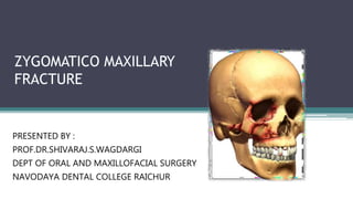
  • 1. ZYGOMATICO MAXILLARY FRACTURE PRESENTED BY : PROF.DR.SHIVARAJ.S.WAGDARGI DEPT OF ORAL AND MAXILLOFACIAL SURGERY NAVODAYA DENTAL COLLEGE RAICHUR
  • 2. CONTENTS • INTRODUCTION • CLASSIFICATION. • EXAMINATION. • SIGNS AND SYMPTOMS. • MANAGEMENT. • SURGICAL APPROACHES. • COMPLICATIONS. • REFERENCES
  • 3. INTRODUCTION  The zygoma or malar complex forms the central support of the cheek and is a strong buttress of the lateral and middle third of the facial skeleton • Zygomatic or malar fracture are the terms commonly used to described fractures that involve the lateral one third of the middle face. Other names for this fracture are:  Zygomaticomaxillary complex  Zygomaticomaxillary compound  zygomatico orbital  Zygomatic complex  Malar  Trimalar  Tripod
  • 4. CLASSIFIACTION Knight and North (1961) Rowe and Killey (1968) Yanagisawa ( 1973) Larsen and Thomson (1978) Rowe and Williams (1985) Spiessl & Schroll (1972) Poswillo’s classification Markus zing classification Manson and colleages Henderssons classification Zingg classification Ozyagzan classification
  • 5. According to Knight and North (1961) Group I – No significant displacement. Group II – Arch fracture. Group III – Unrotated body fracture (downward and backward). Group IV – Medially rotated body fracture (inward and backward). Group V – Lateral rotated body fracture. Group VI – Complex body fractures.
  • 6. According to Larsen and Thomsen (1968)  Stable fractures that shows - minimal or no displacement ( no intervention) Stable fracture – with displacement ( no fixation)  Unstable fracture with great displacement and disruption at frontozygomatic suture and comminuted fractures. Fractures of zygomatic arch are classified again into: Minimum or no displacement. V type in fracture. Comminuted fracture.
  • 7. ROWE’S AND KILLEY (1968)  Type I : No significant displacement  Type II : Fracture of the zygomatic arch  Type III : Rotation around horizontal axis - Inward displacement of orbital rim - Outward displacement of orbital rim  Type IV : Rotation around longitudinal axis - Medial displacement of frontal process - Lateral displacement of frontal process  Type V : Displacement of the complex enbloc - Medial -Inferior - lateral (Rare)  Type VI : Displacement of orbitoantral partition - Inferiorly -Superiorly  Type VII : Displacement of orbital rim segments  Type VIII : Complex comminuted fractures
  • 8. ROWE & WILLIAMS CLASSIFICATION Fractures unstable after elevation a)Arch only (inferiorly displaced) b)Rotation around horizontal axis i)medially ii)Laterally c)Dislocations en bloc i) inferiorly ii)medially iii)Postero-laterally Fractures stable after elevation a)Arch only (medially displaced) b)Rotation around the vertical axis i)medially ii)Laterally
  • 9. SPIESSLAND SCHROLL (1972) • TYPE 1- zygomatic arch fracture. • TYPE 2- zygomatic - complex fracture with no significant displacement. • TYPE 3- zygomatic complex fracture - partial medial displacement. (Kinking at F-Z suture) • TYPE 4 - Z.C with total medial displacement(complex fracture of F-Z suture). • TYPE 5- Z.C dorsal displacement (2 fracture sites in zygomatic arch). • TYPE 6- Z.C, inferior displacement. • TYPE 7- Z.C, comminuted fracture.
  • 10. YANAGISAWA(1973) • GROUP 1- non displaced fracture- no treatment. • GROUP 2- arch fracture(pure) • GROUP 3- medial/lateral rotation around vertical axis. • GROUP 4- medial/ lateral rotation around longitudinal axis. • GROUP 5-medial/ lateral displacement without rotation. • GROUP 6- isolated rim fracture. • GROUP 7- all complex fracture.
  • 11. EBERHARD KRUGER(1986) FRACTURE OF ZYGOMA • No displacement. • Partial medial • Total medial • Dorsal displacement. • Inferior displacement. • Comminuted fracture.
  • 12. MANSON& COLLEAGUES(1990) Based on - Amount of energy dissipated by the facial bones. - Secondary to the traumatic force. - Findings in CT scans High energy fractures Lower energy fractures • Extreme displacement. Displacement • Comminution of articulations. NIL • Segmentation of bones. Treatment- Treatment- • Require extensive exposure Less agressive and Aggressive fixation.
  • 13. ANATOMY PRINCIPAL STRUCTURE OF MIDFACE MAJOR ROLE IN FACIAL CONTOUR 4 PROCESSES -ORBITAL -FRONTAL -TEMPORAL -MAXILLARY 4 ARTICULATIONS -FRONTAL -TEMPORAL -MAXILLARY -SPHENOID MUSCLE ATTACHMENTS - MASSETER -TEMPORAL FASCIA
  • 16. FRACTURE PATTERNS • Depends on- DIRECTION & MAGNITUDE OF FORCE • Fracture lines pass through lines of greatest weakness • Malar bone represent strong bone on fragile suppport • Force appiled on zygoma - distributed through four processes to adjacent articulating bone –weaker than zygoma • Three lines of fracture extend from IOF Anteromedial – orbital floor Superolateral - Inferior direction
  • 17. Clinical examination 1.NEUROLOGICAL STATUS. • First step is to assess neurological status……. • Associated neurologic injury was encountered in 57% of patients. 2.VISUAL STATUS 3.EXAMINATION OF ZYGOMA
  • 18. INSPECTION Symmetry of arches. Pupillary level. Presence of orbital oedema. Sub-conjunctival ecchymosis. Ant & lat projection of zygoma. Intra-oral. • PALPATION -Systematic & thorough. -Compare one side with another.
  • 19. SIGNS AND SYMPTOMS • Periorbital ecchymosis and edema. • Flattening of the malar prominence. • Flattening of the zygomatic arch. • Pain. • Ecchymosis of the maxillary buccal sulcus. • Deformity of the zygomatic buttress of the maxilla. • Deformity of the infra-orbital margin. • Trismus. • Abnormal nerve sensibility. • Epistasis. • Subconjunctival ecchymosis. • Crepitation from air emphysema. • Displacement of palpebral fissure. • Diplopia. • Enopthalmos.
  • 20. Orbital signs and symptoms Periorbital Oedema Circumorbital ecchymosis Subconjunctival haemorrhage Orbital Emphysema Diplopia Unequal pupillary levels
  • 21. Flattening of malar prominence • Characteristic sign • Presence of edema flattening may be difficult to discern Flattening of zygomatic arch • Loss of normal convex curvature
  • 22. Displacement of palpebral fissure Deformity of orbital margin
  • 23. ORBITAL EXAMINATION FORCED DUCTION TEST • To determine presence of mechanical obstruction to globe movement. • Test done to assess the cause of reduced movement of extraocular muscles following trauma to mid-face region. • It helps to determine if the cause is mechanical/ neurological. DIPLOPIA
  • 24.
  • 25. RADIOGRAPHIC EXAMINATION  Postero-anterior oblique view (OM/PNS view): excellent assessment of sinuses and their walls, zygoma and its processes and rims of orbit  Submentovertex view is specific for zygomatic arch fractures
  • 28. STEPS IN TREATING ZMC FRACTURE 1. Prophylactic antibiotics. 2. Anesthesia. 3. Clinical examination and forced duction test. 4. Protection of the globe. 5. Antiseptic preparation. 6. Reduction of the fracture. 7. Assessment of reduction. 8. Determination of necessity for fixation.
  • 29. 9. Application of fixation device. 10. Assessment of ocular motility. 11. Bone graft for extra orbital osseous defects. 12. Post surgical ocular examinations. 13. Post surgical images.
  • 30. NEED FOR FIXATION Indications for fixation 1. Comminuted fracture fragments. 2. Doubt regarding the stability Role of MASSETER in displacement: • Albright and McFarland recommended IMF following fracture reduction helps to reduce the pull of the masseter muscle on the repositioned ZMC. • Ellis et al reviewed series of isolated ZMC fractures treated by different approaches and fixation schemes and found no evidence of post reduction instability
  • 31. TREATMENT MODALITIES NO TREATMENT INDIRECT REDUCTION- • Gillies approach. • Keens approach. • Lateral coronoid approach. • Eyebrow approach. • Percutaneous approach. • Transnasal approach. DIRECT REDUCTION FIXATION. Coronal approach/bicoronal. Upper eyelid/ blepharoplasty. Supraorbital eyebrow approach. Transconjunctival . Subcilliary. Infraorbital. Maxillary vestibular approach
  • 32. TEMPORARY SUPPORT INDIRECT FIXATION- • Internal pin fixation. • Transfixation with kirshner wire. DIRECT FIXATION- • Transosseous wiring. • Bony platting.
  • 33. NO TREATMENT NON-DISPLACED FRACTURES-9-50 % OF ZMC # MEDICAL CONTRAINDICATIONS MINIMAL DEGREE OF DISPLACEMENT. UNLIKELY TO RESULT IN COSMETIC DEFORMITY DISTURBANCE OF VISION PARESTHESIA IMPAIRED MANDIBULAR MOVEMENTS.
  • 34. DECISION TO INTERVENE DEPENDS ON SIGNS & SYMPTOMS. FUNCTIONAL IMPAIRMENT. PRESENCE OF OPHTHALMIC INJURIES. PROGRESSIVE PROPTOSIS. VISUAL ACUITY DETERIORATION. VISUAL INTEGRITY ON UNAFFECTED SITE. OTHER RELATED FACIAL INJURIES. MEDICAL CONDITION OF PATIENT.
  • 35. DELAYED TREATMENT 1. MANIPULATION OF ORBITAL BONES 2. RISK OF EYEBALL INJURY. 3. EXISTING HAEMORRGHAGE. 4. PROGRESSIVE PROPTOSIS. 5. PRE-EXISTING BLINDNESS IN OTHER EYE. 6. GENERAL MEDICAL CONDITION. 7. NEUROLOGICAL STATUS QUESTIONABLE. 8. GROSS OEDEMA. 9. CRITICAL PERIOD - 5-10th DAY
  • 36. DELAYED TREATMENT ADVANTAGES OEDEMA RESOLVES- DETAILED EXAMINATION OF EYE. ANTRUM CLEARS - BETTER RADIOGRAPH. HAEMATOMA STILL NOT ORGANISED –DISSECTION EASIER. >5-10 DAYS DIFFICULTY IN DISIMPACTING & REDUCING. PHYSIOLOGICAL RESORPTION OF # MARGINS. INTERDIGITATION LESS ACCURATE. INELASTICITY OF FIBROUS TISSUE AT SITE OF MALUNION.
  • 37. IMMEDIATE TREATMENT • IN THE ABSENCE OF COMPLICATIONS. • NO OEDEMA. ADVANTAGES EXCELLENT REDUCTION -NOT COMPROMISED NO SOFT TISSUE SCARRING NO CHANGE IN MORPHOLOGY SUPERIOR FACIAL SOFT TISSUE CONTOUR.
  • 38. APPROACHES:  Temporal approach –  Buccal sulcus approach – Gillies (1927) Keen (1909), Balasubramanium (1967)  Lateral coronoid approach – Quinn (1977)  Eyebrow approach -  Percutaneous approach - Dingman & Natvig (1964) Stroymeyer (1844) INDIRECT REDUCTION
  • 39. TEMPORAL APPROACH Popular approach First described by Gillies & coworkers in 1927 – arch fractures ADVANTAGES :  Allows application of greater amount of controlled force to disimpact even the most difficult zygomatic fracture.  For treatment of fractures which are consolidated already  Quick and simple method DISADVANTAGE:  Encountered temporal vessels- hemorrhage
  • 40. Incision – 2.5cm, through skin & subcutaneous tissue White glistening surface Temporalis muscle bulge
  • 41. PLACEMENT OF ROWES ZYGOMATIC ELEV ATOR AND ELEVATION.
  • 42. BUCCALSULCUS APPROACH Keen’s Technique (1909) ADVANTAGES: Avoidance of any external scar. Used for both arch and ZMC fracture DISADVANTAGES: Unstable fractures – external incision for fixation TECHNIQUE: •A small incision (approximately 1 cm) is made in the mucobuccal fold, beneath the zygomatic buttress of the maxilla. •Sharp end of periosteal elevator – inserted & swept in a sweeping motion to contact infratemporal surface of maxilla & zygoma. •A heavier instrument inserted behind the infratemporal surface of the zygoma, using superior, lateral & anterior force to reduce the bone.
  • 43. LATERAL CORONOID APPROACH Quinnin1977–described Usedonlyforarchfractures TECHNIQUE : •3 to 4 cm incision -anterior border of the ramus. •Insision made - to the depth of the temporal muscle insertion (not to the bone) •Instrument between the temporal muscle and the zygomatic arch - readily palpable. •A flat-bladed instrument- inserted into the pocket& arch is elevated while palpating the arch extraorally.
  • 44. ELEVATION FROM EYEBROWAPPROACH •Used for both ZMC & arch fracture ADVANTAGE: •Fracture at the orbital rim is visualized directly •Fixation can be done with same incision. DISADVANTAGE: •Difficult to generate a large amount of force, especially in the superior direction. DINGMAN ZYGOMATIC ELEVATOR
  • 45. PERCUTANEOUS APPROACH •Most simple of all techniques as no soft tissue dissection is necessary •Direct route to elevate the depressed zygoma is through the skin surface of the face overlying the zygoma. ADVANTAGE: Produces forces anteriorly, laterally, and superiorly in a very direct manner DISADVANTAGE: Scar on the face in a very noticeable location.
  • 46. ELEVATION OF THE ZYGOMA WITH A BONE HOOK •Strohmeyer in 1844 •Poswillo`s intersecting lines. •Stab incision made and hook inserted. •Apply strong traction. Stacey bone hook
  • 47. Carrol-Girard bone screw Elongated corkscrew with a T- bar handle. Contains threads on its working end. Screw is threaded into body of zygoma following a hole placement & is used as a handle to reduce fracture Can control ZMC position in all 3 planes of space. LARGE BONE SCREW
  • 48. SURGICALAPPROACHES TO ZMC  Maxillary vestibular approach  Supraorbital eyebrow approach  Upper eyelid approach  Lower eyelid approach  Transconjunctival approach  Coronal approach DIRECT REDUCTION
  • 49. MAXILLARY VESTIBULAR APPROACH • Most useful when performing any of wide variety of procedures in the midface. • Safe access to the entire facial surface of the midfacial skeleton- from the zygomatic arch to the infraorbital rim to the frontal process of the maxilla. ADVANTAGE: • Hidden intraoral scar. • Rapid and simple approach • Complications are few.
  • 50. TECHNIQUE Subperiosteal Dissection V –Y Closure Incision through the mucosa, submucosa, facial musculature,and periosteum (3-5mm superior to mucogingival junction) Infraorbital rim, anterior maxilla & ZMC Buttress - treated
  • 51. LATERAL BROW APPROACH Popular approach - Access to the lateral orbital rim and the frontozygomatic suture ADVANTAGE: Simple, safe and rapid approach Scar is usually hidden within the confines of the eyebrow No important neurovascular structures are involved in this approach.
  • 52. DISADVANTAGE :  In individual who has no eyebrows extending laterally and inferiorly along the orbital margin, this approach is undesirable.  Incisions made along the lateral orbital rim outside of the eyebrow are very conspicuous in such individuals, and another type of incision may be indicated.  Extremely limited access.
  • 53. TECHNIQUE •Incision – parallel to the eyebrow hair. •The incision is made through skin and subcutaneous tissue to the level of the periosteum in one stroke. (2cm) •Incision through periosteum along lateral orbital rim and subperiosteal dissection into lacrimal fossa. •Because of the concavity just behind the orbital rim in this area, the periosteal elevator is oriented laterally as dissection proceeds posteriorly. Closure: The incision is closed in two layers, the periosteum and the skin.
  • 54. UPPER EYELID APPROACH  The upper eyelid approach to the superolateral orbital rim is also called UPPER BLEPHAROPLASTY, UPPER EYELID CREASE, & SUPRATARSAL FOLD APPROACH.  In this approach, a natural skin crease in the upper eyelid is used to make the incision. Advantage: ADVANTAGE: Inconspicuous scar it creates - best approaches to the region.
  • 55. TECHNIQUE Closure:The wound is closed in two layers, periosteum and skin/muscle. To facilitate retraction of the skin/muscle flap, it can be widely undermined laterally and retracted with small retractors. Periosteum is divided 2-3mm posterior to the orbital rim with a scalpel. Sagittal section through orbit and globe showing dissection between orbicularis oculi muscle and the orbital septum Incision – begins 10mm superior to upper lid margin & lies 6mm above lateral canthus as it extends laterally. The initial incision is made through skin and muscle.
  • 56. LOWER EYELID APPROACH SUBTARSAL approach: • Frequently employed – access to infraorbital rim & orbital floor • Along natural skin crease at/below the level of tarsus, approx half distance between lash margin and orbital rim ADVANTAGE: • Relatively easy • Scar is imperceptible • Minimal complication
  • 57. SUBCILIARY approach: • Also called INFRACILIARY APPROACH/ BLEPHAROPLASTY • Incision is - approx 2 mm below the eyelashes and can be extended laterally as necessary (top dashed line) along the natural crease inferior to lateral canthal ligament. ADVANTAGE: • Imperceptible scar • Simultaneously exposes infraorbital & F-Z areas. DISADVANTAGE: • Technically difficult • Risk of postoperative ectropion
  • 58. TECHNIQUE •Skin incision •Dissect b/w skin & muscle until orbital rim is reached •2nd incision – through muscle & periosteum to bone •Incision – skin & muscle at same level •Dissect down anterior to orbital septum to orbital rim •Combination of these – STEPPED incision •Subcutaneous dissection toward rim – few mm •Followed by muscle incision at lower level & then following orbital septum to rim. •Fine scissors are used during dissection –spreading motion. •Incision through periosteum – 3-4mm below rim
  • 59. TRANSCONJUNCTIVALAPPROACH  Originally described by Bourguet in 1928.  Also called INFERIOR FORNIX APPROACH.  2 types:  Preseptal (Tessier)  Retroseptal (Tenzel&Miller) approaches.  Converse & colleagues added a lateral canthotomy to transconjunctival retroseptal incision for improved lateral exposure.
  • 61. TECHNIQUE Sagital section through orbit showing preseptal and retroseptal placement of incision. Initial incision for lateral canthotomy Initial canthopexy incision to dissect in the subconjunctival plane. The dissection should be just below the tarsal plate and extend no farther medially than the lacrimal punctum. Closure of transconjunctival incision and inferior canthopexy
  • 62. ADVANTAGE :  Produce excellent cosmetic results because the scar is hidden in the conjunctiva.  If a canthotomy is performed in conjunction with the approach, the only visible scar is the lateral extension, which heals with an inconspicuous scar.  Rapid and no skin or muscle dissection is necessary. DISADVANTAGE : • medial extent of the incision is limited by the lacrimal drainage system.
  • 63. CORONALAPPROACH  The coronal or bi-frontal incision is a versatile surgical approach to the upper and middle regions of the facial skeleton, including the zygomatic arch ADVANTAGE: •The surgical scar is hidden within the hairline. •Excellent acess to orbits, zygoma & arch.
  • 64. TECHNIQUE Incision placement: •Hairline of patient •Amount of inferior access •Incision – skin, subcutaneous tissue & galea •Lateral aspect of skull- superficial layer of temporal fascia incised 2cm supr to zygomatic arch •Dissect inferiorly - zygomatic arch •Periosteal incision – along the superior aspect of arch •Pericranium is incised along lateral orbital rim – FZ suture •Wound is closed in layers – fascia , galea, skin.
  • 66. MODIFICATION OF HEMICORONAL APPROACH •The anterior arm of the incision is curved downward toward the superior wall of the orbit before it reaches the vertex of the skull within the hairline. •The ‘backcut’ provides excellent exposure of the entire zygomatic complex and the arch. • Aesthetic and is less invasive thereby being quite acceptable by patients. Journal of Maxillofacial and Oral Surgery 2010 Volume 9, Number 3, 270-272
  • 67. Common methods include wire osteosynthesis and rigid fixation by plates Less common methods include external pin fixation and maxillary antral support IMMOBILIZATION
  • 68. PIN FIXATION  External pin fixation Can be used for fractures that demonstrate an intact body of the zygoma but severe communition at the junction with the surrounding bones  Internal pin fixation Was introduced by Fryer and results in stable entity and relatively free of complications Techniques make use of K-wire placement
  • 69. SINUS PACKING SUPPORT  Gauze or balloon can be used to provide inferior support to the zygoma  Lateral wall is approached through a Caldwell-Luc  ½ inch gauze dipped in antibiotic of choice is placed along the floor anteroposteriorly  Antral balloon can be used by it is relatively imprecise and cannot adapt to the topography
  • 71. INDIRECT FIXATION • Zygomatic bone - rigidly secured to facial skeleton. INDICATION • Gross loss of bone in FZ region and infra orbital rim.  ZYGOMATICO-ZYGOMATIC  NASO-ZYGOMATIC  ZYGOMATICO-PALATAL  MAXILLO-ZYGOMATIC  FRONTO-ZYGOMATIC  CRANIO-ZYGOMATIC
  • 73. EXTERNAL FIXATION Accomplished with wires suspended from plaster head caps, head frames and by pins connected to one another with universal joints and cold cure acrylic. ADVANTAGES- 1. Three dimensional stability. 2. Minimal scarring. 3. Adjustability of the reduction. DISADVANTAGES- 1. Patient comfort is compromised. 2. Need for specific hardware. 3. Lack of usefulness in comminuted fractures.
  • 74. DIRECT FIXATION • TRANS-OSSEOUS WIRING/ OSTEOSYNTHESIS FZ SUTURE INFRA ORBITAL MARGIN • BONE PLATES
  • 75. FIXATION TECHNIQUES - PRINCIPLES 1. Use self-threading bone screws. 2. Use hardware that will not scatter postoperative CT scans. 3. Place at least two screws through the plate on each side of the fracture. 4. Avoid important anatomic structures. •Use Y,L,T shaped plates where fracture line in the zmc buttress region is low. •Prevents damage to the roots and nerve bundle.
  • 76. 5. Use a thin plate as possible in the periorbital areas. 6. Place as many bone plates in as many locations as necessary for ensuring stability. 7. If concomitant fractures of other midfacial bones exist, it will be necessary to apply fixation devices more liberally. 8. In areas of comminution or bone loss, span the gap with the bone plate.
  • 77. BONE PLATES FOUR POINT FIXATION-  COMMINUTED ZMC FRACTURES SITES OF FIXATION- 1. F-Z SUTURE. 2. INFRAORBITAL RIM. 3. ZYGOMATIC ARCH. 4. MAXILLARY BUTTRESS.
  • 78. THREE POINT FIXATION-  NON-COMMINUTED ZMC FRACTURES  SITES OF FIXATION- •F-Z SUTURE • INFRAORBITAL RIM • ZYGOMATIC ARCH (OR) MAXILLARY BUTTRESS.
  • 79. TWO POINT FIXATION-  SIMPLE NON-COMMINUTED ZMC FRACTURES  SITES OF FIXATION- F-Z SUTURE  INFRAORBITAL RIM OR MAXILLARY BUTTRESS
  • 80. PLACEMENT OF FIRST PLATE FIXATION OF FIRST PLATE
  • 81. PLACEMENT OF SECOND PLATE PLACEMENT OF THIRD PLATE
  • 83. Complications of periorbital incision 1. Minor - dehiscence hematoma /seroma lymphedema 2. Vertical shortening of lower lid prevention - superior support of lower lid for several days( best achieved with frost sutures). 3. Ectropion – associated with subciliary incision and trans conjunctival incision(mild /moderate/severe) 4. Entropion – inward curl of lower eyelid -occurs less commonly but more distressing (subciliary incision )
  • 84. 5.Infraorbital nerve injury  Either direct injury to nerve due to trauma or iatrogenic  Mostly these injuries are temporary(neuropraxia) due to stretching or compression of infraorbital nerve.  Markedly displaced fractures - neurotmesis can occur  Patient may complain of numbness , different sensation and pain on heat /cold / light touch .  ZMC fracture which are treated with rigid fixation – early recovery of neurosensory deficit .
  • 85. 6.Persistent Diplopia  Binocular diplopia initially present with ZMC fracture should resolve within 5-7 days after fracture treatment  Result of  edema or hematoma of one or more extraocular muscles or their nerves  Introrbital edema  Ocassionally muscle entrapment  If persists, it may be due to scar contracture and adhesions either within the ocular muscles or between them.
  • 86. 7.Enopthalmos  Most commonly caused by increased volume of orbit  Difficult to correct secondarily, however improvement is possible.  Surgery can be done to reduce orbital volume by – reconstructing the internal orbit - by placing a space occupying material behind the globe ( glass beads , silicon sheets , sponges , teflon beads , cartilage graft, hydroxyapatite, metallic mesh or plate)
  • 87. 8.Blindness Occasionally reported after ZMC fracture  Causes- direct damage to optic nerve - hemorrhage into optic sheath - intraocular edema - retrobulbar hemorrhage 9.Maxillary sinusitis  Caused by inflammation of sinus membrane and occlusion of ostium.  Usually respond to antibiotic and decongestant therapy.
  • 88. 10.Ankylosis of zygoma to coronoid process - very rare - when noted usually fibrous. Causes- - improper reduction of zygoma leaving arch in close proximity to coronoid process. - untreated zygomatic fracture - post-operative infection
  • 89. 11. Malunion of the zygoma Signs and symptoms – •Flattening of malar prominence • Enopthalmos •Altered pupillary level • Limitation of mandibular movements Treatment: - camouflaging the defect with implant or transplant - repositioning of malpositioned bone
  • 90. CONCLUSION  Surgical intervention is an effective treatment modality of depressed ZMC fracture, whereas a nonsurgical approach is often used for nondisplaced fractures.  Most of them can be treated solely by an intraoral approach & rigid fixation at ZM buttress.  Further exposure of FZ junction or infra orbital rim is necessary for severely displaced fractures, requiring additional fixation.
  • 91. REFERENCES • ORAL AND MAXILLOFACIAL TRAUMA-FONSECA • PETERSON’S PRINCIPLES OF ORAL AND MAXILLOFACIAL SURGERY • ROWE AND WILLIAMS MAXILLOFACIAL TRAUMATOLOGY • TEXTBOOK OF ORAL AND MAXILLOFACIAL SURGERY