1. Off-track 2 in embryonic motor axon guidance
David Robinson1 and Samantha Alsbury1
1University of Greenwich, Department of Life and Sports Science, Medway Campus, Central Avenue, Chatham Maritime, Kent, ME4 4TB.
Through bioinformatic analysis a number of
uncharacterized proteins were identified as potential
axon guidance cues from their protein structure
(Dolan et al., 2007). One of these candidates was
CG8964, now known as off-track 2, as it is related
to the known motor axon guidance gene off-track.
Winberg et al. (2001) reported that, in off-track (otk)
mutants, the ISNb fuses to the ISN (Figure 1), and
that the mutation is lethal.
Investigating whether otk is implicated in PCP,
Linnemannstöns et al. (2014) created a new otk null
allele (otkA1). They also identified an otk paralog,
off-track 2 (otk2), and created an otk2 null allele
(otk2C26). The new otk allele is viable, suggesting a
second site mutation contributed to the lethality and
axon guidance defects previously reported.
Using an antibody for Otk2, we investigated the
protein’s distribution in stage 16 wild type embryos
(Figure 2). 1D4 (anti-FasII) was used to examine
motor neurons in filleted late stage 16 and stage 17
embryos.
In addition to the ISNb fusion phenotype, the muscle 12-innervating motor neurons
(MN12s) project anteriorly in otkA1 mutants (Figure 4).
MN12s project anteriorly in otkA1 mutants
otkA1 null mutants exhibit previously described
phenotypes (ISNb fusion, MN13s absent, RP3
absent; Winberg et al., 2001). These results do not
support the hypothesis that the axon guidance
phenotypes described by Winberg et al. (2001) were
due to a second-site mutation.
otk mutants also display previously unreported
phenotypes: MN12s and MN13s project anteriorly.
Winberg et al.’s (2001) model of Otk suggests that it
binds to PlexA, the receptor for the neuronally-
expressed repellant Sema1a, allowing axons to
defasciculate at precise points. This does not account
for anterior projections, highlighting the need for a
more sophisticated model of otk’s role in motor axon
guidance.
The same phenotypes, with the exception of the ISNb
fusion, were observed in otk2C26 mutants. This is the
first time that an axon guidance role has been
attributed to Otk2, which might form part of a
receptor complex with Otk and PlexA.
This suggestion is supported by the presence of
phenotypes in otkA1/+, otk2C26/+ transheterozygotes
and that interactions between the proteins have been
observed previously (Linnemannstöns et al., 2014).
We provide evidence of genetic interactions between
the off-tracks and fz2, raising the possibility that otk
and otk2 are required for axons’ responses to the
repellent Wnt4 (Inaki et al., 2007).
Dolan, J. et al., 2007. The extracellular Leucine-Rich Repeat superfamily;
a comparative survey and analysis of evolutionary relationships and
expression patterns. BMC Genomics 8:320.
Inaki, M. et al., 2007. Wnt4 is a local repulsive cue that determines
synaptic target specificity. Current biology : CB, 17(18), pp.1574–9.
Linnemannstöns, K. et al., 2014. The PTK7-related transmembrane
proteins off-track and off-track 2 are co-receptors for Drosophila Wnt2
required for male fertility. PLoS genetics, 10(7), p.e1004443.
Winberg, M.L. et al., 2001. The transmembrane protein Off-track
associates with Plexins and functions downstream of Semaphorin
signaling during axon guidance. Neuron, 32(1), pp.53–62.
We thank S. Richardson, G. Tear, and E. Thompson for their support and
provision of apparatus, K. Linnemannstöns and colleagues for flies and
antibodies, the BSDB for supporting our attendance at the conference, and
the University of Greenwich for funding the project.
Introduction
MN12s project anteriorly in otk2C26 mutants
Figure 1. (from Winberg et
al., 2001). The ISNb fuses to
the ISN (fat arrow), thereby
failing to innervate muscles
6/7 (arrow with *), 12 and 13
(arrowheads with *).
Methods and Results
Discussion
Acknowledgements
References
Figure 2. Otk2 is expressed in the VNC (red arrow) and in
the roots of the motor neurons.
The ISNb fusion phenotype is present in the new
otkA1 allele (Figure 3).
Figure 3. ISNb fusion in otkA1 mutants. A. Wild type. ISNb
indicated. B. otkA1 mutant. Absence of ISNb indicated. Graph
shows percentage of hemisegments with phenotype.
The ISNb fusion phenotype is not seen in otk2C26 mutants, though the MN12s
project anteriorly (Figure 5).
Both otkA1 and otk2C26 mutants exhibit defects in regions of M13 and M6/7 (RP3
axon) (examples from otk2C26 in Figure 6). These are also present in several
transheterozygotes and otk2 gain-of-function lines (Table 1).
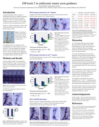
MN13 and RP3 phenotypes
Figure 6. MN13 and RP3 phenotypes in otk2C26. A. Wild type. MN13s project posteriorly (blue
arrow); RP3 axon projects posteriorly (green arrow). B. otk2C26. MN13s fail to project (blue arrow). C.
otk2C26. MN13s project anteriorly (blue arrow). D. otk2C26. RP3 axon fails to project (green arrow).
Table 1. MN13 and RP3 phenotypes in mutants,
transheterozygotes, and otk2 gain-of-function lines. Percent
of abnormal hemisegments on left of cells; number of
hemisegments in brackets. *significant at p < .05 (also in red).
The 2nd, 3rd, and 4th columns correspond to B, C, and D,
respectively, in Figure 6.
Figure 4. MN12s project anteriorly in
otkA1 embryos.
A. Wild type (“Normal”). MN12s project
dorsally from the M13 innervation site
(blue arrow), then posteriorly (red arrow).
B. otkA1 (“Mild”). MN12s project
dorsally from the M13 innervation site
(blue arrow), then anteriorly (red arrow).
C. otkA1 (“Moderate”). MN12s project
anteriorly (red arrow) at the M13
innervation site (blue arrow).
D. otkA1 (“Severe”). MN12s project
anteriorly (red arrow) ventral to the M13
innervation site (blue arrow).
Figure 5. MN12s project anteriorly in
otk2C26 embryos.
A. Wild type (“Normal”). MN12s project
dorsally from the M13 innervation site
(blue arrow), then posteriorly (red arrow).
B. otk2C26 (“Mild”). MN12s project
dorsally from the M13 innervation site
(blue arrow), then anteriorly (red arrow).
C. otk2C26 (“Moderate”). MN12s project
anteriorly (red arrow) at the M13
innervation site (blue arrow).
D. otk2C26 (“Severe”). MN12s project
anteriorly (red arrow) ventral to the M13
innervation site (blue arrow).
Phenotype distribution differs
between genotypes, χ2 (3, n = 180)
= 19.53, p < .001.
Phenotype distribution differs
between genotypes, χ2 (3, n = 168)
= 13.46, p < .01.