Reflex and Voluntary Control of Movement
•Als PPT, PDF herunterladen•
30 gefällt mir•14,979 views

An introductory lecture on neural organization of reflexes and voluntary control of movement.

Empfohlen
Weitere ähnliche Inhalte
Was ist angesagt?
Was ist angesagt? (20)
Ähnlich wie Reflex and Voluntary Control of Movement
Ähnlich wie Reflex and Voluntary Control of Movement (20)
Mehr von Csilla Egri
Mehr von Csilla Egri (14)
Kürzlich hochgeladen
Kürzlich hochgeladen (20)
Reflex and Voluntary Control of Movement
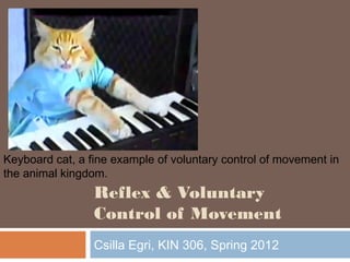
- 1. Keyboard cat, a fine example of voluntary control of movement in the animal kingdom. Reflex & Voluntary Control of Movement Csilla Egri, KIN 306, Spring 2012
- 2. Outline Locomotion reflexes Central pattern generators (CPGs) Descending tracts Pyramidal tracts Extrapyramidal tracts Cortical control of movement Motor cortex 2
- 3. Motor Control 3 There are three levels in the hierarchy of motor control: Spinal cord Locomotion reflexes CPGs Brain stem Talked about these during the vestibular and visual system lectures Postural reflexes Locomotor regions Voluntary control of movement Cortical motor areas Voluntary control of movement These different areas are highly interdependent and movements result from the coordinated action of these three regions.
- 4. Spinal cord: locomotion reflex example 4 Stepping reflex in newborns Disappears around 6 weeks, gradually replaced by voluntary walking behaviour Simplified half-center model for alternating rhythm generation B&B Figure 14-8
- 5. Spinal cord: central pattern generators (CPGs) 5 Neuronal network capable of generating a rhythmic pattern or motor activity in the absence of sensory input Walking, swimming, respiration Simplest CPGs contain spontaneously bursting or reciprocally innervated neurons Basic firing pattern modified by sensory or descending inputs
- 6. Spinal cord: CPG example 6 Transect afferent input, and decerebrate cat: With support, walking motion is reproducible with stimulation of brainstem Each limb is controlled by its own CPG
- 7. Spinal Cord: organization of motor tracts 7 Motor neurons of ventral horn organized topographically Descending upper motor neurons control lower motor neuron firing Also influenced by the activity of interneurons & peripheral sensory receptors B&L Figure 9-12
- 8. Spinal Cord: organization of motor tracts 8 Medial reticulospin al tract Direct (pyramidal) pathway lateral and anterior corticospinal tract corticobulbar tract Indirect (extrapyramidal) pathway rubrospinal tract tectospinal tract vestibulospinal tract reticulospinal tract (lateral & medial)
- 9. Direct (pyramidal) pathways 9 Pyramidal tracts originate primarily in motor, premotor and supplementary motor areas of cortex ~ 90% of descending axons cross in the medulla and descend in lateral columns of spinal cord (lateral corticospinal tract) Control distal muscles; precise, agile, skilled movements ~ 10% cross over in spinal cord (anterior corticospinal tract) Control proximal trunk muscles; coordinate movements of axial skeleton Corticolbulbar tracts also originate in motor cortex, descend to brainstem Axons terminate in motor nuclei of cranial nerves Control precise, voluntary movement of head, neck and tongue
- 10. Indirect (extrapyramidal) pathways 10 Complex, polysynaptic circuits involving motor cortex, basal ganglia, thalamus, cerebellum, and reticular formation Rubrospinal: from red nucleus to contralateral muscles controlling precise movements of distal parts of upper limbs Tectospinal: from superior colliculus to contralateral muscles controlling reflexive movements of head and neck to auditory or visual stimuli Medial reticulospinal tract Vestibulospinal: from vestibular nucleus to ispsilateral skeletal muscles of trunk and proximal parts limbs for maintaining posture and balance Medial and lateral reticulospinal: from reticular formation to ipsilateral skeletal muscles of trunk and proximal parts of limbs for maintaining posture and regulating muscle tone in response to body movements.
- 11. Motor areas of cerebral cortex 11 Primary motor cortex Main motor area involved in executing voluntary movements Movement elicited with least amount of electrical stimulation Pre motor cortex Sensory guidance of movement (receive input from posterior parietal cortex Contributes to extrapyramidal pathways Lesion impairs ability to develop strategy for movement Supplementary motor cortex Involved in planning of complex and two handed movements Coordinates posture
- 12. Motor areas of cerebral cortex: somatotopy 12
- 13. Specialized cortical motor areas 13 Broca’s area (area 44, 45) Coordinated movements of tongue and vocal cords for word formation Lesion results in expressive aphasia Voluntary eye movement field Also controls voluntary blinking Area for hand skill Lesions result in motor apraxia: uncoordinated, nonpurposeful hand movements Guyton Figure 55-3
- 14. Voluntary movement: simplified linear sequence of events 14 Command must be organized by the brain 1. Identify target in space a) objective is identified in posterior parietal cortex which receives input from somatosensory, visual, vestibular and auditory systems b) Sense of body position in relation to target also required 2. info transmitted to supplementary & premotor areas where the motor plan is developed a) Choice of muscles, sequence of contractions, required force and trajectory computed 3. Motor plan transmitted to primary motor cortex and down descending pathways to interneurons & motor neurons
- 15. Motor Plan 15 Sensory feedback provided through ascending afferent pathways Transmitted to motor cortex either directly from thalamus or indirectly thru connections between somatosensory & visual cortex Motor cortex has bidirectional connections with thalamus, cerebellum & basal ganglia Important in planning & execution of movement (more in next lecture)
- 16. Objectives After this lecture you should be able to: Give an example of a locomotion reflex and an activity governed by a CPG Discuss the organization of the spinal cord and how it relates to voluntary control of movement Include both pyramidal and extrapyramidal descending tracts Describe the organization of the motor cortices Outline the sequence of events involved in initiation of voluntary movement 16
- 17. 17 Test your knowledge 1. A lesion to Broca’s area results in _____________________ 2. Precise, voluntary movements of the head, neck and tongue are controlled by descending inputs via the __________________________ tract 3. Motor neurons descending in the pyramidal tracts originate in ________________________ whereas motor neurons descending in the extrapyramidal tracts originate in _______________________________________________.
Hinweis der Redaktion
- Gutyon is best
- Vestibulospinal Reflexes Senses falling/tipping contracts limb muscles for postural support Vestibulocollic Reflexes acts on the neck musculature to stabilize the head if body moves Vestibulo-ocular Reflexes stabilizes visual image during head movement causes eyes to move simultaneously in the opposite direction and in equal magnitude to head movement Locomotor regions: midbrain locomotor regions: when stimulated, leads to sustained locomotion. Involved in initation of movement.
- When one motor neuron is active, the other is inhibited
- Modification by afferent input ensures can adapt to certain situations (terrain for example) can see CPG activity in transected cat The simplest movements are reflexes (knee jerk, pupil dilation), which are involuntary, stereotyped and graded responses to sensory input, and have no threshold except that the stimulus must be great enough to activate the relevant sensory input pathway. Fixed action patterns (sneezing, orgasm) are involuntary and stereotyped, but typically have a stimulus threshold that must be reached before they are triggered, and are less graded and more complex than reflexes. Rhythmic motor patterns (walking, scratching, breathing) are stereotyped and complex, but are subject to continuous voluntary control. Directed movements (reaching) are voluntary and complex, but are generally neither stereotyped nor repetitive. Rhythmic motor patterns comprise a large part of behaviour. They are also complex (unlike reflexes) yet stereotyped (unlike directed movements) and, by definition, repetitive (unlike fixed action patterns). As a consequence of this combination of behavioural importance and experimental advantage, rhythmic motor pattern generation has been studied extensively. Key to understanding rhythm generation in this (and many other network-based CPGs) is the concept of a half-centre oscillator. A half-centre oscillator consists of two neurons that individually have no rhythmogenic ability, but which produce rhythmic outputs when reciprocally coupled. Several types of interacting processes can support this rhythm generation (see
- showed that when a portion of the brain stem of a cat was cut across the middle—thus severing any connections between the brain and the spinal cord—the cat was still capable of standing. Furthermore, if a specific region of the brain stem was stimulated, the cats could be induced to walk on a treadmill, and alternating bursts of muscle activity could be recorded in extensors and flexors in conjunction with walking (Shik et al., 1966). These series of experiments led to the conclusion that each limb is controlled by a central pattern generator (CPG) in the spinal cord, which controls rhythmic motor activity, including walking. Shik and colleagues experimented with a cat whose brain stem was severed but that was still able to walk on a treadmill when a specific region of the brain stem was stimulated. The top of the figure shows the brain and the spinal cord. The muscle activity recorded from the flexors and extensors demonstrates that they are contracting and relaxing at opposite times from each other, consistent with normal function.
- Refer to overhead slide # 1 regarding these notes: interneurons located in medial part of spinal cord make bilateral synaptic connections with motoneurons on axial muscles interneurons located more laterally make ipsilateral synaptic connections with motoneurons of girdle muscles (shoulder and pelvis) interneurons located in lateral part of spinal cord make synaptic connections with distal limb muscles The Propriospinal system is a series of neurons whose axons run up and down (rostral-caudal direction) the spinal cord. They connect different segmental levels of the spinal cord together by synapsing with interneurons and motoneurons. Medial propriospinal neurons have long branching axons – some extend the length of the spinal cord – to coordinate movements of the neck and pelvis allowing the axial muscles to be coordinated. They are located in the ventral and medial columns. Lateral propriospinal neurons interconnect a smaller number of segments and have more focused connections. This explains the greater interdependence of action of more distal muscles at the hand and wrist. The shoulder and elbow have more stereotyped movements and thus are more interconnected than the hand and wrist but less than the axial muscles. They are located in the dorsal and lateral columns.
- Direct: input to lower motor neurons via axons that extend directly from the cerebral cortex. Indirect pathway: input to lower motor neurons from motor centers in the brainstem. These brain stem centers in turn receive input from the basal gangli, thalamus, brainstem nuclei, reticular formation, cerebellum and cerebral cortex.
- Some axons of corticobulbar tract cross over, some do not
- Rubrospinal red nucleus located in midbrain Lateral Vestibulospinal Tract primarily excites proximal extensor motoneurons & inhibits flexor motoneurons of both upper and lower limbs through interneurons & propriospinal neurons Medial Vestibulospinal Tract makes synaptic connections with medial motoneuron groups (neck and back muscles) and with nearby interneurons and propriospinal neurons some monosynaptic excitatory connections to ipsilateral neck motoneurons and monosynaptic inhibitory connections to contralateral neck motoneurons Both are important in the reflex control of balance and posture in response to head movements. Medial Reticulospinal Tract axons from pontine reticular formation descend in ventral columns on ipsilateral side of spinal cord make excitatory synaptic connections with axial muscles and proximal limb extensor muscles Functions to support posture Neurons in the reticulospinal tract rise from a cluster of nuclei in the reticular formation in the pons and medulla. Lateral Reticulospinal Tract axons from medullary reticular formation descend bilaterally in ventral part of lateral columns make inhibitory synaptic connections with neck and back motor neurons has widespread polysynaptic inhibitory connections with extensor motor neurons & excitatory connections with flexor motor neurons Tectospinal Tract originates in deep layers of superior colliculus axons cross to contralateral side of body just below periaqueductal gray matter project to medial interneurons in upper cervical segments regulates contralateral movements of head in response to visual, auditory and somatic stimuli Receives inputs from the cortex via a cortico-tectospinal pathway
- The motor cortex itself is subdivided into three areas, each with its own topographical representation of muscle groups and specific motor functions; M1 is primary motor cortex, PMA is premotor area, and SMA is supplementary motor area, initially subdivided based on electrophsyiological experiments: which area can evoke movement with the lowest stimulation
- Motor cortex is plastic: meaning the representation of body parts can change with practice or disuse. Important for rehabilitation after stroke. motor regions of the cortex contain somatotopic motor map of body (mapping from cortical activation site to muscles on contralateral side of body) - found in primary motor cortex, premotor cortex, and supplementary motor areas. orderly arrangement of control areas for face, digits, hand, arm, trunk, leg and foot fingers, hands and face (used in tasks requiring greatest precision and finest control) have disproportionately large representation
- Voluntary eye movement controls tracking or moving eyes voluntarily to different objects. Damage results in locking involuntarily on certain objects
- commands from the brain got to spinal motor neurons or interneurons carried by descending pathways (e.g. corticospinal tract as discussed earlier) posterior parietal cortex = association area motor plans include: choice of muscles, strength of contraction and sequence of contraction
- This is also true for correction of errors (more later with basal ganglia and cerebellum).