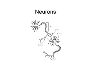
Neurons are the basic building blocks of the nervous system and communicate with each other via electrical and chemical signals. They have dendrites that receive information, a cell body that contains genetic material, and an axon that sends information to other neurons. Neurons transmit signals through electrical impulses and the release of neurotransmitters across small gaps between neurons called synapses. Experiences and stimuli shape the development of neural pathways and connections in the brain throughout childhood and adolescence as the brain undergoes pruning and myelination.
3 ; some can be up to 3 feet long (motor-sensory axons extending down the spinal cord). 4 This phenomenon is generated through the flow of positively charged ions across the neuronal membrane
Momentarily stops the impulse
Neurotransmitters stored in the axon’s terminals are released into the synaptic gap by Neuron #1 and received by Neuron #2 Although the process is complex, the end result is they “turn Neuron #2 on or off” - telling it to ‘transmit’ or ‘don’t transmit’ the message 2 Activity with electric cords and bodies…
You tube 1. 3D animation of a neuron and an action potential. Ion channels in the soma open and allow an increase in the intracellular voltage. Once 1the voltage reaches a threshold, an action potential is generated, which travels the length of the axon and passes into the terminals. The voltage in the terminals stimulate the release of neurotransmitters which cross the synapse, opening ion channels in dendrites of the post-synaptic neuron and increasing the voltage of the second neuron. By Stephen Hicks, 2006. www.metope.org 2 cartoon explaining action potential and ions You tube 2 discovery channel start 1:09 Have them sculpt a neuron with materials
Until recently it was believed that we will not grow more…now new neurons have been found in areas of the brain such as the hippocampus (an area of the brain associated with learning and memory it is the connections more than the number of neurons, that count read: you hold your newborn so his sky-blue eye are just inches from the brightly patterned wallpaper. ZZZZZT: a neuron form his retina makes an electrical connection with the one in his brain’s visual cortex You gently touch his palm with a clothespin: he grasps it, drops it, and you return it to him with soft words and a smile. CRACKLE: neurons from his hand strengthen their connection to those in his sensory-motor cortex. He cries in the night: you feed him, holding his gaze because nature has seen to it that the distance from a parent's crooked elbow to his eyes exactly matches the distance at which a baby focuses. ZAP: neurons in the brain’s amygdala send pulses of electricity through the circuits that control emotion. You hold him on your lap and talk…and neurons from his ears start hardwiring connections to the auditory cortex And you thought you were just playing with your kid” from Your Child’s Brain , Newsweek You tube .44sec Growth of axons in the brain You tube This time lapse video shows development of a normal neuron (left) and a mutated neuron that does not express the Ena/VASP proteins. Cultured for two days, the normal one extends an axon and many dendrites, while the mutated neuron fails to make such extensions.
Demonstrate with example page 9: What wires a child’s brain is repeated experiences in which she is actively engaged. The brain becomes “hard wired” to respond along established pathways. Skills are developed and refined, habit patterns are formed.
1 Axons sprout new terminals, dendrites grow new spines, and the neuron grows additional dendrites - all in preparation to process and remember more information , more efficiently. 2. Example con’d page 11
Baby grabs the ball, she’s making the connection that the action she just did got her what she wanted. Do brain gym of cross crawl and leg stretch
Demonstrate pruning with people up and those not connected to sit…they have been pruned…notice remaining neurons are less cluttered and more organized. Pruning is as important as branching…like pruning a tree, it removes weaker, less used synapses which might decrease the effectiveness of the brain By tow the toddler can recognize and name the object as a ball and say simple sentences like “ball go”…imagine the thoughsands of additional neural pathways that would be involved in this new skill!
The brain changes it’s architecture day by day, possibly even minute by minute, reshaping itself continually to cope with experience. Synapses in the brain create pathways that build functional architecture that lets us become who we are and will be ….most plastic in the first 3 years becomes less plastic with age although healthy brain continues to grow throughout life A baby’s brain Is a work in progress 3. Genes (nature) are the blueprint and experience (nurture) are the carpenter’s the create a unique human being…..genes are on/off switch and environment are the fine tune buttons What do you do to keep your brain healthy? Share with a neighbor. What more could you do?
New connections in an infant’s brain are forming at the rate of 3 billion a second This chart shows the rates of glucose consumption by various regions of the cerebral cortex, as a function of age. Note the rapid increase between birth and age three to a level that far exceeds that of adults and the gentle downward slope from around age 10. The brain has decreased synapses but increased in power It is thought that we start out with twice s many synapses to help assure that the brain will have a maximum capability to develop accordin to the environment it is born into. This gives the young brain exceptional flexibility and resilience
2. If not those parts of the brain may never completely develop their potential 3. Children need the re However new connections can be formed at ANY time!….study of convent in Minnesota. Of the 150 retired nuns in this convent, 25 are older than 90. They do not seem to suffer from dementia as early or as severely as the general population. Found that those nuns who stay active, who constantly challenge their minds live longer and stay alert. Critical periods represent a narrow window of time during which a specific part of the body is most vulnerable to the absence of stimulation or to environmental influences. Vision is a good example: Unless an infant sees light during the first 6 months, the nerves leading from the eye to the visual cortex of the brain that processes those signals will degenerate and die. Prenatal development, the period before a baby is born, also includes critical periods. Remember the drug thalidomide and its effects on prenatal development? Women who took the drug between the 38th and 46th days of pregnancy gave birth to infants with deformed arms, or no arms, Women who took the drug between the 40th and 46th days of pregnancy gave birth to infants with deformed legs or no legs. Women who took the drug after the 50th day of pregnancy gave birth to babies with no birth defects or problems. Sensitive periods are the broad windows of opportunity for certain types of learning. Sensitive periods represent a less precise and often longer period of time when skills, such as acquiring a second language, are influenced. But, if the opportunity for learning does not arise, these potential new skills are not lost forever. Individuals learn new languages at many different times in their lives. The skills acquired during sensitive periods are those that some people are better at than others. They include the social, emotional and mental characteristics that make us interesting people. Individuals who work with children need to be aware of the sensitive period concept so that they can provide learning opportunities that benefit children in many ways. The early brain research highlights birth through age 3 as a sensitive period for development and learning in all areas.
Watch 10 things every child needs and brain map it. Have participants brainstorm what enriches and environment by age group. How do caregivers help with that? What is the provider role?
brain nerve cells, or neurons, are initially produced in the center of the developing brain. To function normally, neurons must migrate to the brain's cortex, or outer layer, and other structures. How do neurons know where to migrate? During the early 1970s, researchers examining the developing brains of monkeys discovered that neurons often clung to long fibers of cells called glia. Neurons use these glial fibers, which radiate from the brain's inner to outer surfaces, as a highway to carry them through the brain to their destination. Some neurons also use the axons of other nerves to migrate from one brain area to another. You tube 1: Granule cell migrate along radial glial invitro Your tube 2: Neuronal precursors born in the subventricular zone (SVZ) of the neonatal and adult rodent brain migrate 3--8 mm from the walls of the lateral ventricle into the olfactory bulb. This tangentially oriented migration occurs without the guidance of radial glia or axonal processes. The cells move closely associated, forming elongated aggregates called chains, which are ensheathed by astrocytes. We have developed a culture system in which postnatal mouse SVZ neuronal precursors assemble into chains with ultrastructural and immunocytochemical characteristics equivalent to those in vivo but without the astrocytic sheath. Time-lapse videomicrography revealed that individual cells migrate along the chains very rapidly (~122 µm/hr) in both directions. Periods of cell body translocation were interspersed with stationary periods. This saltatory behavior was similar to radial glia--guided migration but ~4 times faster. Neuronal precursors isolated from embryonic cortical ventricular zone or cerebellar external granule layer did not form chains under these conditions, suggesting that chain migration is characteristic of SVZ precursors. This study directly demonstrates that SVZ neuronal precursors migrate along each other without the assistance of astrocytes or other cell types.
Myelin coats the neurons axons much like the insulated coating protects the electrical wires of a cord. Insulation is important for electrical wiring or it would short out. With an unisulated or non-myelinated cell, the electrical impulse does not short out, but ravels down the axon much more slowly and inefficiently. The nodes of Ranvier, located between the myelinated sections of the axon, are areas of low electrical resistance where almost all of the axon’s sodium channels are concentrated. These nodes are where action potentials can regenerate after the ion currents associated with them have propagated passively down the insulating myelin sheath between one node and the next. Schwann cells produce the fatty insulin called myelin that surrounds some nerve fibers. unmyelinated neurons do not possess an active neuregulin gene and that myelinated neurons do. they inserted the neuregulin gene into the unmyelinated axons. Instead of just sitting on the axons, the Schwann cells now produced thick myelin sheaths around them. So it appears that the gene instructs the Schwann cells to build the myelin wrap. Shaken Baby Syndrome - in newborns, only the most primitive systems, such as those needed for sucking, have been coated with myelin. Their brain is mushy. Therefore shaking an infant whips this mushy brain around in the skull and provides stretching forces that can rip apart areas of the brain. This leads to “Shaken baby Syndrome” which can kill or leave a child deaf, blind and/or mentally retarded.
It’s growth corresponds to the ability to use increasingly higher-level mental abilities. The process of myelination in human brains is not completed at least until most of us are in our 20’s and 30’s or even longer The system is overall remarkably responsive to stimulation from the environment, the schedule of myelination appears to put some boundaries around “appropriate” forms of learning at any given age” Jane Healy
![Description of a Neuron ,[object Object],[object Object],[object Object],[object Object]](data:image/gif;base64,R0lGODlhAQABAIAAAAAAAP///yH5BAEAAAAALAAAAAABAAEAAAIBRAA7)