SlideShare ist ein Scribd-Unternehmen logo
1 von 20
Chelsea Hayes
12/5/2014
1
Traumatic Brain Injury: Metabolic Perspective
The metabolic response to acute traumatic brain injury (TBI) involves a myriad of undesirable
corporeal and cerebral alterations. Some of those alterations include a modified immune function,
severely disrupted, unchecked metabolism, and a hypercatabolic state. A number of critical pathways,
hormones and inflammatory mediators contribute to this deranged metabolic picture, leading to shifts
in fuel sources and nutrition demands. Based on the supporting studies in the following ten annotated
bibliographies (nine controlled experimental and one review study), this overview identifies four main
pathways affected by acute TBI, which include: glycolysis, gluconeogenesis, glycogenolysis and
proteolysis. These pathways and their regulatory influences can be further understood by their roles in
and effects on specific tissues.
In the brain, the primary injury induces the release of cytokines, most commonly TNF-alpha, IL-
1, and IL-6, as part of the immediate inflammatory response. This severe local inflammation and direct
disruption of the mitochondrial membrane result in a demand for energy to repair the damaged area.
Hyperglycolysis ensues, followed by suppressed cerebral glucose uptake and increased net lactate
uptake, along with a massive outpour of counter-regulatory hormones like cortisol, glucagon and the
catecholamines (1, 2). The degradation of brain glycogen attempts to attend to the emergency energy
need for local tissue repair, while peripheral carbohydrate stores are mobilized via lactate to hepatic and
renal gluconeogenesis to support brain glucose (1). Endogenous fueling of the brain post TBI shifts to
the mobilization of total body glycogen reserves and the production of lactate, which is thought to act as
a brain fuel through the astrocyte-neuron lactate shuttle (3).
In the heart, hypermetabolism is attributed to the increase in levels of catabolic hormones and
cytokines known to elevate cardiac output and induce tachycardia and mild hypertension. The
downstream effects of this include increased oxygen consumption and caloric requirements (4).
In the liver and kidney, catecholamines and glucagon stimulate the breakdown of glycogen into
glucose (5). They also stimulate glucagon secretion and inhibit insulin secretion after injury, which has
been linked to a hyperglycemic response (5). When the supply of oxygen is limited, pyruvate is reduced
to lactate. The increased lactate levels and subsequent acidosis is thought to contribute to secondary
neuronal damage (5). However, studies have found that nutritive needs are supported by large,
coordinated increases in lactate shuttling throughout the body, largely regulated by gluconeogenesis.
Gluconeogenesis in the liver and kidneys is the main lactate clearance pathway; thus blood glucose
elevations following TBI are mostly due to lactate mobilization from body glycogen reserves (1).
Chelsea Hayes
12/5/2014
2
Within 24 hours following injury, hepatic glycogen stores are rapidly depleted through
glycogenolysis. This rapid glycogen depletion in the liver prompts gluconeogenesis in the kidneys and
liver to become the primary energy provider for the brain, blood cells, and bone barrow (6). The liver
also utilizes glycolysis as a source of intermediates mainly from amino acid breakdown in skeletal muscle
and fatty acid breakdown in adipose tissue (proteolysis and lipolysis, respectively). Ultimately, whole-
body catabolism ensues, characterized by a marked increase in protein turnover (2). The amino acid
depletion by way of proteolysis leads to a negative nitrogen balance and enhanced whole body protein
breakdown continually stimulated by inflammation and the sympathetic nervous system (2). It is
important to note that because TBI results in a stressed-starvation state as opposed to a nonstressed-
starvation state, glucose requirements are much higher and the breakdown of skeletal muscle proteins
are the primary carbon-skeleton source for gluconeogenesis to make glucose. In fact, the adaptation to
the use of ketone bodies may not occur (6).
This profound hypercatabolic response with an excessive metabolic state often leads to severe
malnutrition, especially protein calories, and it is often suggested that TBI patients receive around two
times (2.0g/kg/d) the normal recommended amount of protein intake. Moreover, specialized enteral
diets with both higher protein content and immune-enhancing supplements may be beneficial to
maintain anabolism and reduce complications (7).
Accordingly, the Nutrition Risk Screening 2002 tool should be utilized to identify patients at risk
for malnourishment and appropriate nutrition protocols should be implemented. Appropriate protocols
for early nutrition and reduced likelihood of nutrition-related complications include guidelines for
determining energy expenditure, changes in REE and protein needs, preferred routes and timing of
enteral feeding, and measures for monitoring tolerance and nutrition adequacy (7).
More information on the metabolic effects of TBI described above can be found in the following
annotated bibliographies, which include: two randomized controlled laboratory studies that explore
glucose metabolism in head-injured rats, two clinical studies that investigate the role of lactate
metabolism in TBI patients, one randomized controlled laboratory study that assesses the altered
nutritional state in brain-injured rats, two randomized controlled laboratory studies on TBI rats that
consider treatments to improve dysimmunity, and two randomized controlled laboratory studies and
one review study that evaluate nutritional support in TBI.
Chelsea Hayes
12/5/2014
3
References:
1. Glenn TC, Martin NA, McArthur DL, et al. Endogenous nutritive support following traumatic brain
injury: peripheral lactate production for glucose supply via gluconeogenesis. J. Neurotrauma 2014.
2. Moinard C, Neveux N, Royo N, et al. Characterization of the alteration of nutritional state in brain
injury induced by fluid percussion in rats. Intensive Care Med. 2005;31(2):281-288.
3. Jalloh I, Carpenter KLH, Grice P, et al. Glycolysis and the pentose phosphate pathway after human
traumatic brain injury: microdialysis studies using 1,2-C glucose. J. Cereb. Blood Flow Metab. 2014.
4. Yanko JR, Nurse AP, Hospital AG, Mitcho K, Manager TP, Hospital AG. Acute Care Management of
Severe Traumatic Brain Injuries. 2001;23(4):1-23.
5. Ly L, Dq C. A correlation study of the expression of resistin and glycometabolism in muscle tissue after
traumatic brain injury in rats. 2014;17(3):125-129.
6. Pepe JL, Barba CA. The metabolic response to acute traumatic brain injury and implications for
nutritional support. J. Head Trauma Rehabil. 1999;14(5):462-474.
7. Vizzini A, Aranda-Michel J. Nutritional support in head injury. Nutrition 2011;27(2):129-32.
Chelsea Hayes
12/5/2014
4
Glenn TC, Martin NA, McArthur DL, et al. Endogenous nutritive support following traumatic brain injury:
peripheral lactate production for glucose supply via gluconeogenesis. J. Neurotrauma 2014.
doi:10.1089/neu.2014.3482.
12 non-penetrating moderate to severe head-injured males aged 16 or older and 6 healthy, non-
smoking, weight-stable, 28±8.22 year old females were enrolled in this controlled clinical trial to study
the metabolic fates of glucose and lactate in patients suffering from TBI, using two stable, non-
radioactive isotope tracers. The relationships among lactatemia, glycemia and cerebral substrate supply
and metabolism in TBI-suffering individuals were also explored in this study. Head-injured patients with
a terminal illness, severe neurologic illness, or an acute complete spinal cord injury were excluded along
with control subjects that were taking medications, had abnormal lung function or were diseased and/or
injured.
Cerebral blood was monitored daily to assess brain metabolism, while stable non-radioactive D2-glucose
and 3-13 𝐶 lactate isotope tracers were infused 2-10 days after ICU admission for 90 minutes. Arterial
and jugular blood samples were collected prior to isotope infusions and every hour for 3 hours following
infusion to measure the patients’ background isotope glucose and lactate enrichments. Blood lactate
concentrations and enrichments and blood glucose concentrations and enrichments were determined
for final evaluation in whole body metabolism calculations. Metabolic data was only taken from 0-5 days
post-injury and not all studies were completed due to the patients’ clinical status.
2 groups were compared:
 6 healthy controls receiving infused glucose and lactate isotope tracers
 12 moderate to severe head injured males receiving infused glucose and lactate isotope tracers
Both populations showed significant cerebral net glucose uptake from low jugular bulb glucose
concentrations compared to arterial values; however, the increase in body glucose rates of appearance
and disappearance post TBI were not significantly different. 71% higher lactate appearance and disposal
rates in TBI patients were observed. The glucose rates of appearance and disappearance post TBI from
GNG significantly increased to 67.1% due to greater hepatic and renal conversion of lactate to glucose
compared to controls. Therefore, the role of lactate as a gluconeogenic precursor is markedly elevated
following TBI. Accordingly, these findings suggest that lactate is a major contributor to hepatic and renal
glucose production post TBI, and since lactate flux rates were 40% greater than glucose flux rates,
lactate is a more important carbohydrate-derived carbon source than glucose overall.
Chelsea Hayes
12/5/2014
5
Charrueau C, Belabed L, Besson V, Chaumeil J, Cynober L, Moinard C. Metabolic response and nutritional
support in head injury: evidence for resistance to renutrition. J Neurotrauma. 2009; 26: 1-10. doi:
10.1089/neu.2008.0737.
To identify the need for specific nutritional support in head injury (HI) patients by testing if conventional
nutritional support in HI patients restores nutritional status, the standard enteral nutrition of 7 healthy
and 14 gastrostomy HI (fluid percussion-induced) male Sprague-Dawley rats were evaluated in this
randomized controlled laboratory trial.
The rats were separated into three groups—an adlibitum fed group, a standard chow diet HI group
(receiving NaCl 0.9% at a constant rate by the enteral route), and a standard enteral HI group (receiving
enteral nutrition of 290kcal/kg/day and 3.29g N/Kg/day at a constant rate). All rats were housed in
metabolic cages and assessed over a 4 day period for anorexia, body weight loss and muscular atrophy
via the following measurements: body and organ weight, food and energy intake, 3-methylhistidine
(MH)/creatine ratio, amino acid concentration, tissue protein, and albumin and fibrogen concentrations.
3 groups were compared:
 7 Adlibitum fed standard chow diet healthy rats (AL control group)
 8 Gastrostomy HI with free access to standard chow diet rats (HI experimental group)
 6 Gastrostomy HI rats receiving the standard polymeric enteral diet (HI-EN experimental group)
Significant decreases in body and organ weights were not mediated by enteral nutrition. In conclusion,
due to nutritionally unimproved significant increases in muscular atrophy and significant decreases in
nitrogen balance in the HI-EN group compared to the HI and AL groups, standard enteral nutrition is
ineffective in restoring HI-associated nutritional alterations and reductions in intestinal mass.
FIG. 1. Body weight variation between day 0 and day 4 in
the following groups: rats fed ad libitum (AL), head-injured
rats (HI), and HI rats fed the standard enteral nutrition (HIEN).
The results are expressed in g. Values are expressed mean
standard error of the mean (SEM). Analysis of variance
(ANOVA)þDuncan test: *p<0.05 versus AL
FIG. 2. Cumulated nitrogen balance from day 0 to day 4 in the following
groups: rats fed ad libitum (AL), head-injured rats (HI), and HI rats fed the
standard enteral nutrition (HIEN). The results are expressed in g. Values are
expressed as mean_standard error of the mean (SEM). Analysis of variance
(ANOVA)þDuncan test: *p<0.05 versus AL; **p<0.05 versus AL and HI.
Chelsea Hayes
12/5/2014
6
Moinard C, Neveux N, Royo N, et al. Characterization of the alteration of nutritional state in brain injury
induced by fluid percussion in rats. Intensive Care Med. 2005;31(2):281-288. doi:10.1007/s00134-004-
2489-9.
24 male Wistar rats were used in this randomized controlled laboratory study to characterize their
hypercatabolism level and to evaluate their nutritional status after moderate severity fluid percussion
TBI.
Rats were allowed to acclimatize and then were randomized into three groups of eight and examined for
10 days in environmentally-controlled metabolic cages following TBI.
Their body weight, food intake, and urine volume were assessed daily. Their glutamine and arginine
concentrations in plasma and tissues were also measured.
3 groups were compared:
 TBI group (n = 8)
 Healthy pair-fed (PF) group (n = 8)
 Healthy, ad libitum fed (AL) control group (n = 8)
TBI induced severe anorexia, revealing a 78% decline in food intake. Additionally, TBI induced a decrease
in whole body weight starting on day one. The TBI induced-anorexia reduced the hepatic protein
content by -47% and increased muscular proteolysis to 50% in the first two days of TBI. In conclusion,
the above findings support that TBI impairs nutritional status through its association with prolonged
anorexia and its enhancement of proteolysis, distal intestinal atrophy, and altered renal function.
Chelsea Hayes
12/5/2014
7
Ly L, Dq C. A correlation study of the expression of resistin and glycometabolism in muscle tissue after
traumatic brain injury in rats. 2014;17(3):125-129. doi:10.3760/cma.j.issn.1008-1275.2014.03.001.
Few studies explore the relationship between resistin (RSTN) and hyperglycemia after TBI. Thus, this
study sought to evaluate the muscular RSTN expression in TBI rates and its relationship with
glycometabolism to characterize the role it plays in hyperglycemic post-TBI rats. 78 clean SD male rats
(300-350g) were the chosen subjects in this randomized controlled correlation study.
All rats were randomly divided into a sham operation group (bone window procedure), a traumatic
group (fluid percussion), and a RSTN antibody injection group (fluid percussion and RSTN antibody
injection). Six rats were respectively sacrificed every 12, 24 and 72 hours and 1, 2 and 4 weeks after
venous blood collection. Thereafter, the right hind leg skeletal muscle tissue in all rats was sampled and
stored.
RSTN gene (Retn) expression via real-time PCR, RSTN expression via western blot, total blood glucose
and serum insulin via ELISA, and calculation of insulin sensitivity check index were assessed.
Comparisons were made among three groups:
 Sham operation group (n=6)
 Traumatic group (n=36)
 RSTN antibody injection group (n=36)
RSTN gene expression, RSTN protein expression, and blood glucose and serum insulin levels were
significantly increased (P<0.05) in the traumatic group and RSTN group when compared to the sham
operation group at each time point. While the blood glucose and serum insulin levels were increased in
the two TBI groups, the Q value was much lower in the RSTN group at each interval. Comparison of
muscle tissue Retn expression and insulin sensitivity (Q value) in the traumatic group revealed a strong
negative correlation (-0.978). In conclusion, serum RSTN is higher in skeletal muscle tissue of rats post
TBI, but insulin sensitivity is lower, suggesting stronger insulin resistance. After RSTN antibody injection,
insulin resistance was improved and blood glucose level was significantly decreased in the RSTN group
further suggesting that RSTN is involved in insulin resistance. Further investigation is required before a
direct causality between RSTN and hyperglycemia can be made.
Figure 1. Relationship between Q value and Retn expression.
Chelsea Hayes
12/5/2014
8
SI D, LI J, LIU J, et al. Progesterone protects blood-brain barrier function and improves neurological
outcome following traumatic brain injury in rats. Experimental and Therapeutic
Medicine 2014;8(3):1010-1014. doi:10.3892/etm.2014.1840.
90 adult male Sprague-Dawley rats were studied to explore the effect of progesterone on the expression
of prostaglandin E2, cyclooxygenase-2 (COX-2), nuclear factor KB and tumor necrosis factor-α in the
brain, BBB permeability, cerebral edema, and neurological outcome after TBI.
Rats were randomly assigned to one of three groups:
 A sham-operated group (SHAM; n = 30)
 A TBI group (TBI; n = 30)
 A progesterone treatment group (TBI-PROG; n = 30)
TBI was induced via the weight drop method in the TBI groups, while the sham group underwent skull
fenestration absent of brain injury. The TBI-PROG group received 16mg/ kg progesterone injections (IP)
at 6 and 12 hours post-injury. All rats were sacrificed 24 hours after TBI and COX-2 and NF-kB
expression, PGE2 and TNF-α, BBB permeability, brain water content and neurological outcome were
measured via respective immunohistochemistry, ELISA, detection of EB dye extravasation and
determination of brain water content and modified neurological severity score (mNSS).
Immunohistochemical analysis revealed significant decreases in COX-2 and NF-kB expression levels in
the cortex of the TBI-PROG group compared to the TBI group. Progesterone treatment also showed
decreases in the PGE2 and TNF- α (4.5 and 1.2ng/g), BBB permeability (8.3μg/g), and brain water
content (78.4%) when compared to the SHAM (respectively 2.3ng/g, 0.6ng/g, 3.2μg/g and 74.3%) and
TBI (7.1ng/g, 1.7ng/g, 14.6μg/g, 82.6%) groups. Progesterone also increased (lower score =
improvement) neurological outcome (TBI-PROG 10.5 versus TBI group 14.8 mNSSs) following TBI.
Accordingly, progesterone treatment inhibits the inflammatory response, reduces the BBB dysregulation
and brain edema, and improves neurological function post-TBI.
Chelsea Hayes
12/5/2014
9
Figure 1. Immunohistochemical analysis of COX‑2 and NF‑κB in the cortex of rats in the SHAM,
TBI and TBI‑PROG groups. Immunohistochemistry of (A‑C) COX‑2 and (D‑F) NF‑κB expression.
(A,D) The SHAM group showed few positive cells, (B,E) The TBI group showed strongly stained
positive cells, and (C,F) the TBI‑PROG group showed fewer positive cells compared with the TBI
group (scale bar, 50 μm; magnification, x400). (G) Administration of progesterone significantly
inhibited the TBI‑induced upregulation of COX‑2 and NF‑κB expression in the cortex
(n=6/group); *P<0.05, compared with the SHAM group; #P<0.05, compared with the TBI
group. COX‑2, cyclooxygenase 2; NF‑κB, nuclear factor κB; TBI, traumatic brain injury, PROG,
progesterone.
Chelsea Hayes
12/5/2014
10
Jalloh I, Carpenter KLH, Grice P, et al. Glycolysis and the pentose phosphate pathway after human
traumatic brain injury: microdialysis studies using 1,2-C glucose. J. Cereb. Blood Flow Metab. 2014.
doi:10.1038/jcbfm.2014.177.
To evaluate glycolysis and the pentose phosphate pathways (PPP) as routes of glucose metabolism in
the brains of TBI patients, 15 male and female severe TBI patients aged 16-59 years, 8 male and female
macroscopically normal brain patients undergoing surgery for benign tumors aged 59-73 years, and 9
male and female patients undergoing surgery for acoustic neuroma resection aged 20-61 years were
enrolled in this controlled clinical trial.
To measure and analyze lactate labeling patterns, 1,2- 𝐶2
13
glucose was perfused into the severe TBI
patients’ brains, the cranial opening at the end of the normal brain subjects’ neurological procedure and
into the quadriceps of the muscle subjects. Glucose, lactate, pyruvate, glutamate, and glycerol
microdialysates were assessed hourly in the TBI patients, while these microdialysates were respectively
assessed at 4 and 2 hour intervals in the normal brain and muscle subjects. All TBI patients, 2 normal
brain subjects, and 5 muscle subjects were evaluated for a baseline period.
Comparisons were made among 4 groups post-CCI:
 Severe TBI patients (median age 27 years old) receiving 1,2-C2-glucose perfusion
 Macroscopically normal-appearing brain patients
 A non-CNS tissue (quadriceps muscle) comparison group
 Plain, unsupplemented perfusion fluid without 1,2-13C2 glucose group
Microdialysate glucose concentrations significantly increased between the baseline and the 1.2-13C2
glucose perfusion period for all groups (TBI: 1.0-3.8mmol/L; 1.9-3.9mmol/L; 2.8-5.3mmol/L). The ratio of
Figure 1. Simplified schematic of steps
in glycolysis and the pentose
phosphate pathway (PPP) showing
13C labeling patterns resulting
from1,2-13C2 glucose substrate. Red
fills indicate 13C atoms. Glc-6-P,
glucose-6-phosphate; 6PGL, 6
phosphogluconolactone; F6P,
fructose-6-phosphate; G3P,
glyceraldehyde-3-phosphate; PYR,
pyruvate. Figure originally published
in Carpenter et al34 under a Creative
Commons Attribution License.
Chelsea Hayes
12/5/2014
11
PPP-derived 3-13C lactate to glycolytic 2,3-13C lactate was a median 6.7% in the normal brain and 4.9%
in the TBI brain. Showing that microdialysis 1,2-13C2 glucose infusion results in 13C-labeling in lactate in
produced microdialysates enables the comparison of lactate-derived glycolysis to that of lactate-derived
PPP. This study found that glycolytic lactate production was significantly greater in the TBI brain than in
the normal brain and that PPP-derived lactate production was not significantly different between the
two. Further studies are needed to explore the roles of PPP and glycolysis after TBI.
Chelsea Hayes
12/5/2014
12
Xing G, Ren M, Watson WD, et al. Traumatic brain injury-induced expression and phosphorylation of
pyruvate dehydrogenase: a mechanism of dysregulated glucose metabolism. Neurosci. Lett.
2009;454(1):38-42. doi:10.1016/j.neulet.2009.01.047.
48 male Sprague-Dawley rats were studied in this randomized controlled laboratory trial to determine
the relevance of PDHE1α’s mRNA and protein expression (PDHE1α1) and phosphorylated PHDE1 α
protein (p- PDHE1α1) in PDH activity and glucose metabolism after induction of controlled cortical
impact (CCI).
Using PDHEα1 mRNA and protein expression and phosphorylated PDHE1α protein as dependent
variables in PDH activity and glucose metabolism, 48 healthy male Sprague-Dawley rats were randomly
assigned to either a naïve control, craniotomy, or controlled cortical impact traumatic brain injury
group.
Brain samples were collected at 4 and 24 hours and 3 and 7 days post CCI in all groups (n = 4 for each
group and time point) and were treated with specific antibodies against each protein to allow for
visualization of immunoreactive bands in western blots. Additionally, real-time PCR determination of
PDHE1a1 mRNA and western blotting determination of p-PDHE1a1 protein and PDHE1a1 protein in
naïve control, craniotomy and contralateral CCI and ipsilateral CCI groups were analyzed.
Comparisons were made among 4 groups post-CCI:
 PDHE1α1 protein and mRNA and p- PDHE1α1 in naïve controls (n = 16)
 PDHE1α1 protein and mRNA and p- PDHE1α1 in craniotomy (n = 16)
 PDHE1α1 protein and mRNA and p- PDHE1α1 in contralateral and ipsilateral sides of brain in CCI
(n = 16)
At 4h, 24 h, 3, and 7 days post-CCI PDHE1α1 protein significantly decreased ipsilateral to CCI
(respectively 62%, 75%, 57% and 39%) and contralateral to CCI (77%, 78%m, 78% and 36%) when
compared to naïve controls (100%). Additionally, at 4h, 24h, 3d and 7d, p- PDHE1α1 protein decreased
significantly ipsilateral (31%, 102%, 64%, and 14%) and contralateral (35%, 74%, 60%, and 20%) to CCI
with like reductions in PDHE1α1 and p- PDHE1α1 protein found in the craniotomy CCI group. In
conclusion, PDHE1α1 expression and phosphorylation post-TBI may play an important role in altered
PDH activity and glucose metabolism in the brain. Future research is still needed, however.
Chelsea Hayes
12/5/2014
13
Fig. 1. (A) Western blotting of p-PDHE1_1 and PDHE1_1 protein in the homogenates of rat brain
samples collected at 4 h, 24 h, 3- and 7-day post-CCI. Forty micrograms of total protein of rat brain
hemisphere tissue extract were resolved on SDS-PAGE gel and incubated with the specific primary
antibodies against each protein. N, naïve control; S, craniotomy (sham CCI); C, contralateral CCI
hemisphere; I, ipsilateral CCI hemisphere. (B). Real-time PCR determination of PDHE1_1 mRNA (a)
and semi-quantitative determination of western blotting p-PDHE1_1 protein (b) and PDHE1_1
protein (c) in the homogenates of the naïve control, craniotomy, contralateral CCI and ipsilateral
CCI hemisphere. PDHE1_1 mRNA increased moderately in the CCI group at 4 h and 24 h post-CCI,
increased significantly in the contralateral CCI but decreased significantly in the ipsilateral CCI at 3
days post-CC; p-PDHE1_1 and PDHE1_1 protein, and PDHE1_1:p-PDHE1_1 ratio (d) reduced
significantly in ipsilateral and contralateral CCI at 4 h, 24 h, 3 and 7 days post-CCI when compared
with the naïve group (=C). (*) p < 0.05; (**) p < 0.01, (+) P < 0.09, versus the naïve group,
respectively.
Chelsea Hayes
12/5/2014
14
Belabed L, Charrueau C, Besson V, et al. Impairment of lymphocyte function in head-injured rats: effects
of standard and immune-enhancing diets for enteral nutrition. Clin. Nutr. 2006;25(5):832-41.
doi:10.1016/j.clnu.2006.02.003.
In order to explore the effects of HI on lymphocyte function and to ascertain the effects of an enteral
immune-enhancing diet (IED) compared to standard enteral nutrition, 25 male Srague-Dawley rats were
studied in this randomized controlled laboratory experiment.
Animals underwent a 6 day acclimatization period, followed by an overnight fast. They were randomized
into 4 groups, an AL fed group and three other groups that underwent gastronomy on day 7. Post-
gastronomy, rats were allowed a 6 day recovery period in metabolic cages. Thereafter, HI was induced
by way of fluid percussion and rats were separated into a control group and two experimental groups.
The enteral nutrition diet groups were infused 24h/24h (290kcal/kg/d and 3.29g of N/kg/d) at a constant
rate over a 4 day period until 2 hours prior to being sacrificed.
Their plasma fibrinogen and albumin, thymus, white blood cells, and lymphocyte receptor expression
and densities were evaluated.
4 groups were compared:
 Standard chow diet ad libitum fed healthy group (AL; n = 7)
 HI control group receiving a constant rate of 0.9% NaCl via the enteral route along with free
access to the standard chow diet (HI; n = 6)
 Experimental group receiving the enteral standard diet Sondalis HP (HIS; n = 6)
 Experimental group receiving the enteral IED Crucial (HIC; n=6)
A significant increase in plasma fibrinogen was noted in the HI group (6.2g/l) versus the AL group
(2.6g/l), but not in the HIS (4.5g/l) and HIC (5.0g/l) versus AL group. The HI group (16.6g/l) also showed
hypoalbuminemia, which was corrected in the HIC group (20.4g/l) compared to the AL group (25.6g/l).
HI induced an uncorrected significant thymus atrophy (296mg) compared to the AL group (552mg),
although this atrophy was not exhibited in the HIC group (386mg). A significant increase in white blood
cell count was observed in the HI (12.3 103
/𝑚𝑚2
) versus AL (5.5 103
/𝑚𝑚2
) group. CD25 receptor
density in the blood, spleen and mesenteric lymph nodes in the AL group showed a marked increase
after Con A stimulation versus the HI group. Lymphocytes in Peyer patches showed a significant increase
in CD25 receptor density after stimulation only in the HIC group.
Chelsea Hayes
12/5/2014
15
Thus, this model indeed characterized hypercatabolism via fibrinogen and albumin changes. Due to the
thymus atrophy, it also characterized a reorganization of lymphocytes to the lymphoid organ and GI
tract. Additionally, its model of HI dysimmunity revealed that Crucial helped reduce thymus atrophy.
Finally, following HI in rats, EN appeared to efficiently restore blood lymphocyte stimulation capacity,
while the IED formula provided added benefits to attune lymphocyte stimulation capacity in the PPs and
limit thymus atrophy; although, further research remains concerning the deeper mechanism.
Figure 1 Study design.
Chelsea Hayes
12/5/2014
16
Moinard C, Delpierre E, Loi C, et al. An oligomeric diet limits the response to injury in traumatic brain-
injured rats. J. Neurotrauma 2013;30(11):975-980. doi:10.1089/neu.2012.2707.
To assess the effectiveness of an oligomeric formula improving nutritional status by restoring intestinal
balance in rats with TBI, 26 male Sprague-Dawley rats were selected for study in this randomized
controlled laboratory trial.
All 26 rats acclimatized in metabolic cages for a week and then were randomized into three groups:
 Healthy rats fed a standard polymeric enteral nutrition (control group; n = 8)
 TBI rats fed the polymeric diet (TBIP; n = 9)
 TBI rats fed the oligomeric diet (TBIO; n = 9)
All groups underwent gastronomy on day 7 after fasting for 12 hours. Proceeding a 7 day recovery
period, the non-control group received fluid percussion, inducing TBI. 4 hours post TBI (Day 0), enteral
nutrition was introduced. The day following TBI, the enteral nutrition flow rate was increased to
290kcal/g/d and 2.3gN/kg/d infused at a constant rate for 4 days. Rats were weighed daily starting at
Day 0 and ending on Day 4, when they were sacrificed.
Blood samples were taken and various tissues were removed with organ weights, amino acid
concentrations in plasma and muscles, tissue protein content, intestinal morphometry and
enterbacterial translocation and dissemination being secondarily analyzed.
Results showed that significant decreases in body weight were induced by TBI and were reduced by the
oligomeric diet (TBIP versus TBIO). Moreover, the oligomeric formula appeared to attenuate thymus
weight loss after TBI (TBIP at 0.30g versus control at 0.46g). Tissue protein content showed no significant
difference in any groups compared. Glutamine concentration was the only significant difference found
between groups for plasma and amino acid concentrations, revealing improvement by the oligomeric
diet (Control group plasma 591μmol/L and TBIP 615μmol/L versus TBIO 688μmol/L). While intestinal
morphometry showed no significant changes, enterobacterial translocation in the mesenteric lymph
nodes and dissemination in the spleen and liver of the TBI groups were noted, but were not altered by
the enteral diet. Overall, the oligomeric diet reduced thymus atrophy induced by TBI and showed
promise in being potentially beneficial for restoring glutamine stores; however, further research is
necessary to substantiate these promising results.
Chelsea Hayes
12/5/2014
17
FIG. 1. Body weight variation between day 0 and day
4. Variation in total body weight in control rats,
traumatic brain injury + standard polymeric diet rats
(TBIP), and traumatic brain injury + oligomeric diet
rats (TBIO). Results are given as means – standard
error of the mean (analysis of variance + Newman-
Keuls test). *p < 0.05 vs.control; ¤ p < 0.05 vs. TBIP.
Chelsea Hayes
12/5/2014
18
Vizzini A, Aranda-Michel J. Nutritional support in head injury. Nutrition 2011;27(2):129-32.
This review explored the role of nutritional support in the recovery of patients sustaining a head-injury,
and was separated into eight discrete discussion parts:
Hypermetabolism and hypercatabolsim: Head injury results in a complex cascade of metabolic
alterations. An increase in the levels of cytokines and the counter-regulatory hormones such as cortisol,
glucagon, norepinephrine and epinephrine contribute to this altered metabolism. These counter-
regulatory hormones stimulate an increase in heart rate, cardiac output, oxygen consumption,
glycogenolysis and gluconeogenesis, which all play a role in the development of total body metabolic
derangements, including hyperglycemia and hypercatabolism. Hypercatabolism causes the degredation
of skeletal muscle proteins and an increase in urinary nitrogen secretion, resulting in a severe negative
nitrogen balance (>30g/d) and often malnutrition. Further complications may arise as a consequence of
the malnutrition such as hyperglycemia, difficulty with wound healing, increased risk for infection, and
multiple organ failure.
Energy expenditure: Massive increases to around 40-200% in resting energy expenditure (REE) beyond
that of a non-injured person are noted in head-injured patients. The metabolic rate in head-injured
patients may be attenuated by up to 12-32% if given paralyzing agents, sedatives, or barbiturates;
however, steroid administration and feeding does not follow this same pattern, and thus appears to be
ineffective. To predict REE, the gold standard indirect calorimetry is often the measurement of choice.
Although, due to the high variability in metabolic rates post-TBI, energy needs for individual patients
should not be assessed through predictive equations. Because TBI patients often experience a 15% per
week weight loss due to muscle wasting, the supply of sufficient calories is critical, with caution to
overfeeding.
Protein requirements: Hypercatabolism results in greater urinary nitrogen (UNN) secretion, making the
provision of sufficient protein essential. Protein requirements for head-injured persons generally range
from 2.0-2.5g/kg/d with the Management of Severe Traumatic Brain injury guidelines suggesting 15-20%
of total calories as nitrogen calories. However, nitrogen imbalance usually persists for up to 2-3 weeks
after the injury. The UNN measurement has been helpful in determining the true catabolism of an
individual, excluding those with renal failure or undergoing dialysis, to provide personalized protein
supplementation. While protein supplementation will not decrease hypercatabolism, it can secure
protein replacement to maintain anabolism. Some studies suggest IGF-1 hormone therapy to improve
Chelsea Hayes
12/5/2014
19
nitrogen utilization, but it is not recommended due to its association with increases in morbidity and
mortality in critically ill patients.
Specific nutrients: Because zinc is essential for many processes in the body and because patients with
TBI have been known to have increased zinc urinary excretion and decreased serum zinc levels, zinc
supplementation is often provided. However, optimal zinc supplementation dosing is still unclear.
Vitamin E and C and magnesium are also under consideration as beneficial TBI supplements.
Additionally, arginine, glutamine, nucleic acid, omega-3 fatty acids, and antioxidants are used as
supplements in immune-modulating enteral formulas in trauma patients and critically ill patients.
Glutamine supplementation is still being explored as researchers speculate it plays a role in
hemodynamic instability in severely septic patients. To provide 0.3-0.5g/kg/d glutamine given in two to
three divided doses, recommendations suggest adding standard dosing of glutamine powder to non-
glutamine supplemented enteral formulas for trauma patients.
Nutrition support (timing of feeding): Enteral nutrition provided within 24-72 hours of injury have shown
to be beneficial in a plethora of studies. Infection rates and overall complications can be improved by
early, adequate nutritional intervention, which may even improve Glasgow Outcome Scores at 3
months. Recommendations include providing >50-60% of goal calories via enteral nutrition within the
first week of hospitalization to decrease cognitive recovery time, as well as, attaining full caloric
replacement within 7 days post TBI.
Nutrition Support (Route of feeding): Enteral nutrition is now the preferred method of feeding due to
improved early enteral access procedures. If enteral access or nutrition goals are not achieved within 48-
72 hours after the injury, parenteral nutrition may be necessary. Short-term (< 4 weeks) enteral feeding
requires nasogastric or nasoenteric tubes, whereas long-term enteral feeding requires gastronomy or
jejunostomy tubes (>4 weeks). Enteral feeding comes with the potential for complications such as
diarrhea, delayed gastric emptying, abdominal distention, aspiration, and pneumonitis. SCCM and SPEN
guidelines recommend gastric and small bowel feeding for ICU patients. Across the board, enteral
nutrition should be delivered continuously (20 mL/h, increasing by 10-20mL/h every 6-8 hours) with a
controlled pump to improve tolerance, reduce the potential for infections and promptly achieve
nutritional goals.
Monitoring nutritional status: REE should be measured regularly or whenever changes in a patient’s
metabolic demand are affected, according the ASPEN guidelines. UNN measurements should be taken
Chelsea Hayes
12/5/2014
20
on TBI patients, excluding those undergoing dialysis, once or twice per week until protein balance has
been reached. Prealbumin, albumin and CRP may be monitored weekly for reflection of nutritional
status if inflammatory markers are stable. Monitoring of electrolytes, like potassium and phosphorus,
may be relevant nutrition status indicators in TBI patients as well.
Clinical practice: The Nutrition Risk Screening 2002 tool should be utilized to identify patients at risk for
malnourishment and appropriate nutrition protocols should be implemented. Appropriate protocols for
early nutrition and reduced likelihood of nutrition-related complications include guidelines for
determining energy expenditure, changes in REE and protein needs, preferred routes and timing of
enteral feeding, and measures for monitoring tolerance and nutrition adequacy.

Weitere ähnliche Inhalte

Was ist angesagt?

Intermittent fasting and metabolic syndrome
Intermittent fasting and metabolic syndromeIntermittent fasting and metabolic syndrome
Intermittent fasting and metabolic syndromefathi neana
 
Metabolic respons to injury
Metabolic respons to injuryMetabolic respons to injury
Metabolic respons to injuryNabarun Biswas
 
Metabolic response to injury 14 03-16
Metabolic response to injury 14 03-16Metabolic response to injury 14 03-16
Metabolic response to injury 14 03-16surgerymgmcri
 
Physiological Responses to Surgery & Trauma
Physiological Responses to Surgery & TraumaPhysiological Responses to Surgery & Trauma
Physiological Responses to Surgery & TraumaMuhammad Shoyab
 
The Benefit of Fasting in Neurological Disorders
The Benefit of Fasting in Neurological DisordersThe Benefit of Fasting in Neurological Disorders
The Benefit of Fasting in Neurological DisordersAdeWijaya21
 
Combating malnutrition in ckd
Combating malnutrition in ckdCombating malnutrition in ckd
Combating malnutrition in ckdVishal Bagchi
 
Alterations of Mitochondrial Functions and DNA in Diabetic Cardiomyopathy of ...
Alterations of Mitochondrial Functions and DNA in Diabetic Cardiomyopathy of ...Alterations of Mitochondrial Functions and DNA in Diabetic Cardiomyopathy of ...
Alterations of Mitochondrial Functions and DNA in Diabetic Cardiomyopathy of ...CrimsonPublishersIOD
 
Acs0825 Metabolic Response To Critical Illness
Acs0825 Metabolic Response To Critical IllnessAcs0825 Metabolic Response To Critical Illness
Acs0825 Metabolic Response To Critical Illnessmedbookonline
 
Relationship between trauma and diseases
 Relationship between trauma and diseases Relationship between trauma and diseases
Relationship between trauma and diseasesSaid Dessouki
 
Intermittent fasting and Autophagy
Intermittent fasting and AutophagyIntermittent fasting and Autophagy
Intermittent fasting and Autophagysudharani028
 
Research Paper
Research PaperResearch Paper
Research PaperEdina Shu
 

Was ist angesagt? (20)

247 ketogenic diet and its role in eliminating
247 ketogenic diet and its role in eliminating 247 ketogenic diet and its role in eliminating
247 ketogenic diet and its role in eliminating
 
A
AA
A
 
Intermittent fasting and metabolic syndrome
Intermittent fasting and metabolic syndromeIntermittent fasting and metabolic syndrome
Intermittent fasting and metabolic syndrome
 
Nutrition
NutritionNutrition
Nutrition
 
Metabolic respons to injury
Metabolic respons to injuryMetabolic respons to injury
Metabolic respons to injury
 
Metabolic response to injury 14 03-16
Metabolic response to injury 14 03-16Metabolic response to injury 14 03-16
Metabolic response to injury 14 03-16
 
Physiological Responses to Surgery & Trauma
Physiological Responses to Surgery & TraumaPhysiological Responses to Surgery & Trauma
Physiological Responses to Surgery & Trauma
 
Nutritional strategies for cognitive decline
Nutritional strategies for cognitive decline Nutritional strategies for cognitive decline
Nutritional strategies for cognitive decline
 
Metabolic stress response
Metabolic stress responseMetabolic stress response
Metabolic stress response
 
The Benefit of Fasting in Neurological Disorders
The Benefit of Fasting in Neurological DisordersThe Benefit of Fasting in Neurological Disorders
The Benefit of Fasting in Neurological Disorders
 
Managing Chronic Inflammation
Managing Chronic InflammationManaging Chronic Inflammation
Managing Chronic Inflammation
 
Qphc 1-001 (3)
Qphc 1-001 (3)Qphc 1-001 (3)
Qphc 1-001 (3)
 
Qphc 1-001 (1) (1)
Qphc 1-001 (1) (1)Qphc 1-001 (1) (1)
Qphc 1-001 (1) (1)
 
Diabetes
DiabetesDiabetes
Diabetes
 
Combating malnutrition in ckd
Combating malnutrition in ckdCombating malnutrition in ckd
Combating malnutrition in ckd
 
Alterations of Mitochondrial Functions and DNA in Diabetic Cardiomyopathy of ...
Alterations of Mitochondrial Functions and DNA in Diabetic Cardiomyopathy of ...Alterations of Mitochondrial Functions and DNA in Diabetic Cardiomyopathy of ...
Alterations of Mitochondrial Functions and DNA in Diabetic Cardiomyopathy of ...
 
Acs0825 Metabolic Response To Critical Illness
Acs0825 Metabolic Response To Critical IllnessAcs0825 Metabolic Response To Critical Illness
Acs0825 Metabolic Response To Critical Illness
 
Relationship between trauma and diseases
 Relationship between trauma and diseases Relationship between trauma and diseases
Relationship between trauma and diseases
 
Intermittent fasting and Autophagy
Intermittent fasting and AutophagyIntermittent fasting and Autophagy
Intermittent fasting and Autophagy
 
Research Paper
Research PaperResearch Paper
Research Paper
 

Ähnlich wie C.Hayes_TBI Annotated Bib_Final Draft

critical illness and metabolism ICRC 2012.pptx
critical illness and metabolism ICRC 2012.pptxcritical illness and metabolism ICRC 2012.pptx
critical illness and metabolism ICRC 2012.pptxssuser6aa7a3
 
Ayuno intermitente en la salud, envejecimiento y enfermedad.
Ayuno intermitente en la salud, envejecimiento y enfermedad.Ayuno intermitente en la salud, envejecimiento y enfermedad.
Ayuno intermitente en la salud, envejecimiento y enfermedad.Nicolas Ugarte
 
Ma Li Lucas Cowell Lin 2010
Ma Li Lucas Cowell Lin 2010Ma Li Lucas Cowell Lin 2010
Ma Li Lucas Cowell Lin 2010Elizabeth Lucas
 
Hypoxia neonatal
Hypoxia neonatalHypoxia neonatal
Hypoxia neonatalravensnake
 
Seaman, Palombo, Metabolic Syndrome, JCM 2014
Seaman, Palombo, Metabolic Syndrome, JCM 2014Seaman, Palombo, Metabolic Syndrome, JCM 2014
Seaman, Palombo, Metabolic Syndrome, JCM 2014Adam Palombo DC, ICCSP
 
Research Proposal: The Effect of Leucine Supplementation on Diabetic Athletes...
Research Proposal: The Effect of Leucine Supplementation on Diabetic Athletes...Research Proposal: The Effect of Leucine Supplementation on Diabetic Athletes...
Research Proposal: The Effect of Leucine Supplementation on Diabetic Athletes...Chad Wessinger
 
Nutritional needs and weight loss after brain injury
Nutritional needs and weight loss after brain injuryNutritional needs and weight loss after brain injury
Nutritional needs and weight loss after brain injuryjames young
 
41392_2022_Article_1149.pdf
41392_2022_Article_1149.pdf41392_2022_Article_1149.pdf
41392_2022_Article_1149.pdfRodrigoDalia1
 
Respuesta metabólica al Trauma Resumen.pdf
Respuesta metabólica al Trauma Resumen.pdfRespuesta metabólica al Trauma Resumen.pdf
Respuesta metabólica al Trauma Resumen.pdfCronoMetrizada1
 
Nutrition_and_Metabolism_in_Sports,_Exercise_and_HealthMss_Telegram_050221150...
Nutrition_and_Metabolism_in_Sports,_Exercise_and_HealthMss_Telegram_050221150...Nutrition_and_Metabolism_in_Sports,_Exercise_and_HealthMss_Telegram_050221150...
Nutrition_and_Metabolism_in_Sports,_Exercise_and_HealthMss_Telegram_050221150...Sumni Uchiha
 
Food-Matrix-and-Gherlin-Hormone
Food-Matrix-and-Gherlin-HormoneFood-Matrix-and-Gherlin-Hormone
Food-Matrix-and-Gherlin-HormoneAbdallah M. Ayoub
 
passive heating - DM n Non DM.pdf
passive heating - DM n Non DM.pdfpassive heating - DM n Non DM.pdf
passive heating - DM n Non DM.pdfDrVaishaliMathapati
 
ENHANCED RECOVERY AFTER SURGERY (ERAS)
ENHANCED RECOVERY AFTER SURGERY (ERAS)ENHANCED RECOVERY AFTER SURGERY (ERAS)
ENHANCED RECOVERY AFTER SURGERY (ERAS)fast.track
 
Mechanisms of Glucose Homeostasis Final
Mechanisms of Glucose Homeostasis FinalMechanisms of Glucose Homeostasis Final
Mechanisms of Glucose Homeostasis FinalDan Svedberg
 
Glicemia e fat food ingestion
Glicemia e fat food ingestionGlicemia e fat food ingestion
Glicemia e fat food ingestionRuy Pantoja
 
Aberrancy in CNS Signals and Other Factors Related to Altered Homeostasis, ...
Aberrancy in CNS Signals and Other Factors Related to Altered Homeostasis, ...Aberrancy in CNS Signals and Other Factors Related to Altered Homeostasis, ...
Aberrancy in CNS Signals and Other Factors Related to Altered Homeostasis, ...Vinod Nikhra
 

Ähnlich wie C.Hayes_TBI Annotated Bib_Final Draft (20)

critical illness and metabolism ICRC 2012.pptx
critical illness and metabolism ICRC 2012.pptxcritical illness and metabolism ICRC 2012.pptx
critical illness and metabolism ICRC 2012.pptx
 
Ayuno intermitente en la salud, envejecimiento y enfermedad.
Ayuno intermitente en la salud, envejecimiento y enfermedad.Ayuno intermitente en la salud, envejecimiento y enfermedad.
Ayuno intermitente en la salud, envejecimiento y enfermedad.
 
Ma Li Lucas Cowell Lin 2010
Ma Li Lucas Cowell Lin 2010Ma Li Lucas Cowell Lin 2010
Ma Li Lucas Cowell Lin 2010
 
Hypoxia neonatal
Hypoxia neonatalHypoxia neonatal
Hypoxia neonatal
 
Seaman, Palombo, Metabolic Syndrome, JCM 2014
Seaman, Palombo, Metabolic Syndrome, JCM 2014Seaman, Palombo, Metabolic Syndrome, JCM 2014
Seaman, Palombo, Metabolic Syndrome, JCM 2014
 
Research Proposal: The Effect of Leucine Supplementation on Diabetic Athletes...
Research Proposal: The Effect of Leucine Supplementation on Diabetic Athletes...Research Proposal: The Effect of Leucine Supplementation on Diabetic Athletes...
Research Proposal: The Effect of Leucine Supplementation on Diabetic Athletes...
 
Thompson branch2017
Thompson branch2017Thompson branch2017
Thompson branch2017
 
Nutritional needs and weight loss after brain injury
Nutritional needs and weight loss after brain injuryNutritional needs and weight loss after brain injury
Nutritional needs and weight loss after brain injury
 
41392_2022_Article_1149.pdf
41392_2022_Article_1149.pdf41392_2022_Article_1149.pdf
41392_2022_Article_1149.pdf
 
Respuesta metabólica al Trauma Resumen.pdf
Respuesta metabólica al Trauma Resumen.pdfRespuesta metabólica al Trauma Resumen.pdf
Respuesta metabólica al Trauma Resumen.pdf
 
malnutrition.ppt
malnutrition.pptmalnutrition.ppt
malnutrition.ppt
 
critical care nutrition.ppt
critical care nutrition.pptcritical care nutrition.ppt
critical care nutrition.ppt
 
Nutrition_and_Metabolism_in_Sports,_Exercise_and_HealthMss_Telegram_050221150...
Nutrition_and_Metabolism_in_Sports,_Exercise_and_HealthMss_Telegram_050221150...Nutrition_and_Metabolism_in_Sports,_Exercise_and_HealthMss_Telegram_050221150...
Nutrition_and_Metabolism_in_Sports,_Exercise_and_HealthMss_Telegram_050221150...
 
Food-Matrix-and-Gherlin-Hormone
Food-Matrix-and-Gherlin-HormoneFood-Matrix-and-Gherlin-Hormone
Food-Matrix-and-Gherlin-Hormone
 
passive heating - DM n Non DM.pdf
passive heating - DM n Non DM.pdfpassive heating - DM n Non DM.pdf
passive heating - DM n Non DM.pdf
 
ENHANCED RECOVERY AFTER SURGERY (ERAS)
ENHANCED RECOVERY AFTER SURGERY (ERAS)ENHANCED RECOVERY AFTER SURGERY (ERAS)
ENHANCED RECOVERY AFTER SURGERY (ERAS)
 
FInal Draft
FInal DraftFInal Draft
FInal Draft
 
Mechanisms of Glucose Homeostasis Final
Mechanisms of Glucose Homeostasis FinalMechanisms of Glucose Homeostasis Final
Mechanisms of Glucose Homeostasis Final
 
Glicemia e fat food ingestion
Glicemia e fat food ingestionGlicemia e fat food ingestion
Glicemia e fat food ingestion
 
Aberrancy in CNS Signals and Other Factors Related to Altered Homeostasis, ...
Aberrancy in CNS Signals and Other Factors Related to Altered Homeostasis, ...Aberrancy in CNS Signals and Other Factors Related to Altered Homeostasis, ...
Aberrancy in CNS Signals and Other Factors Related to Altered Homeostasis, ...
 

C.Hayes_TBI Annotated Bib_Final Draft

  • 1. Chelsea Hayes 12/5/2014 1 Traumatic Brain Injury: Metabolic Perspective The metabolic response to acute traumatic brain injury (TBI) involves a myriad of undesirable corporeal and cerebral alterations. Some of those alterations include a modified immune function, severely disrupted, unchecked metabolism, and a hypercatabolic state. A number of critical pathways, hormones and inflammatory mediators contribute to this deranged metabolic picture, leading to shifts in fuel sources and nutrition demands. Based on the supporting studies in the following ten annotated bibliographies (nine controlled experimental and one review study), this overview identifies four main pathways affected by acute TBI, which include: glycolysis, gluconeogenesis, glycogenolysis and proteolysis. These pathways and their regulatory influences can be further understood by their roles in and effects on specific tissues. In the brain, the primary injury induces the release of cytokines, most commonly TNF-alpha, IL- 1, and IL-6, as part of the immediate inflammatory response. This severe local inflammation and direct disruption of the mitochondrial membrane result in a demand for energy to repair the damaged area. Hyperglycolysis ensues, followed by suppressed cerebral glucose uptake and increased net lactate uptake, along with a massive outpour of counter-regulatory hormones like cortisol, glucagon and the catecholamines (1, 2). The degradation of brain glycogen attempts to attend to the emergency energy need for local tissue repair, while peripheral carbohydrate stores are mobilized via lactate to hepatic and renal gluconeogenesis to support brain glucose (1). Endogenous fueling of the brain post TBI shifts to the mobilization of total body glycogen reserves and the production of lactate, which is thought to act as a brain fuel through the astrocyte-neuron lactate shuttle (3). In the heart, hypermetabolism is attributed to the increase in levels of catabolic hormones and cytokines known to elevate cardiac output and induce tachycardia and mild hypertension. The downstream effects of this include increased oxygen consumption and caloric requirements (4). In the liver and kidney, catecholamines and glucagon stimulate the breakdown of glycogen into glucose (5). They also stimulate glucagon secretion and inhibit insulin secretion after injury, which has been linked to a hyperglycemic response (5). When the supply of oxygen is limited, pyruvate is reduced to lactate. The increased lactate levels and subsequent acidosis is thought to contribute to secondary neuronal damage (5). However, studies have found that nutritive needs are supported by large, coordinated increases in lactate shuttling throughout the body, largely regulated by gluconeogenesis. Gluconeogenesis in the liver and kidneys is the main lactate clearance pathway; thus blood glucose elevations following TBI are mostly due to lactate mobilization from body glycogen reserves (1).
  • 2. Chelsea Hayes 12/5/2014 2 Within 24 hours following injury, hepatic glycogen stores are rapidly depleted through glycogenolysis. This rapid glycogen depletion in the liver prompts gluconeogenesis in the kidneys and liver to become the primary energy provider for the brain, blood cells, and bone barrow (6). The liver also utilizes glycolysis as a source of intermediates mainly from amino acid breakdown in skeletal muscle and fatty acid breakdown in adipose tissue (proteolysis and lipolysis, respectively). Ultimately, whole- body catabolism ensues, characterized by a marked increase in protein turnover (2). The amino acid depletion by way of proteolysis leads to a negative nitrogen balance and enhanced whole body protein breakdown continually stimulated by inflammation and the sympathetic nervous system (2). It is important to note that because TBI results in a stressed-starvation state as opposed to a nonstressed- starvation state, glucose requirements are much higher and the breakdown of skeletal muscle proteins are the primary carbon-skeleton source for gluconeogenesis to make glucose. In fact, the adaptation to the use of ketone bodies may not occur (6). This profound hypercatabolic response with an excessive metabolic state often leads to severe malnutrition, especially protein calories, and it is often suggested that TBI patients receive around two times (2.0g/kg/d) the normal recommended amount of protein intake. Moreover, specialized enteral diets with both higher protein content and immune-enhancing supplements may be beneficial to maintain anabolism and reduce complications (7). Accordingly, the Nutrition Risk Screening 2002 tool should be utilized to identify patients at risk for malnourishment and appropriate nutrition protocols should be implemented. Appropriate protocols for early nutrition and reduced likelihood of nutrition-related complications include guidelines for determining energy expenditure, changes in REE and protein needs, preferred routes and timing of enteral feeding, and measures for monitoring tolerance and nutrition adequacy (7). More information on the metabolic effects of TBI described above can be found in the following annotated bibliographies, which include: two randomized controlled laboratory studies that explore glucose metabolism in head-injured rats, two clinical studies that investigate the role of lactate metabolism in TBI patients, one randomized controlled laboratory study that assesses the altered nutritional state in brain-injured rats, two randomized controlled laboratory studies on TBI rats that consider treatments to improve dysimmunity, and two randomized controlled laboratory studies and one review study that evaluate nutritional support in TBI.
  • 3. Chelsea Hayes 12/5/2014 3 References: 1. Glenn TC, Martin NA, McArthur DL, et al. Endogenous nutritive support following traumatic brain injury: peripheral lactate production for glucose supply via gluconeogenesis. J. Neurotrauma 2014. 2. Moinard C, Neveux N, Royo N, et al. Characterization of the alteration of nutritional state in brain injury induced by fluid percussion in rats. Intensive Care Med. 2005;31(2):281-288. 3. Jalloh I, Carpenter KLH, Grice P, et al. Glycolysis and the pentose phosphate pathway after human traumatic brain injury: microdialysis studies using 1,2-C glucose. J. Cereb. Blood Flow Metab. 2014. 4. Yanko JR, Nurse AP, Hospital AG, Mitcho K, Manager TP, Hospital AG. Acute Care Management of Severe Traumatic Brain Injuries. 2001;23(4):1-23. 5. Ly L, Dq C. A correlation study of the expression of resistin and glycometabolism in muscle tissue after traumatic brain injury in rats. 2014;17(3):125-129. 6. Pepe JL, Barba CA. The metabolic response to acute traumatic brain injury and implications for nutritional support. J. Head Trauma Rehabil. 1999;14(5):462-474. 7. Vizzini A, Aranda-Michel J. Nutritional support in head injury. Nutrition 2011;27(2):129-32.
  • 4. Chelsea Hayes 12/5/2014 4 Glenn TC, Martin NA, McArthur DL, et al. Endogenous nutritive support following traumatic brain injury: peripheral lactate production for glucose supply via gluconeogenesis. J. Neurotrauma 2014. doi:10.1089/neu.2014.3482. 12 non-penetrating moderate to severe head-injured males aged 16 or older and 6 healthy, non- smoking, weight-stable, 28±8.22 year old females were enrolled in this controlled clinical trial to study the metabolic fates of glucose and lactate in patients suffering from TBI, using two stable, non- radioactive isotope tracers. The relationships among lactatemia, glycemia and cerebral substrate supply and metabolism in TBI-suffering individuals were also explored in this study. Head-injured patients with a terminal illness, severe neurologic illness, or an acute complete spinal cord injury were excluded along with control subjects that were taking medications, had abnormal lung function or were diseased and/or injured. Cerebral blood was monitored daily to assess brain metabolism, while stable non-radioactive D2-glucose and 3-13 𝐶 lactate isotope tracers were infused 2-10 days after ICU admission for 90 minutes. Arterial and jugular blood samples were collected prior to isotope infusions and every hour for 3 hours following infusion to measure the patients’ background isotope glucose and lactate enrichments. Blood lactate concentrations and enrichments and blood glucose concentrations and enrichments were determined for final evaluation in whole body metabolism calculations. Metabolic data was only taken from 0-5 days post-injury and not all studies were completed due to the patients’ clinical status. 2 groups were compared:  6 healthy controls receiving infused glucose and lactate isotope tracers  12 moderate to severe head injured males receiving infused glucose and lactate isotope tracers Both populations showed significant cerebral net glucose uptake from low jugular bulb glucose concentrations compared to arterial values; however, the increase in body glucose rates of appearance and disappearance post TBI were not significantly different. 71% higher lactate appearance and disposal rates in TBI patients were observed. The glucose rates of appearance and disappearance post TBI from GNG significantly increased to 67.1% due to greater hepatic and renal conversion of lactate to glucose compared to controls. Therefore, the role of lactate as a gluconeogenic precursor is markedly elevated following TBI. Accordingly, these findings suggest that lactate is a major contributor to hepatic and renal glucose production post TBI, and since lactate flux rates were 40% greater than glucose flux rates, lactate is a more important carbohydrate-derived carbon source than glucose overall.
  • 5. Chelsea Hayes 12/5/2014 5 Charrueau C, Belabed L, Besson V, Chaumeil J, Cynober L, Moinard C. Metabolic response and nutritional support in head injury: evidence for resistance to renutrition. J Neurotrauma. 2009; 26: 1-10. doi: 10.1089/neu.2008.0737. To identify the need for specific nutritional support in head injury (HI) patients by testing if conventional nutritional support in HI patients restores nutritional status, the standard enteral nutrition of 7 healthy and 14 gastrostomy HI (fluid percussion-induced) male Sprague-Dawley rats were evaluated in this randomized controlled laboratory trial. The rats were separated into three groups—an adlibitum fed group, a standard chow diet HI group (receiving NaCl 0.9% at a constant rate by the enteral route), and a standard enteral HI group (receiving enteral nutrition of 290kcal/kg/day and 3.29g N/Kg/day at a constant rate). All rats were housed in metabolic cages and assessed over a 4 day period for anorexia, body weight loss and muscular atrophy via the following measurements: body and organ weight, food and energy intake, 3-methylhistidine (MH)/creatine ratio, amino acid concentration, tissue protein, and albumin and fibrogen concentrations. 3 groups were compared:  7 Adlibitum fed standard chow diet healthy rats (AL control group)  8 Gastrostomy HI with free access to standard chow diet rats (HI experimental group)  6 Gastrostomy HI rats receiving the standard polymeric enteral diet (HI-EN experimental group) Significant decreases in body and organ weights were not mediated by enteral nutrition. In conclusion, due to nutritionally unimproved significant increases in muscular atrophy and significant decreases in nitrogen balance in the HI-EN group compared to the HI and AL groups, standard enteral nutrition is ineffective in restoring HI-associated nutritional alterations and reductions in intestinal mass. FIG. 1. Body weight variation between day 0 and day 4 in the following groups: rats fed ad libitum (AL), head-injured rats (HI), and HI rats fed the standard enteral nutrition (HIEN). The results are expressed in g. Values are expressed mean standard error of the mean (SEM). Analysis of variance (ANOVA)þDuncan test: *p<0.05 versus AL FIG. 2. Cumulated nitrogen balance from day 0 to day 4 in the following groups: rats fed ad libitum (AL), head-injured rats (HI), and HI rats fed the standard enteral nutrition (HIEN). The results are expressed in g. Values are expressed as mean_standard error of the mean (SEM). Analysis of variance (ANOVA)þDuncan test: *p<0.05 versus AL; **p<0.05 versus AL and HI.
  • 6. Chelsea Hayes 12/5/2014 6 Moinard C, Neveux N, Royo N, et al. Characterization of the alteration of nutritional state in brain injury induced by fluid percussion in rats. Intensive Care Med. 2005;31(2):281-288. doi:10.1007/s00134-004- 2489-9. 24 male Wistar rats were used in this randomized controlled laboratory study to characterize their hypercatabolism level and to evaluate their nutritional status after moderate severity fluid percussion TBI. Rats were allowed to acclimatize and then were randomized into three groups of eight and examined for 10 days in environmentally-controlled metabolic cages following TBI. Their body weight, food intake, and urine volume were assessed daily. Their glutamine and arginine concentrations in plasma and tissues were also measured. 3 groups were compared:  TBI group (n = 8)  Healthy pair-fed (PF) group (n = 8)  Healthy, ad libitum fed (AL) control group (n = 8) TBI induced severe anorexia, revealing a 78% decline in food intake. Additionally, TBI induced a decrease in whole body weight starting on day one. The TBI induced-anorexia reduced the hepatic protein content by -47% and increased muscular proteolysis to 50% in the first two days of TBI. In conclusion, the above findings support that TBI impairs nutritional status through its association with prolonged anorexia and its enhancement of proteolysis, distal intestinal atrophy, and altered renal function.
  • 7. Chelsea Hayes 12/5/2014 7 Ly L, Dq C. A correlation study of the expression of resistin and glycometabolism in muscle tissue after traumatic brain injury in rats. 2014;17(3):125-129. doi:10.3760/cma.j.issn.1008-1275.2014.03.001. Few studies explore the relationship between resistin (RSTN) and hyperglycemia after TBI. Thus, this study sought to evaluate the muscular RSTN expression in TBI rates and its relationship with glycometabolism to characterize the role it plays in hyperglycemic post-TBI rats. 78 clean SD male rats (300-350g) were the chosen subjects in this randomized controlled correlation study. All rats were randomly divided into a sham operation group (bone window procedure), a traumatic group (fluid percussion), and a RSTN antibody injection group (fluid percussion and RSTN antibody injection). Six rats were respectively sacrificed every 12, 24 and 72 hours and 1, 2 and 4 weeks after venous blood collection. Thereafter, the right hind leg skeletal muscle tissue in all rats was sampled and stored. RSTN gene (Retn) expression via real-time PCR, RSTN expression via western blot, total blood glucose and serum insulin via ELISA, and calculation of insulin sensitivity check index were assessed. Comparisons were made among three groups:  Sham operation group (n=6)  Traumatic group (n=36)  RSTN antibody injection group (n=36) RSTN gene expression, RSTN protein expression, and blood glucose and serum insulin levels were significantly increased (P<0.05) in the traumatic group and RSTN group when compared to the sham operation group at each time point. While the blood glucose and serum insulin levels were increased in the two TBI groups, the Q value was much lower in the RSTN group at each interval. Comparison of muscle tissue Retn expression and insulin sensitivity (Q value) in the traumatic group revealed a strong negative correlation (-0.978). In conclusion, serum RSTN is higher in skeletal muscle tissue of rats post TBI, but insulin sensitivity is lower, suggesting stronger insulin resistance. After RSTN antibody injection, insulin resistance was improved and blood glucose level was significantly decreased in the RSTN group further suggesting that RSTN is involved in insulin resistance. Further investigation is required before a direct causality between RSTN and hyperglycemia can be made. Figure 1. Relationship between Q value and Retn expression.
  • 8. Chelsea Hayes 12/5/2014 8 SI D, LI J, LIU J, et al. Progesterone protects blood-brain barrier function and improves neurological outcome following traumatic brain injury in rats. Experimental and Therapeutic Medicine 2014;8(3):1010-1014. doi:10.3892/etm.2014.1840. 90 adult male Sprague-Dawley rats were studied to explore the effect of progesterone on the expression of prostaglandin E2, cyclooxygenase-2 (COX-2), nuclear factor KB and tumor necrosis factor-α in the brain, BBB permeability, cerebral edema, and neurological outcome after TBI. Rats were randomly assigned to one of three groups:  A sham-operated group (SHAM; n = 30)  A TBI group (TBI; n = 30)  A progesterone treatment group (TBI-PROG; n = 30) TBI was induced via the weight drop method in the TBI groups, while the sham group underwent skull fenestration absent of brain injury. The TBI-PROG group received 16mg/ kg progesterone injections (IP) at 6 and 12 hours post-injury. All rats were sacrificed 24 hours after TBI and COX-2 and NF-kB expression, PGE2 and TNF-α, BBB permeability, brain water content and neurological outcome were measured via respective immunohistochemistry, ELISA, detection of EB dye extravasation and determination of brain water content and modified neurological severity score (mNSS). Immunohistochemical analysis revealed significant decreases in COX-2 and NF-kB expression levels in the cortex of the TBI-PROG group compared to the TBI group. Progesterone treatment also showed decreases in the PGE2 and TNF- α (4.5 and 1.2ng/g), BBB permeability (8.3μg/g), and brain water content (78.4%) when compared to the SHAM (respectively 2.3ng/g, 0.6ng/g, 3.2μg/g and 74.3%) and TBI (7.1ng/g, 1.7ng/g, 14.6μg/g, 82.6%) groups. Progesterone also increased (lower score = improvement) neurological outcome (TBI-PROG 10.5 versus TBI group 14.8 mNSSs) following TBI. Accordingly, progesterone treatment inhibits the inflammatory response, reduces the BBB dysregulation and brain edema, and improves neurological function post-TBI.
  • 9. Chelsea Hayes 12/5/2014 9 Figure 1. Immunohistochemical analysis of COX‑2 and NF‑κB in the cortex of rats in the SHAM, TBI and TBI‑PROG groups. Immunohistochemistry of (A‑C) COX‑2 and (D‑F) NF‑κB expression. (A,D) The SHAM group showed few positive cells, (B,E) The TBI group showed strongly stained positive cells, and (C,F) the TBI‑PROG group showed fewer positive cells compared with the TBI group (scale bar, 50 μm; magnification, x400). (G) Administration of progesterone significantly inhibited the TBI‑induced upregulation of COX‑2 and NF‑κB expression in the cortex (n=6/group); *P<0.05, compared with the SHAM group; #P<0.05, compared with the TBI group. COX‑2, cyclooxygenase 2; NF‑κB, nuclear factor κB; TBI, traumatic brain injury, PROG, progesterone.
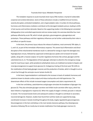
  • 10. Chelsea Hayes 12/5/2014 10 Jalloh I, Carpenter KLH, Grice P, et al. Glycolysis and the pentose phosphate pathway after human traumatic brain injury: microdialysis studies using 1,2-C glucose. J. Cereb. Blood Flow Metab. 2014. doi:10.1038/jcbfm.2014.177. To evaluate glycolysis and the pentose phosphate pathways (PPP) as routes of glucose metabolism in the brains of TBI patients, 15 male and female severe TBI patients aged 16-59 years, 8 male and female macroscopically normal brain patients undergoing surgery for benign tumors aged 59-73 years, and 9 male and female patients undergoing surgery for acoustic neuroma resection aged 20-61 years were enrolled in this controlled clinical trial. To measure and analyze lactate labeling patterns, 1,2- 𝐶2 13 glucose was perfused into the severe TBI patients’ brains, the cranial opening at the end of the normal brain subjects’ neurological procedure and into the quadriceps of the muscle subjects. Glucose, lactate, pyruvate, glutamate, and glycerol microdialysates were assessed hourly in the TBI patients, while these microdialysates were respectively assessed at 4 and 2 hour intervals in the normal brain and muscle subjects. All TBI patients, 2 normal brain subjects, and 5 muscle subjects were evaluated for a baseline period. Comparisons were made among 4 groups post-CCI:  Severe TBI patients (median age 27 years old) receiving 1,2-C2-glucose perfusion  Macroscopically normal-appearing brain patients  A non-CNS tissue (quadriceps muscle) comparison group  Plain, unsupplemented perfusion fluid without 1,2-13C2 glucose group Microdialysate glucose concentrations significantly increased between the baseline and the 1.2-13C2 glucose perfusion period for all groups (TBI: 1.0-3.8mmol/L; 1.9-3.9mmol/L; 2.8-5.3mmol/L). The ratio of Figure 1. Simplified schematic of steps in glycolysis and the pentose phosphate pathway (PPP) showing 13C labeling patterns resulting from1,2-13C2 glucose substrate. Red fills indicate 13C atoms. Glc-6-P, glucose-6-phosphate; 6PGL, 6 phosphogluconolactone; F6P, fructose-6-phosphate; G3P, glyceraldehyde-3-phosphate; PYR, pyruvate. Figure originally published in Carpenter et al34 under a Creative Commons Attribution License.
  • 11. Chelsea Hayes 12/5/2014 11 PPP-derived 3-13C lactate to glycolytic 2,3-13C lactate was a median 6.7% in the normal brain and 4.9% in the TBI brain. Showing that microdialysis 1,2-13C2 glucose infusion results in 13C-labeling in lactate in produced microdialysates enables the comparison of lactate-derived glycolysis to that of lactate-derived PPP. This study found that glycolytic lactate production was significantly greater in the TBI brain than in the normal brain and that PPP-derived lactate production was not significantly different between the two. Further studies are needed to explore the roles of PPP and glycolysis after TBI.
  • 12. Chelsea Hayes 12/5/2014 12 Xing G, Ren M, Watson WD, et al. Traumatic brain injury-induced expression and phosphorylation of pyruvate dehydrogenase: a mechanism of dysregulated glucose metabolism. Neurosci. Lett. 2009;454(1):38-42. doi:10.1016/j.neulet.2009.01.047. 48 male Sprague-Dawley rats were studied in this randomized controlled laboratory trial to determine the relevance of PDHE1α’s mRNA and protein expression (PDHE1α1) and phosphorylated PHDE1 α protein (p- PDHE1α1) in PDH activity and glucose metabolism after induction of controlled cortical impact (CCI). Using PDHEα1 mRNA and protein expression and phosphorylated PDHE1α protein as dependent variables in PDH activity and glucose metabolism, 48 healthy male Sprague-Dawley rats were randomly assigned to either a naïve control, craniotomy, or controlled cortical impact traumatic brain injury group. Brain samples were collected at 4 and 24 hours and 3 and 7 days post CCI in all groups (n = 4 for each group and time point) and were treated with specific antibodies against each protein to allow for visualization of immunoreactive bands in western blots. Additionally, real-time PCR determination of PDHE1a1 mRNA and western blotting determination of p-PDHE1a1 protein and PDHE1a1 protein in naïve control, craniotomy and contralateral CCI and ipsilateral CCI groups were analyzed. Comparisons were made among 4 groups post-CCI:  PDHE1α1 protein and mRNA and p- PDHE1α1 in naïve controls (n = 16)  PDHE1α1 protein and mRNA and p- PDHE1α1 in craniotomy (n = 16)  PDHE1α1 protein and mRNA and p- PDHE1α1 in contralateral and ipsilateral sides of brain in CCI (n = 16) At 4h, 24 h, 3, and 7 days post-CCI PDHE1α1 protein significantly decreased ipsilateral to CCI (respectively 62%, 75%, 57% and 39%) and contralateral to CCI (77%, 78%m, 78% and 36%) when compared to naïve controls (100%). Additionally, at 4h, 24h, 3d and 7d, p- PDHE1α1 protein decreased significantly ipsilateral (31%, 102%, 64%, and 14%) and contralateral (35%, 74%, 60%, and 20%) to CCI with like reductions in PDHE1α1 and p- PDHE1α1 protein found in the craniotomy CCI group. In conclusion, PDHE1α1 expression and phosphorylation post-TBI may play an important role in altered PDH activity and glucose metabolism in the brain. Future research is still needed, however.
  • 13. Chelsea Hayes 12/5/2014 13 Fig. 1. (A) Western blotting of p-PDHE1_1 and PDHE1_1 protein in the homogenates of rat brain samples collected at 4 h, 24 h, 3- and 7-day post-CCI. Forty micrograms of total protein of rat brain hemisphere tissue extract were resolved on SDS-PAGE gel and incubated with the specific primary antibodies against each protein. N, naïve control; S, craniotomy (sham CCI); C, contralateral CCI hemisphere; I, ipsilateral CCI hemisphere. (B). Real-time PCR determination of PDHE1_1 mRNA (a) and semi-quantitative determination of western blotting p-PDHE1_1 protein (b) and PDHE1_1 protein (c) in the homogenates of the naïve control, craniotomy, contralateral CCI and ipsilateral CCI hemisphere. PDHE1_1 mRNA increased moderately in the CCI group at 4 h and 24 h post-CCI, increased significantly in the contralateral CCI but decreased significantly in the ipsilateral CCI at 3 days post-CC; p-PDHE1_1 and PDHE1_1 protein, and PDHE1_1:p-PDHE1_1 ratio (d) reduced significantly in ipsilateral and contralateral CCI at 4 h, 24 h, 3 and 7 days post-CCI when compared with the naïve group (=C). (*) p < 0.05; (**) p < 0.01, (+) P < 0.09, versus the naïve group, respectively.
  • 14. Chelsea Hayes 12/5/2014 14 Belabed L, Charrueau C, Besson V, et al. Impairment of lymphocyte function in head-injured rats: effects of standard and immune-enhancing diets for enteral nutrition. Clin. Nutr. 2006;25(5):832-41. doi:10.1016/j.clnu.2006.02.003. In order to explore the effects of HI on lymphocyte function and to ascertain the effects of an enteral immune-enhancing diet (IED) compared to standard enteral nutrition, 25 male Srague-Dawley rats were studied in this randomized controlled laboratory experiment. Animals underwent a 6 day acclimatization period, followed by an overnight fast. They were randomized into 4 groups, an AL fed group and three other groups that underwent gastronomy on day 7. Post- gastronomy, rats were allowed a 6 day recovery period in metabolic cages. Thereafter, HI was induced by way of fluid percussion and rats were separated into a control group and two experimental groups. The enteral nutrition diet groups were infused 24h/24h (290kcal/kg/d and 3.29g of N/kg/d) at a constant rate over a 4 day period until 2 hours prior to being sacrificed. Their plasma fibrinogen and albumin, thymus, white blood cells, and lymphocyte receptor expression and densities were evaluated. 4 groups were compared:  Standard chow diet ad libitum fed healthy group (AL; n = 7)  HI control group receiving a constant rate of 0.9% NaCl via the enteral route along with free access to the standard chow diet (HI; n = 6)  Experimental group receiving the enteral standard diet Sondalis HP (HIS; n = 6)  Experimental group receiving the enteral IED Crucial (HIC; n=6) A significant increase in plasma fibrinogen was noted in the HI group (6.2g/l) versus the AL group (2.6g/l), but not in the HIS (4.5g/l) and HIC (5.0g/l) versus AL group. The HI group (16.6g/l) also showed hypoalbuminemia, which was corrected in the HIC group (20.4g/l) compared to the AL group (25.6g/l). HI induced an uncorrected significant thymus atrophy (296mg) compared to the AL group (552mg), although this atrophy was not exhibited in the HIC group (386mg). A significant increase in white blood cell count was observed in the HI (12.3 103 /𝑚𝑚2 ) versus AL (5.5 103 /𝑚𝑚2 ) group. CD25 receptor density in the blood, spleen and mesenteric lymph nodes in the AL group showed a marked increase after Con A stimulation versus the HI group. Lymphocytes in Peyer patches showed a significant increase in CD25 receptor density after stimulation only in the HIC group.
  • 15. Chelsea Hayes 12/5/2014 15 Thus, this model indeed characterized hypercatabolism via fibrinogen and albumin changes. Due to the thymus atrophy, it also characterized a reorganization of lymphocytes to the lymphoid organ and GI tract. Additionally, its model of HI dysimmunity revealed that Crucial helped reduce thymus atrophy. Finally, following HI in rats, EN appeared to efficiently restore blood lymphocyte stimulation capacity, while the IED formula provided added benefits to attune lymphocyte stimulation capacity in the PPs and limit thymus atrophy; although, further research remains concerning the deeper mechanism. Figure 1 Study design.
  • 16. Chelsea Hayes 12/5/2014 16 Moinard C, Delpierre E, Loi C, et al. An oligomeric diet limits the response to injury in traumatic brain- injured rats. J. Neurotrauma 2013;30(11):975-980. doi:10.1089/neu.2012.2707. To assess the effectiveness of an oligomeric formula improving nutritional status by restoring intestinal balance in rats with TBI, 26 male Sprague-Dawley rats were selected for study in this randomized controlled laboratory trial. All 26 rats acclimatized in metabolic cages for a week and then were randomized into three groups:  Healthy rats fed a standard polymeric enteral nutrition (control group; n = 8)  TBI rats fed the polymeric diet (TBIP; n = 9)  TBI rats fed the oligomeric diet (TBIO; n = 9) All groups underwent gastronomy on day 7 after fasting for 12 hours. Proceeding a 7 day recovery period, the non-control group received fluid percussion, inducing TBI. 4 hours post TBI (Day 0), enteral nutrition was introduced. The day following TBI, the enteral nutrition flow rate was increased to 290kcal/g/d and 2.3gN/kg/d infused at a constant rate for 4 days. Rats were weighed daily starting at Day 0 and ending on Day 4, when they were sacrificed. Blood samples were taken and various tissues were removed with organ weights, amino acid concentrations in plasma and muscles, tissue protein content, intestinal morphometry and enterbacterial translocation and dissemination being secondarily analyzed. Results showed that significant decreases in body weight were induced by TBI and were reduced by the oligomeric diet (TBIP versus TBIO). Moreover, the oligomeric formula appeared to attenuate thymus weight loss after TBI (TBIP at 0.30g versus control at 0.46g). Tissue protein content showed no significant difference in any groups compared. Glutamine concentration was the only significant difference found between groups for plasma and amino acid concentrations, revealing improvement by the oligomeric diet (Control group plasma 591μmol/L and TBIP 615μmol/L versus TBIO 688μmol/L). While intestinal morphometry showed no significant changes, enterobacterial translocation in the mesenteric lymph nodes and dissemination in the spleen and liver of the TBI groups were noted, but were not altered by the enteral diet. Overall, the oligomeric diet reduced thymus atrophy induced by TBI and showed promise in being potentially beneficial for restoring glutamine stores; however, further research is necessary to substantiate these promising results.
  • 17. Chelsea Hayes 12/5/2014 17 FIG. 1. Body weight variation between day 0 and day 4. Variation in total body weight in control rats, traumatic brain injury + standard polymeric diet rats (TBIP), and traumatic brain injury + oligomeric diet rats (TBIO). Results are given as means – standard error of the mean (analysis of variance + Newman- Keuls test). *p < 0.05 vs.control; ¤ p < 0.05 vs. TBIP.
  • 18. Chelsea Hayes 12/5/2014 18 Vizzini A, Aranda-Michel J. Nutritional support in head injury. Nutrition 2011;27(2):129-32. This review explored the role of nutritional support in the recovery of patients sustaining a head-injury, and was separated into eight discrete discussion parts: Hypermetabolism and hypercatabolsim: Head injury results in a complex cascade of metabolic alterations. An increase in the levels of cytokines and the counter-regulatory hormones such as cortisol, glucagon, norepinephrine and epinephrine contribute to this altered metabolism. These counter- regulatory hormones stimulate an increase in heart rate, cardiac output, oxygen consumption, glycogenolysis and gluconeogenesis, which all play a role in the development of total body metabolic derangements, including hyperglycemia and hypercatabolism. Hypercatabolism causes the degredation of skeletal muscle proteins and an increase in urinary nitrogen secretion, resulting in a severe negative nitrogen balance (>30g/d) and often malnutrition. Further complications may arise as a consequence of the malnutrition such as hyperglycemia, difficulty with wound healing, increased risk for infection, and multiple organ failure. Energy expenditure: Massive increases to around 40-200% in resting energy expenditure (REE) beyond that of a non-injured person are noted in head-injured patients. The metabolic rate in head-injured patients may be attenuated by up to 12-32% if given paralyzing agents, sedatives, or barbiturates; however, steroid administration and feeding does not follow this same pattern, and thus appears to be ineffective. To predict REE, the gold standard indirect calorimetry is often the measurement of choice. Although, due to the high variability in metabolic rates post-TBI, energy needs for individual patients should not be assessed through predictive equations. Because TBI patients often experience a 15% per week weight loss due to muscle wasting, the supply of sufficient calories is critical, with caution to overfeeding. Protein requirements: Hypercatabolism results in greater urinary nitrogen (UNN) secretion, making the provision of sufficient protein essential. Protein requirements for head-injured persons generally range from 2.0-2.5g/kg/d with the Management of Severe Traumatic Brain injury guidelines suggesting 15-20% of total calories as nitrogen calories. However, nitrogen imbalance usually persists for up to 2-3 weeks after the injury. The UNN measurement has been helpful in determining the true catabolism of an individual, excluding those with renal failure or undergoing dialysis, to provide personalized protein supplementation. While protein supplementation will not decrease hypercatabolism, it can secure protein replacement to maintain anabolism. Some studies suggest IGF-1 hormone therapy to improve
  • 19. Chelsea Hayes 12/5/2014 19 nitrogen utilization, but it is not recommended due to its association with increases in morbidity and mortality in critically ill patients. Specific nutrients: Because zinc is essential for many processes in the body and because patients with TBI have been known to have increased zinc urinary excretion and decreased serum zinc levels, zinc supplementation is often provided. However, optimal zinc supplementation dosing is still unclear. Vitamin E and C and magnesium are also under consideration as beneficial TBI supplements. Additionally, arginine, glutamine, nucleic acid, omega-3 fatty acids, and antioxidants are used as supplements in immune-modulating enteral formulas in trauma patients and critically ill patients. Glutamine supplementation is still being explored as researchers speculate it plays a role in hemodynamic instability in severely septic patients. To provide 0.3-0.5g/kg/d glutamine given in two to three divided doses, recommendations suggest adding standard dosing of glutamine powder to non- glutamine supplemented enteral formulas for trauma patients. Nutrition support (timing of feeding): Enteral nutrition provided within 24-72 hours of injury have shown to be beneficial in a plethora of studies. Infection rates and overall complications can be improved by early, adequate nutritional intervention, which may even improve Glasgow Outcome Scores at 3 months. Recommendations include providing >50-60% of goal calories via enteral nutrition within the first week of hospitalization to decrease cognitive recovery time, as well as, attaining full caloric replacement within 7 days post TBI. Nutrition Support (Route of feeding): Enteral nutrition is now the preferred method of feeding due to improved early enteral access procedures. If enteral access or nutrition goals are not achieved within 48- 72 hours after the injury, parenteral nutrition may be necessary. Short-term (< 4 weeks) enteral feeding requires nasogastric or nasoenteric tubes, whereas long-term enteral feeding requires gastronomy or jejunostomy tubes (>4 weeks). Enteral feeding comes with the potential for complications such as diarrhea, delayed gastric emptying, abdominal distention, aspiration, and pneumonitis. SCCM and SPEN guidelines recommend gastric and small bowel feeding for ICU patients. Across the board, enteral nutrition should be delivered continuously (20 mL/h, increasing by 10-20mL/h every 6-8 hours) with a controlled pump to improve tolerance, reduce the potential for infections and promptly achieve nutritional goals. Monitoring nutritional status: REE should be measured regularly or whenever changes in a patient’s metabolic demand are affected, according the ASPEN guidelines. UNN measurements should be taken
  • 20. Chelsea Hayes 12/5/2014 20 on TBI patients, excluding those undergoing dialysis, once or twice per week until protein balance has been reached. Prealbumin, albumin and CRP may be monitored weekly for reflection of nutritional status if inflammatory markers are stable. Monitoring of electrolytes, like potassium and phosphorus, may be relevant nutrition status indicators in TBI patients as well. Clinical practice: The Nutrition Risk Screening 2002 tool should be utilized to identify patients at risk for malnourishment and appropriate nutrition protocols should be implemented. Appropriate protocols for early nutrition and reduced likelihood of nutrition-related complications include guidelines for determining energy expenditure, changes in REE and protein needs, preferred routes and timing of enteral feeding, and measures for monitoring tolerance and nutrition adequacy.