
Empfohlen
Weitere ähnliche Inhalte
Was ist angesagt?
Was ist angesagt? (20)
Ähnlich wie Femoral Access Complications Prevention
Ähnlich wie Femoral Access Complications Prevention (20)
Mehr von Cathy Lewis
Mehr von Cathy Lewis (11)
Kürzlich hochgeladen
Kürzlich hochgeladen (20)
Femoral Access Complications Prevention
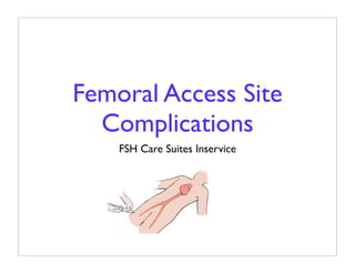
- 1. Femoral Access Site Complications FSH Care Suites Inservice
- 2. Advantages of Percutaneous Femoral Approach • Dominant technique • Does not require arteriotomy and arterial repair • Permits repeated site use for future angiograms • Suture closure of skin not necessary
- 3. Best Treatment - Prevention • Accurate puncture technique • Adequate compression for satisfactory initial hemostasis - Careful attention! • Some complications unavoidable • Early recognition and intervention is key
- 4. Femoral Anatomy Review • Common Femoral Artery (CFA) - Between femoral nerve (medial) and vein (lateral) • Superficial Femoral Artery (SFA) • Profunda (Deep) Femoral Artery (PFA or DFA) - Branches from SFA - Thinner walls; atherosclerotic changes more likely
- 6. Ideal Insertion Site • Below inquinal ligament • Above Common Femoral Artery (CFA) bifurcation
- 7. Seldinger Technique • Anterior and posterior a. wall pierced with needle encased in metal sheath • Both retracted into vessel, needle removed, outer cannula remains inside • Guidewire introduced through cannula • Less chance of intraluminal damage; but produces two holes in artery
- 8. Front Wall Technique for Vascular Access • Most likely route at FSH • Anterior wall pierced; needle immediately centered in artery lumen • Guidewire inserted through needle; needle removed; sheath fed over guidewire • Only one hole in artery; but greater chance for intraluminal damage
- 9. Anticoagulation During Catheterization • Two reasons - Reduce possibility of catheter-related embolic event (surface clots on catheter) - Decrease platelet aggregability to injured plaque • Aspirin - AM of procedure • Heparin - maintain ACT > 300 s • Possible Glycoprotein Inhibitors
- 10. When to Remove Sheath • Diagnostic procedures - immediately • Interventional procedures - 4-6 hr • Why delay for interventional procedures? - Give heparin time to dissipate - Ready access if coronary vessel closes abruptly
- 11. Time of Sheath Removal Important • Short-window of time between subtherapeutic anticoagulation range and rebound thrombin activation • FSH standing orders - remove sheath as soon as PTT < 50 s • Complications increase with cannulation time duration
- 12. Ways to Achieve Hemostasis • Manual Pressure • Sandbags • Pressure Dressing • C-Clamp • Femostop • Vessel Seal/Closure Device
- 13. Pressure Devices FemoStop® CompressAR®
- 14. Vessel Seal/Closure Devices Vasoseal® Perclose® Angioseal®
- 15. Patient Factors that Affect Complication Risk • Hypertension • Peripheral Vascular Disease • Smoking • Diabetes • Obesity • Anticoagulants • Advanced Age • Women
- 16. Procedural Factors that Affect Complication Risk • Size of introducer sheath • Repeated sheath changes • Length of cannulation • Post procedure heparin • Failure to achieve adequate initial hemostasis when removing sheath
- 17. Four Signs of Blood Loss or Hemorrhage at Access Site • Bulging mass in groin or thigh • Pulsatility • Bruit • Tenderness in inguinal area
- 18. Possible Complications • Retroperitoneal Hematoma or Bleed • Hematoma • Pseudoaneurysm • Arteriovenous Fistula • Neuropathy • Arterial Occlusion
- 19. Hematoma • Blood loss may necessitate transfusion • Usually resolves in 2-3 weeks; can take much longer
- 20. Hematoma Physical Findings Nursing Interventions • • Site pain/burning Manual pressure above site • • Difficulty moving hip/leg Notify MD if severe or evolving • Possible tachycardia or • hypotension VS q 15-30 min until hematoma stable, then • Red/purple skin q 2-4 hr discoloration • Outline hematoma • Measure thigh girth q 1 hr until hematoma stable, then q 4-8 hr
- 21. Pseudoaneurysm Often associated with puncture below CFA bifurcation and initial inadequate hemostasis
- 22. Pseudoaneurysm Physical Findings Nursing Interventions • • Groin pain/burning Assess VS, groin site, pedal pules, and bruit • Back pain q 15 min while enlarging, • Swelling at groin site then q 2 hr when stable • • Ecchymosis Thigh girth q 1 hr • • Pulsatile mass CBC, PTT until < 30 s • Bruit
- 23. Ultrasound-Guided Compression • Probe locates tract between artery and pseudoaneurysm; also used for pressure • Surgery when >2 cm 4-5 days after catheterization complicated by significant groin hematoma
- 24. Arteriovenous Fistula • Rare • Usually forms when needle punctures artery and vein - More likely when artery is punctured > 3 cm below inquinal ligament where veins are inferior to arteries
- 25. Arteriovenous Fistula Physical Findings Nursing Interventions • • Swelling at groin site, leg Notify MD pain • Heart and lung sounds • Possible signs high-output q 2 hr heart failure (arterial blood • Check for decreased pedal shunting into venous bed) pulses • Possible tachycardia and • Check for bruit decreased BP
- 26. Neuropathy • Rare • After large hemorrhage or pseudoaneurysm - Pressure exerts on medial and intermediate cutaneous nerve - Usually resolves when cause resolves • Late complication from chronic accumulation of fluid that causes pressure/irritability - Usually resolves when cause resolves
- 27. Neuropathy Physical Findings Nursing Interventions • • Pain, tingling at groin site Notify MD • • Numbness at site or distal Check for altered sensation leg and/or motor ability • • Motor difficulty in affected Compare reflexes and ROM leg to unaffected leg • • Possible decreased patellar In intermediate recovery phases, check VS, groin site tendon reflex and pulses per protocol, • Possible weakness of knee then q 2 hr until symptoms extension resolve • Symptoms may occur as late as 3 months post procedure
- 28. Arterial Occlusion • Very rare • Can occur from large thrombus at puncture site. Use of anticoagulants occurance unlikely. • Large catheter most likely used in a small CFA - Diabetes - Female
- 29. Arterial Occlusion Physical Findings Nursing Interventions • • Pain Notify MD • • Pallor Assess VS, leg, pedal pulses q 15-30 min until • Paresthesia circulation restored • Pulseless • Use Doppler Ultrasound for pulse assessment
- 30. Care Post (4 fr.) Sheath Removal • Site check with VS per unit protocol • Instruct patient to call nurse for any sign of bleeding and apply manual pressure to site - Apply pressure to site while coughing, laughing, or sneezing • BR for 2 hr - Light restraint on affected limb - May elevate HOB 30º
- 31. Site Assessment • Groin - How much ecchymosis and redness? - Is there any bleeding? How much? - Is there a raised mass? Does it pulsate? - Is there a bruit? • Distal extremity - CMS (Color - Motion - Sensory)
- 32. Documentation • Vital signs - HR, BP, RR, rhythm • Neurovascular checks - affected limb • Unexpected outcomes • Nursing interventions/actions taken • Evaluation
- 33. Mayo Study • Implemented a new care standard - BR 3-4 hr (vs. 6) - HOB elevation (vs. flat) - Pressure dressing (vs. sandbag) • 306 retrospective chart audits • Compared complication rates for new vs. old standards McCabe, et.al. (2001)
- 34. Minor Bleeding Mayo Study • Defined as spurting, trickling, or oozing of blood not contained by Band-aid® - Possible redressing of site and/or additional compression (not > 30 min) - BR extended for more than small ooze - No hemodynamic instability - No medical or surgical intervention McCabe, et.al. (2001)
- 35. Major Bleeding Mayo Study • Spurting or brisk bleeding not controlled by site compression - Possible hemodynamic instability - Possible need for diagnostic tests and medical or surgical consultations McCabe, et.al. (2001)
- 36. Minor Hematoma Mayo Study • Collection of extravasated blood under skin that forms a soft raised surface - easily palpable - Controlled by manual compression - No hemodynamic instability - No neurovascular compromise of affected limb - No medical or surgical intervention McCabe, et.al. (2001)
- 37. Major Hematoma Mayo Study • Collection of extravasated blood that may or may not be palpable. May occur under skin, in surrounding tissues, or extend into retroperitoneum - Some classify size > 10 cm - Increased risk for hemodynamic instability - Possible neurovascular compromise - Medical and/or surgical consultation with likely surgical intervention McCabe, et.al. (2001)
- 38. Complication Rate Mayo Study Complication No. % Hematoma - Minor 18 5.9 Hematoma - Major 9 2.9 Bleeding - Minor 13 4.2 Bleeding - Major 1 0.3 Pseudoaneurysm 3 1.0 Arteriovenous fistula 0 0.0 Thorombosis of affected limb 0 0.0 Any major complication 10 3.3 Any complication 15 11.4 McCabe, et.al. (2001)
- 39. Timing After Sheath Removal Mayo Study N = 300 Total Major 15 Number of Patients With 10 End of BR Complications 5 0 0-1 1-2 2-4 4-12 >12 Hours After Sheath Removal McCabe, et.al. (2001)
- 40. Amsterdam Study • Coronary angioplasty, stenting, or both using femoral 6 fr. approach and Heparin 5,000 IU - Also aspirin and Plavix • Manual compression followed by compression bandage • Ambulation at 2 hr
- 41. Amsterdam Study Results • N = 300 (32% stent placements) • Mean time to hemostasis = 9.6 min • 5 (1.7%) bled at ambulation • 9 had 5x5 cm hematoma at 48 hr • All treated conservatively • No late bleeding or vascular complications
- 42. U Minnesota Study (1) • How well does a 4.5 kg (36 cm x 16 cm) sandbag with cross-sectional diameter of 576 cm2 work? - It applies compression force of 3.4 g/cm2 to stop bleeding in artery with intraluminal pressure ≥ 100 mmHg • Randomized study compared complications after angiography with and without sandbags Christensen, et. al. (1998)
- 43. U Minnesota (2) Post Sheath Removal Sandbag Bandage n = 174 n = 176 Rebleeding 22 14 Ecchymosis 13 8 Hematoma 23 20 Christensen, et. al. (1998)
- 44. U Minnesota Study (3) Sandbag Bandage New fem bruit - 6 hr 0 0 New fem bruit - next AM 0 1 Ecchymosis - 6 hr 24 18 Ecchymosis - next AM 41 33 Hematoma - 6 hr 23 20 Hematoma - next AM 13 20 Christensen, et. al. (1998)
- 45. U Minnesota Study (4) Early Complications (< 24 h) Sandbag Dressing Any Bleeding 18 16 -Ooze 8 7 -Brisk Bleeding 10 9 -Other 2 1 Christensen, et. al. (1998)
- 46. U Minnesota Study (5) Late Complications (1-30 days) Sandbag Dressing 2 1 Pseudoaneurysm 1 0 AV Fistula 3 2 Late Bleeding 1 0 Stroke 1 0 Loss of Pulse 1 2 Vascular Surgery 2 2 Other Christensen, et. al. (1998)
- 47. U Minnesota Study (6) • Incidence of vascular complications not statistically significant between groups • Differences in patient satisfaction was statistically significant Sandbag Bandage n = 174 n = 176 Severe 18 4 Discomfort Moderate 6 0 Discomfort Christensen, et. al. (1998)
- 48. Comparison of Physical Findings Overview Bulging Pulsatile Bruit* Tenderness Mass Mass +/- pulse Hematoma varies no yes waves Pseudoaneurysm yes yes yes yes AV Fistula no no yes no * Some elderly adults have femoral bruits - atherosclerosis. It’s a good idea to ascultate the groin pre procedure.
- 49. Patient Scenario - AV Fistula Secondary to Pseudoaneurysm (1)
- 50. Patient Scenario - AV Fistula Secondary to Pseudoaneurysm (2)
- 51. Patient Scenario - AV Fistula Secondary to Pseudoaneurysm (3)