
Empfohlen
Weitere ähnliche Inhalte
Was ist angesagt?
Was ist angesagt? (20)
Ähnlich wie Life cycle of Protozoan parasite
Ähnlich wie Life cycle of Protozoan parasite (20)
Mehr von Al Nahian Avro
Mehr von Al Nahian Avro (20)
Kürzlich hochgeladen
Kürzlich hochgeladen (20)
Life cycle of Protozoan parasite
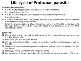
- 1. Life cycle of Protozoan parasite Ichthyophthirius multifiliis: • It is the most pathogenic protozoan parasites of freshwater fishes. • It is also referred as the ‘Ich’. • It is the causative organism of white spot/ Ich disease/ Ichthyophthiriasis. • is an ectoparasite. • It is easily introduced into a fish pond by new fish or equipment which has been moved from one fish-holding unit to another. • When the organism gets into a large fish culture facility, it is difficult to control due to its fast reproductive cycle and its unique life stages. If not controlled, there is a 100% mortality rate of fish. Symptoms: Minute white nodules that look like white grains of salt or sugar of up to 1 mm appear on the skin, fins and gills. Each white spot is an encysted parasite. Affected fishes show irritation, erratic movement and restless with tendency to rub on the sides. Infected fish have small white spots on the skin and gills and produce excess mucus, due to irritation. Some cysts joining together to form irregular patches. Damage caused to the gill tissue of an infected fish can also reduce respiratory efficiency.
- 2. Pathology: Epithelial tissue changes occur in the area where the parasite is lodged and its dislodging causes the epithelial ulcers. Morphology: Cilia Macronucleus
- 4. Prevention and control: Prevention of exposure of fish to the parasites Prompt identification of the disease if it occurs Treatment of infected fishes. Applying quarantine method Control: 7000-20000 ppm NaCl for pond treatment Formalin Potassium per manganate
- 5. Ichthyobodo necatrix It is the causative organism of freshwater Ichthyobodosis. It is an ectoparasite. It is usually attached on dorsal fins and tips of secondary gill lamellae of infected fishes. Symptoms: Small spots appear on the body It may fuse into a greyish-white film from increased production of mucus The more intensely affected areas are reddened and hemorrhagic Infected gills are pale and covered with mucus Loss appetite. Moribund fish rise to the surface and become sluggish and dies.
- 6. Flagellum Contractile vacuole Cytostome Nucleus Flagellum Morphology: Pathology: Goblet cells are not seen in the epidermis Hyperplasia occurs in Malphigian cells Acute hyperplasia and fusion of secondary gill lamellae Clubbing of gill filaments Epithelial cells with parasites become necrotic and blood vessels collapse with proliferation of mucus cells in clubbed filaments.
- 7. Prevention and control: Treatment of infected fishes. Applying quarantine method Control: Formalin 166 ppm for 1 hour (Flash treatment) Nacl 100000 ppm for 15-30 mins Copper sulphate 500 ppm for 1-2 mins Ponds with infected fishes should be drained Should be treated with lime before restocking the new fish
- 8. Chilodonellosis • Chilodonellosis is the important parasitic disease of carp during winter. • The causative agents of this disease are Chilodonella cyprini, Chilodonella piscicola and Chilodonella hexascha • It infect gill, fins and body surface • It causes great loss to the pond fisheries Symptoms: The mass development of parasite causes a greatly higher production of mucus and disturbances in the respiratory function of the skin. The fish is restless, rises to the upper layers of the water. Its entire body covered with the bluish-white coating, particularly in the head region. A smear from the skin surface reveals numerous individuals of the parasites Causes localized hyperplasia of the gill epithelium
- 9. Pathology: It causes localized hyperplasia of gill epithelium, which later becomes more generalized. Proliferating epithelial cells fill the spaces between secondary lamellae, which may fuse together and turn into a single mass Then the respiratory epithelium is covered by the hyperplastic epithelium and then drastically reduce the respiratory surface of the gill. Morphology:
- 10. Trichodiniasis The causative agent of this disease is Trichodina sp. They are common ectoparasites of both freshwater and marine fishes They infect fry, fingerings and adults of Indian major carps and Chinese carps. Symptoms: Colour of the tail turns pale and there is a cream coloured coating due to excessive mucus secretion. Fry and fingerlings show spinning behaviour. Scales become slippery due to excessive mucus. Tinny blood spots develop on fins and body. Fish show abnormal behaviour and colouration. Infected fishes gradually become sluggish and lose weight and become moribund.
- 12. Gyrodactylosis • The causative agent of this disease is Gyrodactylus elegans and Gyrodactylus gracilis • They attack the skin, fins and gills of fish • They are commonly known as gill fluke Symptoms: The colours of the infected fishes become pale The skin becomes more slimy and shows small blood spots The fins becomes droop and fold and gradually become torn Breathing is increased in frequency
- 13. Marginal hook Anchor Developing embryo Morphology Prevention and control: Should be follow quarantine methods Fry and juveniles should be disinfected before stocking Add Methylene blue to the pond water at 1g/10m3 KMno4 at 2 ppm and 3-5 ppm NH4OH at 500 ppm for 5-15 minutes
- 15. Adults on the gills of fish Egg with an oncomiracidium larva Free oncomiracidium After attachment to the gills of a host the oncomiracidium is transformed into the diporpa larva. Fusion of 2 diporpas on the host; Each diporpa attaches its sucker ( VS) to the dorsal papilla ( DP) of the other. Life cycle of monogenetic Trematodes
- 16. Life cycle of Digenean Trematodes Clinostomum compalanatum and Clinostomum marginatum are unsegmented flatworms of the class Trematoda and the order Digenea. They are also called as yellow grub They occur frequently in the skin and the muscle of the freshwater fish. Small cream coloured nodules or cysts ranging from pinhead size up to 2.5 mm depending on their age The number of cysts may vary from 1-100 or more than They have an oval or round shape. The skin of the fish in reaction to the infection produces the cysts, which contain worms. It may take 3 weeks to make clearly visible cysts after the infection and 7 weeks to reach full size
- 17. Metacercaria
- 18. Diplostomum pigmentata It is the causative agent of Black spot disease in fish. The affected fishes are adult, fry and fingerlings of Indian major carps Small brown or black spots on several parts of body and fins. More than 300 cysts may present in a single fish The specific sites of infection are skin and underlying muscles Such spots are also occur in the eyes, the gills and the mouth.
- 19. miracidium egg 1st intermediate host (snail) Free swimming cerceriae Metacerceriae in 2nd intermediate host fish
- 20. Diplostomum spathaceum They are also called as eye fluke They attack in the lens, humour (liquid) or retina of the eye. The extent of the damage ranges from cloudiness and rupture to blindness
- 21. Cestode parasites of fishes • Cestodes are endoparasites of fishes • Complete their life cycle involving two or more hosts • The can infect fishes either in larval and adult condition • A number of species which are parasites on freshwater fishes particularly belonging to Pseudophyllidea, Protocephallidea and Caryophyllidea
- 22. Ligula intestinalis Popular fish tapeworm which may cause heavy mortality Causative agent of the disease ligulosis The immature stage of this tapeworm Infect cyprinid fishes
- 24. Proteocephalus ambloplitis Known as bass tapeworm Can cause serious damage
- 25. The strobila is produced by the development of new proglottids in the neck region, so that the proglottids most distant from neck are the oldest. Proglottids nearest the neck is usually male and those furthest away are female. The life-cycle involve one or more intermediate hosts, mainly various invertebrates but sometimes small vertebrate e.g. Ligula intestinalis Dibothriocephalus latus Scolex Neck Strobila Proglottid
- 26. Zoonosis Is any infectious disease of animals (usually vertebrates) that is naturally transmissible to the humans.
- 27. Clonorchis sinensis Is also called the Chinese liver fluke The causative agent of human opisthorchiasis.
- 28. Dibothriocephalus latus Causative agent of human Dibothriocephaliasis
- 29. Pallisentis ophiocephali Common acanthocephalan worm of murrels. The definitive host is Channa striatus Channa striatus Mature egg Acanthor Acanthella Juvenile with proboscis
- 31. Stress and disease Disease: Disease is defined as an abnormal condition of body and mind of an organism which is expressed with certain symptoms Disease is the end result of the host due to interaction and interrelationship of following 3 factors- Adverse environment Infectious agents (Pathogen) Susceptible host (fish, shrimp) Such adverse/unfavorable environmental conditions of fishes include crowding, temperature fluctuations, Inadequate DO, rough handling, creation of toxic gases, sublethal levels of toxic materials etc. These adverse environmental conditions create stress on fish
- 32. The role of stress in the host-parasite-environment interaction in human medicine has been expressed by Dubos (1955) ‘There are many situations in which the microbe is a constant and ubiquitous component of the environment but causes diseases only when some weakening of the patient by another factor allows infection to proceed unrestrained, at least for a while.’ e.g. Facultative bacterial fish pathogens such as Aeromonas, Pseudomonas are continually present in water supplies, epizootics will usually not occur unless environmental quality and the host defense symptoms of the fish also deteriorate H+P+S2= D
- 33. Stress, infection and disease Host+ Pathogen+ Environmental factors Weak and stress Stage I Infection Stage II Disease Stage III
- 34. Different types of stressors Physical stressors- Temperature, light, turbidity, sounds Chemical stressors- Poor water quality, pollution, Diet composition, Nitrogenous and other metabolic waste etc. Biological stressors- Population density, Parasites and pathogen, predation etc.
- 35. Factors producing diseases in fishes A. Environmental Factors 1. Physical Temperature Light Turbidity: mechanical injury by suspended particles Sounds 2. Chemical DO PH Pollution-mining wastes, mill wastes, sewage pollution Diet composition Salinity Intoxications-Insecticides, pesticides, heavy metal, DDT B. Biological factors: Population density Predation Pathogens and parasites Phytoplankton bloom Inducing agents
- 36. C. Hereditary factors Tumours, albinism, deformities, tuberculosis, dropsy D. Glandular dysfunction 1. Pituitary dysfunction-gill impermeability, suffocation 2. Thyroid dysfunction- decrease iodine 3. Liver dysfunction-metabolic disturbances 4. Pancreas dysfunction-metabolic disturbances 5. Sex gland dysfunction- reproductive inhibition E. Mechanical injury F. Pathogenic organisms G. Atypical cell growth H. Dietary deficiency factors 1. Carbohydrate deficiency 2. Fat deficiency 3. Protein malnutrition 4. Vitamins A-exophthalmia B1- Loss of balance B2- photophobia D-lower haemoglobin E- Anaemia K- blood do notcoagulate I. Procedural Rough handling Transportation Stocking Disease treatment
- 37. Physiological response of fishes to stress In medical usage, stress is the metabolic response of the animal to a stressor Stress is a state produced by an environmental or other factor which extends the adaptive responses of an animal beyond the normal range or which disturbs the normal functioning to such an extend that, in either case the changes of survival are significantly reduced A series of morphological, biochemical and physiological change occur as a result of stress which are called general adaptation syndrome (GAS). It is usually divided into three stages- An alarm reaction A stage of resistance (adaptation to the stress has occurred) A stage of exhaustion (adaptation has been lost because the stress too severe or long lasting)
- 38. These stages are characterized by a variety of physiological and biochemical changes which are not species specific. Some physiological and biochemical changes are- 1. ACTH (adenocorticotrophic hormone) is released from the adrenohypophysis 2. Stress hormones (Cortisone, corticosterone, epinephrine) are released from the adrenal tissue 3. Na+ and Cl- retention increases 4. K+ excretion increases 5. Blood glucose level increases 6. Nitrogen metabolism increases 7. Thyroid output increases 8. Respiration rate increases, cardiac output increases, systolic blood pressure rises
- 39. Host-parasite-environment relationship The actual initiation of disease in fishes is a complex process which involves more than mere contact between the host and the parasite. Host susceptibility, parasite virulence and environmental factors must all interact. Faculatative pathogen e.g. Pseudomonas, Aeromonas, myxobacteria etc. Obligate pathogens e.g. Aeromonas salmonicida, IPNV, VHSV
- 40. PATHOGEN/PARASITE Infectivity Virulence Pathogenecity Viability Strain HOST Species, age Strain Nutritional status Physiological condition DISEASE POTENTIAL ENVIRONMENT Temperature Oxygen concentration Alkalinity Hardness Season Other parameters Reaction of Host, pathogen/parasite and environment
- 41. Relation between stress and culture Extensive- Disease of fish in culture system more or less similar with the wild No fertilizer and feed input-little chance to create stressful condition Semi-intensive- Some amount of fertilizer and feed supplied High dose of fertilizer, improper diet composition can create stressful condition Intensive- Water quality and exchanging Improper amount of feed Introduction of infectious agents Proper sanitation
- 42. Defense against disease Protective barriers Mucus- Act as physical barrier that inhibits the entry of disease organisms from the environment into the fish Act as chemical barrier because it contains enzymes (lysozymes) and antibodies (immunoglobulins) which can kill invading organisms Lubricates the fish which aids movement through the water It is also help in osmoregulation Scales and skin- Act as a physical barrier which protects the fish against injury
- 43. Inflammation- A nonspecific cellular response to an invading protein It is characterized by pain, swelling, redness, heat and loss of function. It is an protective response and is an attempt by the body to wall off and destroy the invader Antibodies- These are specific cellular response which are specifically formed to fight invading proteins or organisms
- 44. Effect of stress on protective barriers A. Mucus- Chemical changes in mucus decrease the effectiveness as a chemical barrier Stress hamper the normal electrolyte (sodium, potassium and chloride) balance which hamper the osmoregulatory function. Handling stress removes mucus and disrupt the physical barrier Chemical stress ( disease treatment) damages mucus B. Scales and skin- Damaged by handling stress C. Inflammation- Hormonal changes decrease the effectiveness of the inflammatory response In cold temperature, the killer cells of the immune system completely stop their works D. Antibody production- Temperature stress- A sudden decrease in temperature, impairs the fish ability to quickly release antibodies against an invading organism Prolonged stress limits the effectiveness of the immune system
- 45. Mechanism of infection into disease The net result of stress on fish is to bring about metabolic changes by their physiological control systems which are designed to aid in surviving the imposed stress An increase in blood glucose (hyperglycemia) A decrease in blood chloride levels (hyperchloremia) An increased number of circulating thrombocytes A decreased inflammatory response Decreased liver glycogen Decrease in serum protein Increased 17-hydroxycorticosteroid levels
- 46. Increased plasma thrombocyte levels provide a more effective hemostatic mechanism Increased fibrinogen help in tissue repair and walling off local infections Increase plasma protein (haptoglobin fraction)serves to bind any haemoglobin released as the result of red cell destruction Thus iron is conserved and renal damage is preserved.
- 47. Increased levels of corticosteroid hormones, with reticuloendothelial deficiencies, combine to reduce the tissue response to microbial invasion and allow infections to spread. Chronic stress significantly reduce the immune response Leucopenia develops due to the hyper-secreation of adrenal corticoids Due to the direct action of increased 17- hydroxycorticosteroids on cell membranes, the fragility of lysosome (phagosome) is decreased. Thus, stress may indirectly result in impaired phagocytosis.
- 48. The non-specific response to invading microorganisms is somewhat dependent on the nutritional state of the host Inanition is synergistic with bacterial infections but antagonism can occur with virus and protozoa. Vitamin A, C or D deficiencies are usually synergistic to infections B-complex vitamin and mineral deficiencies behave variably depending on the host and infectious agent Vitamin C influences the production and maintenance of repair tissue The process infection into disease is thus seem to be a strong function of the physiological state of the host and is actually the sum of a complex series of metabolic interactions between invading microorganism and host-defense mechanism
- 49. The influence of parasite on its host • Every parasite living on or in fish exerts some degree of harmful influence on its host i.e. changes in individual organs or tissues. • Pathogenic and non-pathogenic • However, every parasite is harmful to its host • Sometimes, non-pathogenic parasites show a strongly pathogenic character e.g. Dactylogyrus minutum, a small and allegedly harmless monogenean infesting the gills of the carp, occurs only in small numbers in natural waters but it is capable of prolific reproduction in aquaria, where it might cause the death of its host • Parasite can influence the body of the fish in many different ways.
- 50. Mechanical effects: Damage to the tissue Rupturing of the protective layers Complete or partial atrophy of the internal organs Obstruction of the alimentary canal or vascular system Mass infestation of the skin with ectoparasitic Protozoa (Ichthyobodo, Chilodonella and Trichodina) or monogenetic trematodes (Gyrodactylus) causes excessive mucus. This coating or mucus disturbs the respiratory function of the skin and ionic exchanges of the blood Cause serious mechanical injuries to the gills and the walls of the branchial chamber. Injuries, epithelial hypertrophy and an abnormally high production of mucus occur at the point of attachment of many monogeneans, causing disturbances in the respiratory function of the gills. Cause the partial or complete castration to the reproductive glands. e.g. Ligula
- 51. Toxic effects: Parasite have toxic effects on their fish hosts These effects can be divided into two groups- Due to secretions of special poison glands Due to metabolic activity of the parasite e.g. The mouth armament of Argulus includes a poison gland, the secretion of which may kill small fish in cases of mass infestation. Parasitic toxins might influence the endocrinology of the fish. e.g. The result of disease ligulosis is a considerable lowering in the production of gonadotropic hormones and also the pathological changes in the basophilic cells of the anterior part of that gland. The underdevelopment of the gonads in ligulosis is due to the mechanical suppression by the parasite and also to the toxic effects on the glands
- 52. Consumption of the host’s food: When the parasites are comparatively large or occurs within the host in great numbers e.g. Eubothrium crassum Parasites as vector of other parasites: A parasite is sometimes harmful not only due to its own activity but also because it disseminates agents of some infectious and parasitic diseases e.g. the common leech, Piscicola geometra, is a vector of Trypanoplasma cyprini Parasites as a indirect causes of diseases: By damaging the surface of the body and internal organs of fishes and producing various wounds and ulcerations, parasites favor the penetration of other pathogenic microorganisms, mainly fungi and bacteria. e.g. Great concentrations of Ichthyobodo necatrix and Saprolegnia have been observed around wounds caused by the copepod, Lernaea
- 53. The influence of infestation of non-specific sites: Generally all the parasites inhabit a specific site within their hosts. But sometimes parasites infesting the other organs in where he are not adapted for living and do not cause any observable changes in it e.g. The parasites which are found in kidneys of Rutilus rutilus, are also infest in the gut without causing any observable changes in it The influence of parasite on the growth rate and condition of fishes: e.g. mass infection of Ergasilus sieboldi in fish The influence of parasite on the size of fish populations: The density of fish populations might be considerably affected by parasites, particularly in the event of mass mortality e.g. Ergasilus sieboldi causes 100% mortality of Tinca tinca in the infested ponds
- 54. Reaction of the host fish Cell and tissue reactions: Hypertrophy: • Hypertrophy is the increase in size of an organ or tissue due to the enlargement of its component cells • This response very frequently evoked by the penetration of the parasites, particularly small one. • Abnormal stimulation of the cellular mechanism leads to gigantism of the infected cells • Poorly specialized tissues, such as epithelium and connective tissue, are best suited to this type of reaction e.g. the hypertrophy of gill epithelium due to infestation of various species of Dactylogyrus Inflammation: • Inflammation is a localized physical condition in which part of the body becomes reddened, swollen, hot, and often painful, especially as a biological response of body tissues to harmful stimuli, such as pathogens, damaged cells, or irritants. • The reaction to the presence of parasite is often expressed by the process of inflammation • Inflammation develop at the point of penetration or at the sites of permanent localization of the parasite • Very often a connective tissue capsule is formed around the parasite, more or less isolating it from the surrounding tissue. • The body of the fish responds to all types of injuries by a more or less definite process of inflammation.
- 55. Metaplasia: Metaplasia is the abnormal changes of tissues in their structure and function The tissues of the host respond to the presence of the parasite by metaplasia e.g. Mucoid transformation of epithelial tissue in fish when they are infested with Chilodonella and Trichodina, Gyrodactylus. Epithelial tissues of fishes contain single cells or group of cells which secrete mucus. Harmful influences on the epithelial tissue result in the appearance of increasing numbers of such cells Immunity: The ability of the host to resist infection by the pathogen of a definite disease. Acquired immunity in parasitic diseases is only partial, not totally preventing but diminishing the chances of reoccurrence. It can affect retardation of development and of fertility of the parasite
- 56. Snieszko’s disease production equation by interaction of environmental factors H(A+S2)=D H= Host A=etiological agents S= environmental stressors D=Disease
- 57. Symptoms of the stressed fish Fish run erratically in the surface of the water Fish stays near the surface gasping for breath Fish would not eat, or does not eat as aggressively as past Changes color Rapid breathing Gulfing to the surface Jumping out of the water Motionless, sluggish and can stay in a standstill position Fish stays hidden continuously and would not come out where it can be seen. Fish has nicked fins, open wounds that don’t seem to heal The mucus of fish skin is less or extreme
- 58. Changes in fish during stress condition External changes Internal changes- Hormonal changes: Excess excretion of adrenaline hormone Increasing the rate of sugars in blood Rising of blood pressure Metabolic changes: Significant increase of blood glucose Hyperglycemia accelerated the raising of temperature and blood glucose disturbance Osmotic changes: Induce the mechanism of osmoregulatory disturbances Create water and mineral imbalance
- 59. Stress Adrenaline Fish Water Diffusion Fresh water Water inhibition Gain of weight Hemo dilution Sea Water Loss of Water Loss of weight Hemo concentration Fig.: Interpretation of some stress effects on osmotic balance
- 60. Stress mediated fish diseases Diseases Predisposing environmental factors Columnaris Crowding, handling, seining, adverse temperature and infectious disese Bacterial gill disease Crowding, unfavorable environmental conditions, presence of bacteria, elevated ammonia and particulate matter in water Cold water disease 10 to 15°C – 7 to 13°C Motile Aeromonas Septicemia Injury to skin, fins and gills, excessive handling, low oxygen, pesticides, crowding, improper nutrition
- 61. Prevention of stress Good Management Good water quality Handling and transportation High quality diet Proper sanitation Prevention of disease
- 62. Diagnosis Diagnosis is the art of determination of the nature of the cause/ causative agent of the disease and problems assigned by the disease Importance- For proper health management Detect the possible cause of disease and take the possible measures to protect the fish stock from disease appropriately To prevent and control of fish disease Diagnosis can be performed by two methods 1. Primary or gross diagnosis 2. Laboratory diagnosis Primary or gross diagnosis- Fishes are diagnosed by observation of its external features, clinical signs, physiological change etc. without laboratory tests
- 63. Visit the pond site: Basic pond site information Water supply details Production cycle Stocking density Feeding Waste disposal Diseases and other problems To check the environment: Physical parameters- Temperature Pollution-Affects the nervous system of fish Turbidity-Respiratory problem and mechanical injury Chemical parameters- DO pH of water Presence of insecticides, pesticides, heavy metals etc. Biological parameters- Presence of phytoplankton and zooplankton
- 64. O X y Mortality(%) Days Stage I (Due to toxicity of pathogen) Stage II (Due to infection) Stage III (Due to stress, nutritional factors or environmental factors) Checking of fish by sampling: Changes in appearance and behaviour Prevalence of diseased fish- Incidence of infection-the percentage of fish infected in a population Prevalence of disease = Percentages of diseased fish/ Percentages of fish Fish Mortality Pattern
- 65. Difference between healthy and diseased fish Changes in behaviour Changes in color Fin disorder Skin disorder Gill disorder Locomotion disorder Physical changes
- 66. Observation Healthy fish Diseased fish Possible causes Swimming Easy movement in water Disturbance in locomotion such as- Jerky swimming Movement in a small circle Jumping movement Fin and tail rot disease Swim bladder stress syndrome Nutritional disease Eye disease Nutritional disease Normal Swimming on side and exposing ventral side to the surface Swim bladder stress syndrome Gas bladder disease Don’t show rubbing activity Rubbing bodies against bottom or sides of container or with hard objects External protozoans External crustaceans Argulus sp. Feeding Show normal appetite Reduction of feeding activity Viral and Bacterial infection Poor water quality
- 67. Colour No dark colouration Dark body colouration over the whole body Viral disease-VHS Bacterial disease Nutritional deficiency Normal colouration, not white covering on the body A hazy white covering over the body Columnaris disease Fin Normal colour of fin Redness at the base of their fins Bacterial Hemorrhagic Septicemia Normal appearance of fin Fish’s fin appear to be slowly eaten away and edged in white Fin and tail rot disease Skin Skin with normal slime A slimy covering on their skin in certain spots Protozoan infestation No cottony appearance on the skin White thread-like or cottony puffs on the skin Infestation of Saprolegnia
- 68. Gill Bright red colour Pale pink or white or yellowish or brown Viral, bacterial or trematode infection Excess nitrates in water Fungal infection Normal Gill clubbed and abroded External protozoans Bacterial gill disease Columnaris disease Movement Normal Many fish active at surface, gulping air Lack of O2 in water Protozoan gill parasite Gill traumaNormal Many fish crowding water inlet or crowding air supply Abdomen Not distended Abdomen distended, including oedema Viral, bacterial or fungal infection Cestodes, nutritional imbalance Eye Normal Exophthalmia Viral, bacterial or fungal disease Protozoan parasites Nutritional deficiency
- 69. Laboratory Diagnosis: By primary diagnosis, we know only whether the fish is diseased or not One can find the specific pathogen which cause that disease or the causative agent of the disease The laboratory techniques which are used for laboratory diagnosis- A. Histopathological observation B. Isolation and identification of pathogen C. Pathogenic test D. Rapid diagnosis
- 70. A. Histopathological observation Histology-The study of structure and chemical composition of animal and plant tissue Pathology-The study which deals with the structural and functional changes in tissues due to disease Aim- To know about the structure of cells and tissue To separate the normal cells and tissues from abnormal tissues To investigate the causative agents of the disease
- 71. Anaesthetization of fish Collection of sample Fixation (Formalin, Bauin’s fluid) Dehydration (Methylated spirit) Clearing (Xylene) Infiltration (Wax or paraffin) Embedding Trimming Sectioning Staining (Clearing, rehydration) Drying and mounting (DPX, Canada balsam)
- 73. B. Isolation and identification of pathogen Isolation Viral cell-line culture/tissue culture Bacterial Selective medium culture Fungal-GP (Glucose-peptone) Agar medium Identification Based on characterization Bio-chemical Physiological Pathogenecity test, Virulence test
- 74. C. Pathogenic test: Pathogenic test is a method by which the fish is experimentally infected in the laboratory. Experimental infection is possible through- I. Immersion technique II. Oral treatment III. Intraperitonial injection IV. Intramuscular injection Following test are used for pathogenic test- Pathogenecity test/Challenge test- Pathogenecity study of bacteria isolates to detect their pathogenic ability in fish Virulence test- Determine the degree of pathogenecity of the infectious organisms
- 75. D. Rapid diagnosis: I. Immunodiagnosis- It is based on antigen antibody reaction Techniques of immunodiagnosis are- Agglutination- 1. Slide agglutination 2. Microplate agglutination FAT (Fluorescent antibody technique)- 1. Direct Fluorescent antibody technique 2. Indirect Fluorescent antibody technique Blot technique- 1. Western blot, 2. Dot blot ELISA (Enzyme Linked Immunosorbent Assay) ELAT (Enzyme Labelled Antibody Test)
- 76. Immunoprecipitation (IP) or Radioimmunoassay (RAI)- IP- the binding of antibodies to antigens in solution permits both the purification of antigens RAI- Identification/ quantification of antigens within a given sample II. Immunohistochemistry III. DNA Technique- PCR (Polymerage Chain Reaction) Molecular Biological Technique
- 77. Fish health Management Health-a state of harmony where all conditions of organs i.e. physiological, metabolic condition etc. are in well condition. Health management- Maintenance of proper health condition of any organisms. Fish health Management Preventive measures Control measures
- 78. Prophylaxis Prophylaxis is a measure taken to maintain health and prevent the spread of disease There are two ways for prophylaxis of fish from infection. 1. Protection: a. Provision of pathogen free water by: i. Filtration: Prevent the entry of infective stage of larvae but inadequate to prevent the entrance of microorganisms. ii. Ultraviolet irradiation: The best effective method against micro-organisms in water but cannot applied on large scale. iii. Chemical treatment: Produced unsuitable environment & need experience to apply. b. Provision of pathogen free food. c. Hygiene of the fish farm: i. Disinfection of the habitat by: a) Disinfection of fish farms every 3-4 year Drying and added quick lime. b) Dead fish must be removed. c) Aquatic vegetation must be controlled.
- 79. d. Control of the wild fish. e. Vector must be controlled (snails and birds). f. Quarantine: Fish which are moved form suspected area or infected area to non-infected area must be help in detention for a period of time at least as long as the incubation period of the disease. g. Regular prophylactic survey. h. Random samples must be taken from fish farm for different examination to ensure that the farms free from infectious diseases. i. Independent water supply. j. Age segregation. ii. Disinfection of the equipments and utensils to prevent spread of infection from one farm to another. iii. Disinfection of fish twice yearly by: a) Sodium chloride; b) Malachite green; c) Potassium per-manganate
- 80. 2. Prevention: a. Water: fish need water that is not only pathogen free but also meets the species-specific requirements of temperature, oxygen content & purity. b. Food: fish must be provided with right kind of food with sufficient quantity. Nutrition deficiencies produced disease & also make the fish less able to resist the pathogen. c. Vaccination: vaccination can be by injection, immersion, bath, spray & the oral route. The efficacy of vaccination is influenced by the quality of the vaccine, the ability of the fish to develop immunity and the conditions under which the fish are vaccinated.
- 81. Vaccination should be carried out under the following rules: i. Do not vaccination diseased fish. ii. Starve the fish for 48 hours before vaccination. iii. Vaccine must be applied under aseptically condition as possible. iv. When using the injection method, be sure to apply the vaccine correctly. v. Vaccinate small number of fish & observe for a week before the rest of the population. d. Genetic manipulation to produce species that are resistant to disease. e. Stress factors must be removed or avoided. f. Population density: Accurate calculation of the number of fish /unit is necessary. Suitable number of fish should be put in suitable unit to ovoid the stress factors. Overstocking increase competition among individual fish (10.000 to 50.000 fry per hectare).
- 82. Preventive measures Pre-stocking measures Stocking measures Post-stocking measures
- 83. Pre-stocking measures Disinfection of hatchery and nursery Disinfection of different equipment such as container, net etc. Maintain optimum level of water quality Control of weed and predatory fish and aquatic insects Controlling of undesired bush and branched trees of ponds embankment for sunlight exposure Removal of excessive bottom clay Drying of old pond Application of appropriate dose of lime
- 84. Stocking measures Transportation of fingerling in well-oxygenated disinfected and closed water container Applying of quarantine method for fry and fingerlings before stocking Disinfection of fingerling before stocking Acclimatisation of fry and fingerling with pond water Maintain proper stocking density and species composition of cultured species
- 85. Post-stocking measures Removal of toxic gases periodically by manual dredge on the pond Applying feed in proper amount Periodic checking of fish growth and health condition by sampling of fish stock Control on entry of birds, animals and insects which carry fish pathogen Periodic monitoring of water quality
- 86. Control measures 1. Test and slaughter 2. Quarantine and restriction of movement 3. Immunization and disease resistance 4. Destruction or reduction of intermediate host 5. Limitation or control of the release of toxic substances 6. Systemic treatment 7. Hatchery sanitation 8. Drug therapy
- 87. Drug Drugs are chemical compound which have specific antagonistic effect on the pathogenic agent causing disease Major sources of drugs: Medicinal value of drugs are obtained from- Mineral source- Iodine, Cobalt, Copper, Nickel, Iron etc. Animal source- Thyroid gland, pituitary gland, bones Vegetable/Plant/Herbal source- Roots, leaves, flower, seeds etc. Synthetic drugs- produced in the chemical industry Antibiotics- Antibiotics are chemical substances produced from living cells of various microbes (bacteria, fungi etc.) that suppress the growth and reproduction of other organisms and may eventually destroy them without affecting the host’s tissue. Other sources-Alkaloids, fixed oil,Glycocoids, Resins, Tannins, Volatile oils
- 88. Substances Used In Treatment Substances which are used against fish disease in aquaculture of Bangladesh are broadly classified as- Antibiotics/ antibacterial agents: About 60 antibiotics are available Mode of action: Activates the enzyme which disrupt the bacterial cell wall Act directly on the cell membrane of microbes Affect the function of bacterial ribosome May be bacteriostatic or bacteriocidal Uses: Effective against common group of bacteria, fungus etc. Terramycin Streptomycin Furacin Oxilinic acid etc.
- 89. Antibiotics Water Food Oxytetracycline 10-100mg/L 3.5 gr. /100 1b fish /day Erythromycin 4-8 mg/L 4.5 gr./1001b fish/day Chloramphenicol 10 mg/L 4 gr. /100 1b fish /day Sulphadimidine 0.025 mg/L ? Penicillin 8000-12000 U.I/L 2.5 gr./100 1b fish/day
- 90. Common drugs other than antibiotics Malachite green: Mode of action- Highly lipophilic. Bind to nucleic acid and exhibit RNA synthesis Distrub the production, performance of intracellular respiratory enzyme Use: It is used against fungal infections especially Saprolegnia sp. Infections on eggs and fish Effective against white spot in combination with formalin Dose: Pond treatment-0.25-0.30ppm once in a week for 3 weeks Bath treatment- 0.5-1 ppm Sodium chloride (Salt, Nacl) Formalin Lime Potassium permanganate (KMno4) Iodine Organophosphorus compounds Methylene Blue
- 91. Herbal therapeutics Neem Action- Effective against EUS Dose- After cutting the stem of neem into small pieces, the pieces are applied into the pond Akanda Action- Effective against bacterial and fungal infection Shotihalud Action- Effective against bacterial and fungal infection Banana tree Action- It is used to reduce the alkalinity of water Extracts of various types of tree and plant seed, roots and leaves Action- Having medicinal value for treatment of diseases
- 92. Therapy Therapy means treatment Is that process or phenomenon which deals with the treatment of disease after its occurrence and which is applied to cure it Types of therapy: I. Chemotherapy or drug therapy II. Herbal therapy III. Surgical therapy IV. Physiotherapy
- 93. Chemotherapy or drug therapy Chemotherapy is defined as a particular way or means of preventing or correcting disease condition by using pure chemical substances (drugs). Chemotherapy or drug therapy may be defined as the treatment of disease by the use of drugs. Principles of Chemotherapy: Chemotherapeutants seldom kill or completely eradicate all the disease causing organisms They simply reduce the level of infection, prevents reproduction or replication or retards the growth of pathogens. They dilute the toxins or harmful enzymes produced by the pathogens
- 94. Herbal therapy Herbal therapy is the use of herbs i.e. bark, fruit, leaves, roots etc. of natural plants, having medicinal value for treatment of the diseases It is specially used to control acidity and alkalinity condition of water Surgical therapy Usually surgical technique is used to overcome the disease i.e. removal of tumor, necrosis etc. Physiotherapy It deals with the control or treatment by the physical means over the various body organs If growth of fish is hampered or disturb in swimming, then physiotherapy is used
- 95. Drug therapy The treatment of disease by therapeutic agent or drugs Interplay of three factors-pathogen, fish and therapeutic agent or drugs Both the drug and the dosage must be carefully determined because many therapeutic agent which kill pathogen, may have adverse effects on life Therapeutic agent or drugs should be- The difference between the lethal dose of the substance to the pathogen and to the fish must be at least 1:4. Effective at low dose or concentration Cheap Easily available and easy to handle Safe for user, the environment and aquaculture organism Having no/less side and residual effect Easily soluble Minimum side effects on the productivity of the pond Rapidly biodegradable
- 96. General precautions before chemotherapy To know the common diseases in fish- I) Infectious disease II) Non-infectious disease; III) Epidemic or non-epidemic disease etc. Gross diagnosis of disease Actual diagnosis of disease-to know the actual causative agent of disease Where to apply- I)Open water ; II) Closed water To know the culture system-I) Polyculture; II) Monoculture III) Extensive method; IV) Semi-intensive method; V)Intensive method To know about the fish-I) Species of fish; II) Age of fish; III) Size of fish To know the water-I) Volume; II) Water chemistry-alkalinity, pH and temperature
- 97. Stop the feeding of fish for 24 h before treatment Using plastic buckets for mixing the drugs. Ensure the proper dosages of drug Apply treatment during the day when the temperature is lowest Carry out a trial for treatment on few fish as control before making the treatment on large scale. Wait 12-24h after the trial treatment Watch the fish continuously during treatment and be ready to interrupt proceedings Only repeat the treatment if absolutely necessary but after 30h
- 98. Techniques of drug treatment 1. Addition of chemicals to water A. Flowing treatment- To apply the required concentration of the drug in the water by adding it continuously in predetermined quantities over a definite length of time e.g. Formalin B. Flushes- Diluted chemicals are added at intervals to the water intake C. Dispensing the therapeutic chemical from a boat D. Spraying E. Hanging baskets F. Bath- small, confined and easily controlled volumes of water Dips-less than 5 minutes. Lysol dip in 0.2% or 2000ppm for 5-15 seconds, is used to control ectoparasitic Protozoa and Gyrodactylus Short baths- 5-60 minutes and concentration of the used chemicals are less than the dips. Long baths-treatments for longer period in a low concentration of chemical solution. e.g. Quinine baths (for ectoparasitic protozoa), DDT baths (for crustacean parasites)
- 99. 2. Addition of chemicals to feed: Advantages- used to treat the endoparasites Less substance is required for feed additives Causes less environmental pollution Disadvantages- Fish tend to stop feeding as a result of infection. Even if they eat feed, it is not easy to ensure that they will ingest enough medication to produce the desired effects
- 100. 3. Application of medication directly to fish A. Injection: There are two common places to inject into a fish. a. Intraperitoneal injection (within the body cavity)-the ventral (bottom) part of the fish behind either the pelvic or pectoral fin. b. Intramuscular injection (within the muscle) are the dorsal (upper) part of the fish above the lateral line and below the anterior part of the dorsal fin.
- 101. B. Oral or anal introduction of medication- By syringes of suitable sizes and plastic catheters C. Swabbing and dusting- Swabbing is the application of a therapeutic liquid to the affected area of the skin by painting it with a swab or brush Dusting is the application of a powdered insoluble solid to the affected fish either liberally sprinkled with the powder or rolled in it. e.g. Talcum powder for eradication of Argulus
- 102. Mode of transmission of fish diseases Vertical transmission: Pathogens are transmitted from one or both parents to offspring through sex cell which for offspring. Horizontal transmission: the spread of the pathogen from one individual to another through direct contact, air, or water.
- 103. 1. Direct contact: The etiological agent is transmitted from disease fish to healthy one. When both infected and healthy fish are found in the same pond or when both fish are moving in a restricted to obtain the natural food stuffs or moving for artificial feeding. 2. Indirect contact by: a. Migration of fishes: In natural water resources as rivers, lakes, seas, the infection is transmitted through migration of infected fish, either during laying egg in breeding seasons or during spawning. b. Water: May act as mechanical carrier. The infection is transmitted form one to other through water (up to down stream). c. Soil: Many pathogens of fishes are kept alive and active in the bottom of the ponds especially when it contains high amounts of organic material. e.g. Aeromonas salmonicida and Saprolegina spp.
- 104. d. Feeding stuffs: Transmission of the disease also occurred through infected feeding stuffs. This occurs when the meat of infected fish used as sources of protein for artificial feeding of fishes. e.g. furunculosis or bacterial kidney disease. e. Egg: Some of fish diseases are transmitted though egg such as bacterial kidney disease, viral haemorrhagic septicemia and furunculosis. f. Aquatic birds: Play on important role in spreading of fish diseases especially parasitic diseases. g. Tools and fishing articles: The transmission of the disease may occur also by using contaminated and fishing articles during transportation, catching of the fish or carrying it from one pond to another.
- 105. Types of infections in fish diseases 1. Primary infection: a. Simple infection: It is the infection, which caused by one causative agent, e.g. bacterial kidney disease. b. Mixed infection: It is the infection, which caused by two or more infectious agent, e.g. infectious dropsy of carp. 2. Secondary infection: In which the primary infection considered as predisposing factors, which help the appearance of another infection. Presence of protozoa infections such as Costa leads to appearance of motile haemorrhagic septicaemia.
- 106. Bacteria Any of the unicellular prokaryotic microorganisms that commonly multiply by binary fission and whose cell is typically contained within a cell wall
- 108. Classification of Fish Pathogenic Bacteria 1. Gram-positive bacteria: a. Cocci: i. Staphylococcus epidermis. ii. Streptococcus spp. b. Rods: i. Aerobic a) Renibacterium salmoninarum. b) Lactobacillus spp. c) Mycobacterium spp. d) Nocardia spp. ii. Anaerobic a) Clostridium botulinum b) Eubacterium spp. 2. Gram-negative bacteria: a. Aeromonas group i. Non-motile Aeromonas a) Aeromonas Salmonicida spp. ii. Motile Aeromonas a) A. Hydrophila b) A. Caviae c) A. sobria. b. Enterobacteriaceae i. Yersinia ruckeria ii. Edwardsiella tarda. iii. Edwardsiella ictaluri. iv. Proteus spp. c. Pseudomonas group i. Pseudomonas angilliseptica ii. Pseudomonas fluorescens. iii. Pseudomonas chlororaphis. iv. Pseudomonas putida.
- 109. d. Vibrios i. Vibrio anguillarum. ii. V. cholerae. iii. V. Carcharie. e. Photobacteria i. Photobacterium damselae subsp. piscicida. ii. Photobacterium damselae subsp. damelae. f. Cytophagceae or yellow pigmented bacteria i. Cytophaga ii. Flexibacter iii. Flavobacterium
- 110. Major bacterial diseases in aquaculture Motile Aeromonas Septicemia Columnaris disease Edwardsiellosis Furanculosis Bacterial Haemorrhagic Septicemia Bacterial gill dusease Bacterial diseases in marine fish- Columnaris disease-Flavobacterium maritimus Bacterial cold water disease Bacterial kidney disease Mycobacteriosis Edwardsiellosis Vibriosis
- 111. Motile Aeromonas Septicemia (MAS) It is acute or subacute or chronic infectious disease of all freshwater fishes caused by motile aeromonas bacteria, characterized by rapidly fatal septicemia with few gross signs, exophthalmia, ascitis and ulcer formation. Also called as Haemorrhagic septicemia, infectious dropsy, infectious abdominal dropsy, red pest, red disease, red sore Distribution: World-wide in fresh and brackishwater, specially Asia, Europe and the US Susceptible species: Most cultured and wild fish are susceptible to infection with A.hydrophila such as carp, channel catfish, eel, goldfish snakehead fish, rainbow trout, brown trout and tilapia.
- 112. Etiology: This disease is caused by motile Aeromonas groups. Aeromonas hydrophila, Aeromonas caviae and Aeromonas sorbia. All motile aermonads are gram negative, motile, non-spore-forming. The optimum growth temperature is 28oC, but growth can occur at 37oC. Colonies on nutrient agar are white to pale pink, round and convex, with entire margins. It is a facultative anaerobe, fermenting carbohydrates to acid or acid and gas. Epizootology: The etiological agent is transmitted horizontally i.e. transmission from fish to fish via water 1. high temperature. 2. overcrowding. 3. reduction of oxygen. 4. malnutrition. 5. heavy infestation with parasites. 6. organic pollution. 7. high ammonia and nitrite level. 8. injuries or damage of the skin and gills. 9. rough handling and transportation of fish.
- 113. Pathology: 1. Acute form: Exophthalmia reddening of the skin and accumulation of the fluid in the scales pockets and the abdominal cavity. 2. Chronic form or ulcerative form: dermal ulceration with focal hemorrhages and inflammation. Both dermis and epidermis are eroded and the underlying musculature becomes severely necrotic. Also severe tail and fin rot showed on the infected fish. Histopathology: Necrosis of renal and spleen hematopoietic tissues, intestinal mucous membrane, liver and pancreas Severe edema of dermis from skin lesions
- 114. Prevention: Improve pond management system and follow the quarantine method Reduce over crowding Prevent the mixing of water from other pond specially polluted water Disinfected net and equipment should be used Control: Oxytetracycline- 50-60 mg/kg body weight Sulphamethoxazole- 60-70 mg/kg body weight Oxilinic acid - 2-5 mg/kg body weight (oral treatment) KMno4 -3-5 ppm in the pond Diagnosis: Definitive diagnosis by Isolation and identification Primary isolation on BHA or TSA at 25-30°C.
- 115. Columnaris disease- caused by Flavobacterium columnaris
- 116. Furunculosis Furunculosis is an acute, subacute, chronic or latent disease, primarily among salmonid fishes characterized by formation furuncle or boil-like lesions in various tissues of the body. Etiology: The disease is caused by gram-negative, short bacilli called Aeromonas salmonicida, which is classified into two strains. 1. Typical A. salmonicida, isolated from salmonids only. 2. Atypical A. salmonicida, isolated from salmonids & non salmonids species It is aerobic but is capable growth as facultative anaerobe and not spore-forming. Non-motile A. salmonicida produce brown-pigment on culture media as trypticase soya agar, furunculosis agar and brain heart infusion agar. The microorganism was isolated from skin lesions, liver, Kidney and blood of infected fish. Colonies develop within 48 hours at 22-25 °C as small circular, raised and translucent. Colonies will not grow at 37°C.
- 117. Susceptible species: All species of the family salmonidae are considered to be susceptible to furunculosis. A. salmonicida has been isolated from fishes other than the salmonids such as carp, catfish, and other fish species. Young fish are more susceptible to the disease than old fish. Epizootology: 1. Physical damage of the skin or gills. 2. Poor water quality. 3. Presence of ectoparasites and other diseases. 4. Both smolting and high temperature. 5. High stock density. 6. Rough handling Mode of transmission: 1. The infection was transmitted by ingestion of contaminated food. 2. Water was found to be vehicle for spreading the infection. 3. A carrier is important sources for the infection. 4. The infection occurs through eggs. 5. The infection may occur through injuries of the skin.
- 118. Pathology: 1. Per-acute form: rapid death of fish especially young fish severe bacterial septicemia poor Darkening of skin, rapid breathing and slight exophthalmia Cardiac damage is a possible cause of death. 2. Acute form: hemorrhagic septicemia, including body and vents. Skin lesions may be haemorrhagic patches along the side or on the dorsal body surface, hemorrhages at base of fins. Darkening of skin and sluggishness in movement. Hemorrhages scattered over abdominal walls, viscera and heart. Soft and friable or liquefied kidney is observed.
- 119. Enlarged spleen with round edges. Pale liver with subcapsular haemorrhages. Stomach & intestine may contain bloody mucous. Swim bladder is hyperaemic. Raised furuncles, which usually develop in the dermis due to localization of bacteria rather than the hypodermis. Fish may die within 2-3 days. 3. Sub-acute and chronic form: mortality rate is low & more common in older fish. Slight darkening of skin, lethargy and congested blood vessels at base of fins. Slight exophthalmia fish may have pale or congested gills. The furuncle may be small and compact or large & soft. They contain dark red pus with numerous bacteria. The mature furuncle bursts leaving deep ulcer or healed furuncle may leave scar tissue. The furuncle may be found in liver, kidney and spleen. 4. Intestinal form: This form is associated with low mortality. Prolepses of anus and intestinal inflammation may occur.
- 120. Diagnosis: 1. Case history. 2. Clinical signs and postmortem findings. 3. Isolation and identification of the causative agent. 4. Serological identification of the etiology by: a. Agglutination test. b. Precipitation test. c. Fluorescent antibody technique. d. ELISA. Treatment/ Control: 1. Sulfamerazine: 150-220 mg/kg fish weight/day for 10-14 days. 2. Oxytetracycline: 50-75 mg/kg fish weight/day for 10 days. 3. Furazolidone: 25-100 mg/kg fish weight/day for 10 days. 4. Oxolinic acid: 10-mg/kg fish weight/day for 10 days.
- 121. Prevention: 1. Test and slaughter. 2. The pond is drained and bottom disinfected with quick lime. 3. All utensils used around the fishes and equipment must be disinfected. 4. Transportation of fishes from infected areas to other must be prevented. 5. Fish eggs should be obtained from fishes free from pathogen. 6. Movement of the water stream from infected area to non-infected must be prevented. 7. Immunization of fish against A. Salmonicida has been studied. Laboratory result indicated that fish can produce protective antibodies against the bacterium. (Formalin killed bacterial & mineral oil adjuvant). 8. Stress factors must be removed.