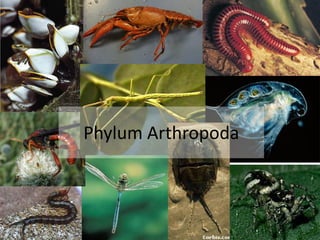
Insects, spiders, crabs, shrimp, millipedes, and centipedes are all arthropods. Arthropods have jointed feet, a segmented body, and an exoskeleton, a cuticle on the outside of their body. Arthropods have by far the greatest number of species of any animal group, at around 900,000 species
2. Phylum Arthropoda
Gr., arthros, joint and
podos, foot
It is the largest phylum of
animal kingdom which
constitute about 83% of all
known species of animals
Arthropods inhabit in all
ecosystems.
Body bilaterally
symmetrical, triploblastic
and metamerically
segmented
Jointed appendages,
usually one pair to a somite
and with varied functions
as jaws, gills, legs etc.
3. All have a hard cuticle/ exoskeleton which is composed of
protein and chitin. It is shed at intervals, called ecdysis or
moulting for growth and development.
Body divisible into head, thorax and abdomen. Head and thorax
often fused to form a cephalothorax.
True coelom reduced and largely replaced by a blood-filled
haemocoel.
Digestive system complete with mouth and anus. Mouth parts
adapted for various modes of feeding.
Respiration by general body surface, gills, tracheae or book-lungs.
Sensory organs comprises of eyes (simple and compound),
chemo- and tactile receptors, and balancing and auditory organs.
Sexes are Dioecious. Fertilization usually internal. Oviparous or
ovoviparous
4. Classification
Subphylum 1. Trilobitomorpha
Gr., tri, three +lobos, lobe + morphe, form
Extinct Trilobites. Mostly marine and bottom-dwellers
Body 3-lobed, due to 2 longitudinal furrows
Head distinct. Probably 1 pair of antennae
e.g. Triarthrus
5. Subphylum 2. Chelicerata
Gr., chele, claw and keros, horn and ata, group
Body is divided into an anterior cephalothorax (prosoma) and a posterior
abdomen (opisthosoma)
Head and thoracic region are fused to form prosoma.
6 pairs of appendages are attached to prosoma
Pair of preoral chelicerae with claws
Pair of postoral pedipalps (usually sensing or feeding)
four pairs of walking legs (5 in horseshoe crabs)
Antennae and true jaws absent
Divided into 3 classes- Merostomata and Arachnida
Horseshoe crabs, spiders, ticks, mites, scorpions
6. Class 1. Merostomata
Gr., meros, thigh and stoma, mouth
Marine with median simple and lateral compound
eyes
5 to 6 pairs of abdominal appendages with book-
gills
Abdomen ending in a sharp telson or spine
Excretion by coxal glands
No Malpighian tubules
e.g. Limulus (horseshoe or king crab)
7. Class 2. Arachnida
Gr., arachne, spider
Eyes simple. No compound eyes
Cephalothorax (prosoma) with 2 chelicerae, 2 pedipalps and 4 pairs
of walking legs
Abdomen without appendages
Respiration by tracheae, book-lungs or both
Excretion by coxal glands and malpighian tubules
e.g. Buthus (Scorpion), Lycosa (wolf spider)
Scorpion
8. Subphylum 3. Mandibulata
(L., mandibula, mandible + ata, group)
Body divisible into head, thorax and abdomen
Head appendages are 1 or 2 pairs of antennae, 1 pairs
of jaws or mandibles and 1 or 2 pairs of maxillae
Compound eyes
9. Class 1. Crustacea
• (L., crusta, shell)
• Head often joined with thorax to form cephalothorax
• Exoskeleton chitinous, hard and calcareous
• Head 5-segmented, bearing 2 pairs of antennae, 1 pairs of
mandibles and 2 pairs of maxillae
• Appendages typically biramous
• Respiration by gills or body surface
• Lobster, crayfish, shrimp, crab, water flea, barnacles
e.g. Macrobrachium rosenbergii, Penaeus monodon, Artemia
crabs
lobsters
euphausids
(krill)
Daphnia
10. Class 2. Myriapoda
• (G., myrios, ten thousand and podos, foot)
• Body worm-like, made of head and elongated trunk with many similar
leg-bearing segments
• Antennae 1 pair, jaws 3 pairs, legs more than 11 pairs
• Respiration by tracheae
• Excretion by 1 or 2 pairs of Malpighian tubules
e.g. Scutigerella
11. Class 4. Insecta
L., insectus, cut or divided
Body made of head (6 fused segments), thorax (3 segments)
and abdomen (up to 11 segments)
Head with compound eyes (1 pair), antennae (1 pair),
mandibles (1 pair) and maxillae (2 pairs)
Mouth parts modified for different feeding habits
Thorax with 2 pairs of jointed legs and 1 or 2 pairs of wings
which may be absent
12. Subphylum 4. Onychophora
G., onychos, claw and phoros, bearing
Worm-like and unsegmented
Single pairs of antennae, eyes and jaws
Numerous stumpy, unjointed clawed legs
e.g. Peripatus
13. Subphylum 5. Tardigrada
Minute, aquatic and segmentation indistnict
No antennae
Mouth retractile with a pair of horny stylets
Four pairs of stumpy and unjointed clawed legs
No respiratory, circulatory and excretory organs
e.g. Macrobiolus (Water bear)
14. Subphylum 6. Pentastomida
Vermiform, unsegmented,
parasitic worms
No antennae
Two pairs of ventral retractile
hooks near mouth
No respiratory, circulatory and
excretory organs
e.g. Linguatula (Tongue worm)
15. Subphylum 7. Pycnogonida
Small, marine, spider-like
and abdomen vestigial
Mouth on a long
proboscis. 4 simple eyes
Appendages include
chelicerae, pedipalps,
ovigerous legs (1 pair)
and long walking legs
No respiratory and
excretory systems
e.g. Nymphon (Sea spiders)
16. Commercially Important Shellfishes of
Bangladesh
Group Bengali name English name Scientific name
Prawn Golda chingri Giant River Prawn Macrobrachium rosenbergii
Chatka chingri Monsoon River
Prawn
Macrobrachium
malcolmsonii
Shrimp Bagda chingri Giant tiger Shrimp Penaeus monodon
Baghtara chingri Green tiger Shrimp Penaeus semisulcatus
Baghtara chingri Kuruma Shrimp Penaeus japonicus
Chaga chingri White Shrimp Penaeus indicus
Bagha chama Blue-tail Shrimp Penaeus merguiensis
Horney chingri Speckled Shrimp Metapenaeus monoceros
Honni/Nona chingri Yellow Shrimp Metapenaeus brevicornis
Ghora chingri Kadal Shrimp Metapenaeus dobsoni
Gura chingri Roshma Shrimp Palaemon styliferus
Ruda chingri Rainbow Shrimp Parapenaeopsis sculptilis
17. Commercially important Lobsters species in
Bangladesh
Group Bengali name English name Scientific name
Spiny
Lobsters
Chhoa Icha Mud Spiny Lobster Panulirus polyphagus
Chhoa Icha Ornate Spiny
Lobster
Panulirus ornatus
Nilcontok Lobster Painted Spiny
Lobster
Panulirus versicolor
18. Economically important crab species
Scientific name Family Bengali name English name
Scylla serrata* Portunidae Shila Kankra Giant Mud Crab
Portunus pelagicus* Sataru Kankra Swimming Crab
Portunus sanguinolentus* Tin Fota Kankra Three-spot Swimming
Crab
Metopograpsus thukuhar Grapsidae Gasho Kankra Paddler Crab
Metopograpsus messor Gasho Kankra Paddler Crab
Episesarma versicolor Kankra Violet Vinegar Crab
Potamon woodmasoni Potamoniade Kata Kankra Freshwater Crab
Potamon martensi Chimta Kankra Spiny Crab
Paratelphusa lamellifrons Pati Kankra Sartotina Crab
Uca urvillei Ocypodidae Holdepa Kankra Fiddler Crab
Uca annulipes Kankra Ghost Crab
Ocypode ceratophthalmus Lal Kankra Horned-eyed Ghost
Crab
19.
20. Macrobrachium rosenbergii
also known as the giant river prawn, giant freshwater prawn
Classification:
Kingdom: Animalia
Phylum: Arthropoda
Sub-Phylum: Crustacea
Class: Malacostraca
Order: Decapoda
Family: Palaemonidae
Genus: Macrobrachium
Species: M. rosenbergii
21. External morphology
Habits and habitat:
Inhibits freshwater rivers, ponds and lakes
Nocturnal, hiding at the bottom during the day and coming to the
surface at night in search of food
It walks slowly at the bottom with the help of its 10 walking legs and
swim actively to the surface with the help of its 10 pleopods
When distrubed, it suddenly springs backwards with the help of a pair
of uropods, attached to the last abdominal segment
To escape from the enemy’s grasp, it can shed off one or more of its
appendages, which phenomenon is called autotomy
Shape and size:
• More or less spindle-shaped, elongated and bilaterally symmetrical
• can grow to a length over 30 cm (12 in)
22. Segmentation and body divisions
The bodies of freshwater prawns are divided into twenty segments
(or somites), all bearing jointed appendages
The segments are arranged into two main regions- an anterior
cephalothorax and a posterior abdomen
Cephalothorax:
Cephalothorax is large, rigid and more or less cylindrical in shape.
It is formed by the union of two segments- Head and thorax and
invisible under a large dorsal and lateral shield, known as the
carapace.
The carapace is hard and smooth, except for two spines on either
side; one (the antennal spine) is just below the orbit and the other
(the hepatic spine) is lower down and behind the antennal spine.
It consists of 13 segments. Head-5 segments, Thorax- 8 segments
These segments bearing jointed appendages
23.
24. Abdomen:
• It is composed of 6 distinct movable segments and a terminal conical
piece, telson
• Each segment carries a pair of jointed appendages called pleopods or
swimmers
• Abdominal segments are dorsally rounded, laterally compressed and
normally bent under the cephalothorax
External apertures:
Mouth- slit-like, open mid-ventrally at the anterior end of cephalothorax
Anus- longitudinal aperture lying ventrally at the base of telson
Paired renal apertures-open on raised papillae on the inner surface of
coxae of antennae
Paired female genital apertures- situated on the inner surface of coxae of
the third pair of walking legs
Paired female genital apertures- situated on the inner surface of coxae of
the fifth pair of walking legs
Exoskeleton:
Body and appendages are covered by a hard, protective and calcareous
shell
It is composed of chitinous cuticle
The exoskeleton comprises several hardened plates, called sclerites
25. Cephalothoracic sclerites:
The anterior and triangular region of dorsal shield is termed dorsal plate
It extends forward over the head as a laterally compressed and serrated
vertical process, called rostrum
An orbital notch, which accommodates a stalked, jointed and movable
compound eye situate on either side of the base of rostrum
There are two spine-like outgrowths just behind and below each orbital notch-
antennal spine and hepatic spine
The posterior region of dorsal shied is termed carapace
Abdominal sclerites:
The sclerite of each abdominal segment is separate and ring-like
Adjacent sclerites are connected by thin, soft, uncalcified cuticle called
arthrodial membranes which provide movable joints
In each abdominal sclerites, its dorsal broad plate –tergum, ventral narrow
transverse bar-like plate- sternum and two lateral flap like plates- pleura
An appendage is connected with the pleuron of its side by a smallplate-
epimeron
26.
27. Tergum and pleura of an abdominal segments overlaps the succeeding
segment, which is called imbricate arrangement
Pleura of sixth abdominal segment are greatly reduced
Two adjacent abdominal segments articulate with each other by means of a
pair of hinge joints, one on either side
The hinge joints are lacking between the third and fourth segments
Appendages:
• Each segment of body bears a pair of jointed appendages.
• 19 pairs of appendages
• Each appendages consists of a common base or protopodite and two ramii or
branches- an inner or median endopodite and an outer or lateral exopodite
• Each basal protopodite is composed of two segments- a proximal coxa for
attachment with the body and a dista basis which bears the two ramii
• All appendages are biramous (L., bi-two and ramus-branch) type
28.
29. Cephalic appendages
There are 5 pairs of cephalic or head apendages- antennules,
antennae, mandibles, maxillulae and maxillae
Antennules
• Attached one or either side, below the bases of eye-stalks.
• The protopodite consists of three segments- precoxa, middle coxa
and basis which bears two long and many jointed feelers.
• Outer feeler is further divided into an inner smaller branch and an
outer larger branch
• These feelers bear sensory setae and are tactile in function
Antennae
• Lie one on either side just below the antennules
• The protopodite is greatly swollen due to presence of excretory
organ which opens by a minute renal aperture on the inner margin
of coxa
• The antennae are sensory, excretory and balancing in function
Mandibles: help in masticating foods
Maxillulae: Help in the manipulation of food
Maxillae: Help in respiration and manipulation of food
31. Thoracic appendages
First maxillipedes
Second maxillipedes
Third maxillipedes
Walking legs:
• First 2 pairs of legs are chelate leg and the rest 3 pairs legs are non-chelate
• A female reproductive aperture lie on the inner side of each third walking leg
• A male genital aperture lie on the arthrodial membrane between the fifth leg
and thorax
32. Abdominal appendages
6 pairs of abdominal appendages
1st 5 pairs are the swimming pleopods or swimmerets, used as paddles and the 6th
pair are uropods, along with the telson
Typical abdominal appendages
First abdominal appendages
Second abdominal appendages
Uropods
33. A summary of the segments and the functions of each appendage
BODY SECTION SOMITE APPENDAGE NAMES (PAIRS) FUNCTIONS OF APPENDAGES AND
RELATED STRUCTURES
Cephalon (front portion
of the cephalothorax
1 embryonic segment (not visible in adults)
2 1st antennae tactile and sensory perception (statocyst)
3 2nd antennae tactile
4 mandibles cutting and grinding food
5 1st maxillae (maxillulae) food handling
6 2nd maxillae food handling; water circulation through the gill
chamber (scaphognathite)
Thorax (rear portion of
the cephalothorax)
7 1st maxillipeds feeding/food handling
8 2nd maxillipeds feeding/food handling
9 3rd maxillipeds feeding/food handling
10 1st pereiopods (chelipeds) food capture
11 2nd pereiopods (chelipeds) food capture; agonistic and mating behaviour
12 3rd pereiopods walking; female gonophores between base of legs
13 4th pereiopods walking
14 5th pereiopods walking: male gonophores between base of legs
Abdomen 15 1st pleopods (swimmerets) swimming
16 2nd pleopods (swimmerets) swimming; copulation in males
17 3rd pleopods (swimmerets) swimming
18 4th pleopods (swimmerets) swimming
19 5th pleopods (swimmerets) swimming
20 uropods propulsion, together with the central telson
34. Digestive system of prawn
Alimentary or digestive canal
Foregut-
Mouth
Buccal cavity
Oesophagus
Stomach- Cardiac and Pyloric
Midgut-
Intestine
Hindgut-
Rectum
Foregut and hindgut are lined internally by
cuticle called intima which isshed during the
moulting
Midgut is lined internally by endoderm
Digestive gland-
Hepatopancreas:
As pancreas, it secretes digestive enzymes
As midgut, it absorbs the digested food
materials
As liver, it serves as an important storage
organ for glycogen, fat and calcium
35. Food and feeding
They are omnivorous.
Prawn feeds algae, moss, aquatic weeds, insects, snails, tadpoles, fish and
debris of the bottom
It feeds at night, being more active at dawn and dusk
Chelate legs, aided by the third maxillipedes, capture and convey food to
the mouth
Coxae of second maxillipedes hold the food in a position, while incisor
processes of mandibles cut it into smaller pieces
Then these food pieces are swallowed with the help of maxillipedes,
maxillulae and maxillae
Inside the buccal cavity, molar processes of mandibles masticate the food,
which is then conveyed to the cardiac stomach through oesophagus.
Passage of food through oesophagus is facilitated by the peristaltic activity
of oesophagus and the sucking action of cardiac stomach
36. Digestion and absorption
Digestion is the process of breaking down food by mechanical
and enzymatic action in the alimentary canal into substances
that can be used by the body
Hepatopancreas
Digestive
enzymes
Ventral chamber
of pyloric
stomach
Hepatopanc
reatic ducts
Cardiac
stomach
Mixes
with
food
Expansion and
contraction of
Cardiac
stomach
Churning of food
which facilitate
digestion
Hastate
plate
Moving
spines of
combed
plates
Smaller
particles of
food
Cardiopyloric
aperture
Digested and
liquefied foodVentral chamber
of pyloric
stomach
Filtered
through
pyloric filtering
apparatus
Finest food
particles
Hepatopanc
reatic ducts
Hepatopancreas Hydrolysed and
absorbed
Undigested
and coarser
food
particles
Intestine for
digestion and
absorption
Undigested
matter
RectumEgestion
37. Absorption is the process of
absorbing or assimilating of digested
small and soluble food substances
into cells or across the tissues and
organs through the villi of the small
intestine by diffusion or osmosis
38. Excess water of undigested residual food is
absorbed back into the body in the large
intestine. Then the dry undigested matter
are stored in the rectum, the lower part of
the large intestine and comes out of the
rectum through the anus as faeces. This
process is called egestion.
39. Respiratory system
Respiratory system is well developed
Its respiratory organs are-
1) inner lining of branchiostegites or gill covers,
2) Epipodites (mastigobranchiae) and
3) Branchiae or gills.
These are sheltered in two large and compressed gill-chambers, one on either side of thorax.
Each gill-chambers are bounded internally by epimeron and externally by the curving pleural
side of carapace or branchiostegites or gill covers
Branchiostegites:
The ventral extension of the carapace on either side of cephalothorax is known as
branchiostegite.
Inner lining of branchiostegites or gill covers is thin, membranous and highly vascular containing
minute blood lacunae.
These form large respiratory surfaces which absorb O2 from water and give out Co2
Epipodites:
There are three pairs of epipodites.
They are the outgrowthsof coxae of the three pairs of maxillipedes
They occupy the anterior part of gill chambers beneath the scaphognathites of maxillae
Epipodites of 1st pair are bilobed and larger than others
40.
41. Gills:
8 gills inside each gill chamber
7 of them are exposed on removing the gill cover and 8th gill lies concealed
beneath the dorsal art of the 2nd gill
42. Types of gills:
Podobranch or foot-gill: attach to the coxa of each 2nd maxillipede
Arthrobranch or joint-gill: attach to the arthrodial membrane which joining the
limb with the body.
Each third maxillipede bears two arthrobranchs
2nd arthrobranch is the smallest and remains concealed beneath the 1st
arthrobranch
Pleurobranch or side gill: The last 5 pairs of gills are attached to the lateral wall of
thoracic segments bearing the 5 pairs of walking legs
Structure:
More or less crescentic in shape
Gradually increase in size backwards i.e. each gill is larger than the one in front of it
Each gill is attached in its moddle to the wall of the thorax by a connection called
gill-root, through which nerves and blood channels enter and leave the gill
All the gills are pyllobranchs i.e. each of them consists of two rows of leaf-like gill
plates arranged like leaves of a book to the base of a gill
Gill-plates are largest in the middle and gradually smaller towards the two ends
A gill-plate
43.
44. 1. An open circulatory system, found
in arthropods, pumps blood into a
cavity called a hemocoel where it
surrounds the organs and then
returns to the heart(s) through ostia
(openings).
2. Blood flows through open spaces
called lacunae and sinuses
3. Blood flows at a very slow velocity
4. Body cavity is filled with blood
(Haemocoel*)
5. Internal organs are bathed by blood
6. Blood takes long time to complete
7. Supply and elimination of materials
are very slow
8. Exchange of materials takes place
between blood and sinuses
9. Blood flow cannot be regulated
1. A closed circulatory system, found
in all vertebrates and some
invertebrates, circulates blood
unidirectionally from the heart,
around the body, and back to the
heart.
2. Blood flows through closed vessels
3. Blood flows at a very high velocity
4. Haemocoel is absent
5. Internal organs are not in direct
contact with blood
6. Blood takes short time to complete
7. Supply and elimination of materials
are very rapid
8. Exchange of materials between
blood and tissues takes place
through the capillaries
9. Blood flow can be regulated
Circulatory system
45. Prawn has an open type or lacunar type of blood vascular system
This type of blood vascular system is characterized by the absence
of capillaries so that blood flows through open spaces, the lacunae
or sinuses, in body.
Blood vascular system of prawn includes
I. Pericardium
II. Heart
III. Arteries
IV. Blood sinuses
V. Blood channels
VI. Blood
There are no veins and capiilaries
Circulatory system
46. Pericardium
Heart lies dorsally in the posterior part of
thorax,
It is enclosed in a spacious haemocoelic
chamber, the dorsal sinus or pericardium.
Floor of pericardium is in the form of a thin
horizontal septum, lying just above
hepatopancreas and gonad.
The septum is attached to the dorsal body
wall and to the thoracic wall.
47. Heart
• It is a muscular and triangular organ.
• Its apex is directed anteriorly. Broad base is directed posteriorly.
• Cardio-pyloric strand and two lateral strands will keep the heart in
position inside the pericardium.
• Thick and muscular wall of heart is perforated by five pairs of
valvular, slit-like apertures called ostia.
• Blood from dorsal sinus or pericardium can enter into the heart
through Ostia.
i) First pair of Ostia - Mid dorsally
ii) Second pair of Ostia - Mid ventrally
iii) The third pair - Posteriorly
iv) The fourth pair – Anterio-laterally,
v) The fifth pair-Postero-laterally.
48. Arteries
The heart sends blood to the body through narrow tube-like arteries.
Five of them arise from the anterior end and one from the posterior end of the heart
a) Median ophthalmic arteries:
It arises from the apex of the heart.
It runs forward mid-dorsally to the cardiac stomach, oesophagus and head.
It joins the two antennary arteries above the oesophagus in cephalic region.
b) Antennary arteries: A pair of antennary arteries arises from the apex of the heart
on both sides of the median ophthalmic artery. Each artery runs forwards along the
outer border of the mandibular muscle. Its branches are
1) Pericardial branch of this artery goes to pericardium.
2) Gastric branch goes to cardiac stomach
3) A mandibular branch goes to mandibular muscle.
4) Then it divides into a dorsal branch and a ventral branch. The ventral branch
divides to supply the blood to antennule, the antenna and the renal organ.
5) The dorsal branch sends an optic artery to the eye and then divide.
6) Then it bends to unite with the same opposite branch and the median ophthalmic
to form a circular loop like artery or circulus cephalicus.
7) It gives a pair of rostral arteries to the rostrum.
50. Hepatic arteries
A pair of hepatic or hepatopancreatic arteries arise from heart ventro-laterally
one on each below the antennary artery.
They go to the hepatopancreas within which they divide and subdivide
Median posterior artery :
A short but stout artery arises from the postero -ventral surface of the heart.
It bifurcates into a supra-intestinal artery and a sternal artery.
The supra intestinal is also called dorsal abdominal artery which supplies blood
to the midgut and the dorsal abdominal muscles.
The sternal artery runs downwards through the hepatopancreas and then
pierces the ventral thoracic ganglionic mass and go to the ventral side.
It divides into ventral thoracic branch and ventral abdominal branch.
The ventral thoracic branch supplies blood to the sternal region up to the mouth,
the first three pairs of walking legs, the maxillae, the maxillulae, oesophagus,
gonads etc.. The ventral abdominal branch runs posteriorly up to the anus and
supplies blood to ventral abdomen, the last two pairs of legs, pleopods, uropods,
the hindgut etc.
51. Blood sinuses
True capillaries and veins are absent.
Arteries repeatedly branch in various organs
of body. Arterial branches open freely into
blood sinuses or lacunae of the haemocoel.
All the sinuses of the body open into a pair of
ventral sinuses which are lying below
hepatopancreas on the floor of thorax.
52. Blood channels
These channels are lacunar tubes without proper
walls.
Six afferent bronchial channels carry venous blood
from each ventral sinus to the gills of that side.
As blood flows through gills, it gives off Co2 and
receives a fresh supply of O2 from water in the gill
chamber.
Oxygenated blood from the gills of both sides is
brought to the pericardium by six efferent bronchial
channels.
53. Blood
It is colorless, thin and watery fluid.
It contains amoeboid white corpuscles or leucocytes.
No red blood cells
The respiratory pigment is haemocyanin, hence the
blood is bright blue in color when combined with
oxygen because its metallic base is copper instead of
iron. It is colorless when de-oxygenated.
Blood distributes digested food, oxygen to all body
parts.
Blood has the extensive capacity of clotting.
54.
55. Sexual dimorphism
The sexes of prawn are separate and
show well-marked sexual dimorphism.
Sexual dimorphism is the difference in
morphology such as in colour, shape, size,
and structure between male and female
members of the same species beyond the
differences in their sexual organs.
56. Male Female
Body larger than female Body usually smaller than male
1 The male have a narrower abdomen than
female
The female have a broader
abdomen than male
2 The Cephalothorax of the male is
proportionately larger than female
The head of the mature female is
smaller than male
3 The second pair of chelate legs are longer
and stronger
The second pair of chelate legs are
smaller and thin,
4 The thoracic legs are closely arranged. The thoracic legs are less closely
arranged.
5 Second pair of pleopod has appendix
masculina.
Absent.
6 1st abdominal segment has pointy central lump Female has no central lump
7 The male genital openings are present on
the arthroidal membrane of the 5th pair or
walking legs.
The female genital openings are on
the coxae of the 3rd pair of walking
legs.
8 The male prawn is bigger in size than
female prawn of the same age.
The female prawn is smaller than
the male of same age.
57. Male reproductive system:
a) Testis:
The two testes are soft, white and elongated bodies which fuse at their
anterior ends to form a common lobe.
They are long and narrow.
They enclose between them a gap for the passage of the cardio-pyloric
strand connecting heart to pyloric stomach.
They are present on the posterior half of the hepato-pancreas and beneath
the pericardial sinus and heart. Anteriorly they extend up to the renal sac
and posteriorly they run upto the first abdominal segment.
Each testis consists of a large number of coiled, narrow and thin-walled
seminiferous tubules embedded in a connective tissue.
The cavity of each tubule is lined by a single layer of germinal epithelium,
which undergo spermatogenesis to form spermatozoa
A mature sperm consists of a rounded cytoplasmic body, containing a large,
dark, crescentic nucleus and a tail-like blunt process
58. Vasa deferentia:
• A long, coiled and narrow tube, the vas deferens, arises from each
testis near its posterior ends
• It coils near the hepato-pancreas.
• It runs vertically downwards between the abdominal flexor muscles
on the inner side and thoracic wall on the outer side
Seminal vesicle:
Each vas deferens reaching ventrally near the base of fifth leg and
swells to form a club-shaped structure called seminal vesicle.
It stores the spermatophores.
Each seminal vesicle opens to the exterior through a male genital
aperture situated on the inner side of coxa of 5th walking leg.
Each male genital aperture is covered by a small flap of integument
59. Female reproductive system
It contains a pair of ovaries and a pair of oviducts.
a) Ovaries:
• The two ovaries are whie, compact and sickle-shaped bodies
touching each other at both the ends but leaving a gap in the middle
for the passage of the cardiopyloric strand.
• These are present on the posterior half of the hepatopan-creas
below the pericardial sinus and heart.
• They extend anteriorly up to the renal sac and posteriorly up to the
first abdominal segment.
• In the breeding season the ovaries enlarge and may extend into the
first abdominal segment.
• Each ovary has a large number of ova surrounded by a membrane.
The ova are in the different stages of development in the ovary.
• Immature ova lie towards the centre while mature ova towards the
surface of ovary
60. Oviducts:
These are slender and curved tubes. Each oviduct
starts from the middle of the outer border of the
ovary.
It runs vertically downwards to open through a
female genital aperture
Female genital aperture:
• The oviducts open out through female genital
opening.
• They are present on the coxa of the third pair of
walking legs.
63. Morphotypes
Three different morphotypes of males exist. The first stage is called "small
male" (SM); this smallest stage has short, nearly translucent claws. The
ratio of claw to body length is 0.5 ±0.1.
If conditions allow, small males grow and metamorphose into "orange
claws" (OC), which have large orange claws on their second chelipeds,
which may have a length of 0.8 to 1.4 times their body size.
OC males later may transform into the third and final stage, the "blue claw"
(BC) males. These have blue claws, and their second chelipeds may
become twice as long as their bodies. The ratio of claw to body length is 1
.6 ±0. 1.
Males of M. rosenbergii have a strict hierarchy: the territorial BC males
dominate the OCs, which in turn dominate the SMs.
The presence of BC males inhibits the growth of SMs and delays the
metamorphosis of OCs into BCs; an OC will keep growing until it is larger
than the largest BC male in its neighbourhood before transforming.
64. All three male stages are sexually active, and females that
have undergone their premating moult will co-operate with
any male to reproduce.
BC males protect the female until their shells have hardened;
OCs and SMs show no such behaviour.
The presence of this new BC male then delays the transition
of the next OC to the BC morphotype, causing it to attain a
larger size following its metamorphosis. BC males dominate
OC males, regardless of their size, and suppress the growth
of SM.
65. Reproductive behavior
After ovarian maturation, M. rosenbergii females experience a moult, known as a pre-
spawning or pre-mating moult, which usually occurs at night. After this moulting process,
courting and mating commence. Successful mating can only take place between ripe
females, which have just completed their pre-mating moult and are therefore soft-
sheltered, and hard-shelled males.
Mating of M. rosenbergii is completed in six stages, which are summarized below:
Females approach males 2 to 3 days before their pre-mating moult.
At first the female is chased away but later, after several hours of persistence, is
allowed to remain near the male.
About a day before the pre-mating moult the female is already totally accepted by the
male, positioned below it or between its long second pair of claws.
Once between his chelae, the female may turn to face the male, making contact with
the male’s walking legs. Prior to fertilization, the female re-orients herself in order to
position her dorsal telson under his head region.
The male mounts the female, and begins characteristics rubbing action with pleopods
on the ventral lower cephalothorax and upper abdominal segments of the female. The
female appears to become torpid.
The male grasps the female’s rostrum with his chelae of periopods, and may also grasp
the gill operculum. The female is turned ventral side up with chelae stretched out in
front, and remains torpid. This ensures that the reproductive organs are aligned.
66. Copulation and fertilization
In the natural environment, mating of Macrobrachium takes place all
year round, although, due to environmental reasons, peak mating
takes place only during certain periods of the year- May, June and
July.
About 200-300 mature eggs are laid by the female at one time in slimy
strings
Fertilization is external
During copulation the male deposits the spermatophores near the
female genital openings of the female and the eggs are fertilized as
they come out.
After fertilization, the eggs are fastened to the pleopods by the sticky
secretion of certain tegumental glands
The eggs hanging from pleopods look like berries or bunches of
grapes
She carriesthem whenever she goes and the eggs are kept aerated by
the slow back and forth movements of plepods
The female bends down her abdomen to protect first the eggs and
later the young.
67. Life Cycle
There are four stages in the life of a freshwater prawn, viz, egg, larva, Post
larva/juvenile and adult
Development of prawn from egg is direct as there is no free larval form
The offspring hatching out of the egg resembles the adult except in size
The eggs are hatch in 5-6 weeks and cling to the pleopods of female prawn for
some time.
Larvae hatch during the night.
The newly hatched larvae which are devoid of many segments and appendages of
the adult start swimming in about 5 minutes
Larvae normally swim with their heads down and ‘jump’ when they contact a
surface.
Larvae need brackish water to survive at this stage.
Even if larvae hatch in freshwater, they will not survive if they are not put into
brackish water within two or three days.
Larvae are planktonic and in the wild generally eat zooplankton, small insects and
larvae of other aquatic invertebrates.
Larvae undergo 11 moults over a period of 15 to 40 days before transforming into
post larvae
Larvae take about 45 days to metamorphose into post-larvae (PL).
68. ESTIMATED RATE OF LARVAL DEVELOPMENT1
1st stage takes place from 1st to 2nd day after
hatchin
g
2nd " " " " 2nd to 4th " " "
3rd " " " " 4th to 7th " " "
4th " " " " 7th to 12th " " "
5th " " " " 11th to 16th " " "
6th " " " " 15th to 21st " " "
7th " " " " 18th to 24th " " "
8th " " " " 22th to 28th " " "
9th " " " " 25th to 31st " " "
10th " " " " 28th to 33rd " " "
11th " " " " 31st to 37th " " "
12th " " " " 35th to 41st " " "
13th " " " " 38th to 45th " " "
69. The rate of this transformation depend upon feed quantity and
quality, temperature and other water quality variables.
Post larvae can tolerate a wide range of salinity, but freshwater is
their normal habitat.
And so, two to three weeks after metamorphosis, the PL move
against the current and head towards freshwater canals and rivers.
They abandon the planktonic habit at this stage and become
benthic.
They are omnivorous, feeding on aquatic insects and their larvae,
phytoplankton, seeds of cereals, fruit, small mollusca and crustacea,
fish flesh, slaughterhouse waste and animal remains.
They move by crawling and generally swim with their dorsal side
uppermost. They can swim rapidly.
70.
71.
72. Shrimp
Shrimps belong to the order Decapoda and family Penaeidae
Group Bengali name English name Scientific name
Shrimp Bagda chingri Giant tiger Shrimp Penaeus monodon
Baghtara chingri Green tiger Shrimp Penaeus semisulcatus
Baghtara chingri Kuruma Shrimp Penaeus japonicus
Chaga chingri White Shrimp Penaeus indicus
Bagha chama Blue-tail Shrimp Penaeus merguiensis
Horney chingri Speckled Shrimp Metapenaeus monoceros
Honni/Nona chingri Yellow Shrimp Metapenaeus brevicornis
Ghora chingri Kadal Shrimp Metapenaeus dobsoni
Gura chingri Roshma Shrimp Palaemon styliferus
Ruda chingri Rainbow Shrimp Parapenaeopsis sculptilis
73. Penaeus monodon
Penaeus monodon is commonly known as giant tiger shrimp
It is one of the fastest growing species among the various types of shrimp
Females are about 25–30 cm in length and males are about 20–25 cm in
length.
A long brackish red or brown line present on the ventral side.
The carapace and abdomen are transversely banded with alternative red
and white.
Antennae are longer than antennules
It is a euryhaline species, thrive in both marine and brackish water but
suitable salinity range for its faster growth is 15-30ppt.
Optimum temperature for Penaeus monodon is 18-31ºC
Attain its maturity in about 9 months
First three pairs of thoracic legs are chelate.
Abdomen is laterally compressed and straight
74. Life cycle
Adults Penaeus monodon are bottom-dwellers in the offshore areas at depths
of 20-70m
Sexually mature shrimp spawn in the deep sea.
Each gravid or ripe female lays more than 500000 eggs per spawning
Spermatophores are deposited in the female’s thelycum by the male’s
petasma
Mating can only occur between newly molted females and hard-shelled males
Development is indirect
After hatching, the early larval stages are- nauplius, protozoaea, mysis and
post-larvae
The eggs take about 12-15h to hatch into nauplii, the first larval stage
This stage is characterized by the possession of three pairs of appendages:
the antennules, the antennae and the mandibles and also a single median
eye
The nauplius has no mouth and cannot take in food
It feeds on yolk reserves
There are six nauplius stages (N1-N6) that moult five times within 48 to 56
hours, depending on temperature
75. The nauplius metamorphoses into a protozoaea
There are 3 successive stages of protozoaea (P1-P3), all of which
are completed within 5 to 6 days
These stages are characterized by the presence of functional
mandibles i.e. theyare capable of feeding, a distnict cephalothorax,
an abdomen which terminates in a forked telson and a pair of
stalked and compound eye
it swims vertically and diagonally forward towards the water surface
with the head uppermost.
The protozoaea changes into the mysis stage
There are three successive stages of mysis development, separated
by moults within 4-5 days
The mysis larva has the appearance like small shrimp and swims
in quick darts accomplished by bending the abdomen backwards.
For mysis sub-stages, the most prominent change is the
development of pleopods. The pleopods appear as buds at Mysis I,
which protrude at Mysis II and finally become segmented at Mysis
III.
76. The mysis larva undergoes a true metamorphosis and becomes post-larva
The post larva resembles an adult prawn
The post-larva is characterized by the number and arrangement of the spine
on the rostrum and the fully developed pleopods
After 1-3 weeks, the post-larva metamorphoses into juvenile
Within 4-5 months, the juvenile changes into sub-adult and return to the
offshore water
The postlarva migrate to the shallower estuarine environment where
vegetation provides nursery grounds for their development and here they
grow to the juvenile and sub-adult stages.
Reaching sub-adult, they return back to the sea and becomes sexually
mature adult.
After copulaton, the adult female spawn in deep sea
Then the nauplius, protozoaea and mysis stage occurred in deep sea.
77.
78. Food and feeding
Shrimps are either omnivorous or scavengers or detritus feeders
They start feeding at protozoaea stage
Protozoaea and early mysis stages prefer phytoplankton
At mysis and early post-larvae, food preferences changes to
zooplankton such as rotifer, brine shrimp or other small invertebrates
The foods of shrimp consists of polychaetes, mussels, small crabs
and shrimps, small invertebrates, worms, vegetable matter etc.
79. Artemia or brine shrimp
Artemia is widely-used in aquaculture as an excellent food source for fish
and crustaceans.
Artemia has several characteristics which make it ideal for aquaculture use.
It is easy to handle,
adaptable to a wide-range of environmental conditions,
non-selective as a filter-feeder and
is capable of growing at very high densities.
Artemia also has a high nutritive value (40–60 percent protein, rich amino
acid composition),
an unchanging food requirement,
high conversion efficiency,
short generation time,
high fecundity rate and long lifespan.
The whole animal (even adult stage) may be consumed without previous
processing by many aquaculture organisms.
80. Systematic classification
Phylum: Arthropoda
Class : Crustacea
Sub-class : Brachiopoda
Order : Anostraca
Family : Artemidae
Genus : Artemia
Artemia is a branchiopod crustacean found in salt pans or coastal inland
salt lake where salinity ranges between 100ppt to 200-250ppt
It exists in two forms-
1) Bisexual forms where in both males and females are present
2) Parthenogenetic forms where in only females are resent
In bisexual population, it attains 10mm in length and in parthenogenetic
strains, 20mm in length
Artemia salina is the mot common
It has an elongated body with well-developed antennule
The second antenna of male is modified into large hooked graspers or
claspers, used for holding the female during copulation
The second antenna of female is less developed and sensorial in function
81. 11 pairs of thoracic appendages (thoracopods) used for feeding, locomotion and respiration
Each thoracopod has three functional parts- telopodite acting as filter for filter-feeding, oar like
endopodite are locomotory and exopodite as gills
Two compound eyes
A pair of penis is seen at the posterior part of trunk at male.
At female, an unpaired brood pouch or uterus is seen posterior to 11th pair of appendages.
Ripe eggs are transported from the ovaries into an unpaired brood or uterus.
82. Mating and fertilization
Mating of Artemia starts with the male grasping the female with its antennae between the
uterus and the last
The couples can swim around in this riding position for several hours.
Copulation is very fast, with the male abdomen bent forward and one penis introduced into
the uterus opening.
Modes of reproduction:
Two main modes of reproduction are found in Artemia.
In normal conditions, Artemia are ovoviviparous
In ovoviviparous reproduction, the fertilized eggs develop to free-swimming nauplii which
are released by the mother
In extreme adverse conditions such as high salinity (150ppt) and low oxygen, Artemia are
oviparous
The shell glands located in the uterus secreted a shell around the embryo in gastrula
stage.
The embryo enters into a state of dormancy which is called cysts and are released by the
mother
An adult Artemia reproduce at the rate of 300 cysts/ nauplii in every four days and it can
live upto 50/60 days
83. Life cycle
Cysts:
The dehydrated dormant eggs of Artemia are called cysts. These minute
brown particles (200–300 um in diameter) are found floating in many salt
lakes and brine ponds or carried to the shore by wind and wave action.
The cyst have a 3-layered shell in which thickest layer is chorion which is
made of lipoprotein impregnated with chitin and haematin.
This layer protects the embryo from mechanical stress or radiation
When these cysts are soaked in sea water, embronic development is
resumed and a nauplius larva hatch out within 24-36h
After 15-20 hours later, the cyst's outer membrane bursts (breaking stage)
and the embryo enclosed by the hatching membrane becomes visible.
The embryo comes out of the shell and hangs underneath the empty shell.
This stage is called umbrella stage
Soon after hatching membrane breaks and free swimming nauplii larva
emerges
84. Nauplii:
The newly hatched nauplius is 400-500 micron in length
The first instar larva is brownish orange due to the presence of yolk.
It has three pairs of appendages-antennule, antenna and mandible and an
unpaired red ocellus or nauplius eye in the head region.
Mouth and anus are absent
Do not feed for about 12 hours
After 12 hours, it starts feeding on algal cells, bacteria and detritus.
After 15 moults, it becomes adult.
Juveniles:
The larva grows and differentiates through about 15 molts during which the
trunk and abdomen elongate and the digestive tract becomes functional.
Food particles are collected from the setae of the antennae.
Lateral complex eyes develop at both sides of the ocellus.
Adults
Important changes in the morphology of Artemia take place from the 10th
instar on.
Antennae lose their primitive function and undergo sexual differentiation.
Thoracopods also differentiate into three functional parts: telopodites as filter,
oar-like endopodites for locomotion and membranous expodites as gills.
85.
86. Riding pairs:
• Copulation in Artemia starts with the male grasping the female with its
antennae between the uterus and the last pair of thoracopods.
• The couples can swim around in this position for several days.
• Copulation is very fast, with the male abdomen bent forward and one penis
introduced into the uterus opening.
Food and feeding habit
They are non-selective filter feeders
It feeds mainly on algae, detritus and bacteria
During larval stage, setae on the antenna serve in filtering food particles from
surrounding water
But in juveniles and adults, thoracopods assist in feeding.
When the thoracopods move forward, water is drawn into the teopodites which
act as filter.
The food particles are concentrated in a food groove between the base of the
legs
Glands along the groove secrete the adhesive material that helps in clumping
the minute particles into minute balls
Then the food groove transfer the food particles to the mouth
87. Crab
Crab any of the members of decapod crustaceans
beonging to the suborder Brachyura with a broad,
rather round, upper carapace and a small abdomen
tucked beneath the body living in marine, brackish or
freshwaters
About 133 species of crabs are found in whole earth
4 species of freshwater crabs and 11 species of
marine brackishwater crabs are found in Bangladesh.
Among them, most commercially important crab
species are serrated mud crab, also known as
mangrove crab or Scylla serrata
88. Mud crab
Systematic position:
Kingdom: Animalia
Phylum: Arthropoda
Class: Crustacea
Order: Decapoda
Family: Portunidae
Genus: Scylla
Species: Scylla serrata
• Mud crab are omnivorous and scavangers.
• The food of mud crab consists of small mollusca, trash fish, algae,
decaying animals, decaying matter and other crustaceans