![Presenting complaint(s)
SM [NHN : 52020 2662]
Admission Date : 15/02/16 Discharged Date : 19/02/16
SM, 40 y/o i-T/F admitted with Right Iliac Fossa
Mass/Collection. She had RIF pain, vomiting,
fever and diarrhea. 3rd admission for the same
complaint(s). Planned open appendecectomy
for clinical appendicitis 5/12 ptca but NO appe.
done yet operative diagnosis was RIF abscess
thus <50ml of pus drained from the RIF under
general anesthesia.](data:image/gif;base64,R0lGODlhAQABAIAAAAAAAP///yH5BAEAAAAALAAAAAABAAEAAAIBRAA7)
Empfohlen
Weitere ähnliche Inhalte
Was ist angesagt?
Was ist angesagt? (20)
Andere mochten auch
Andere mochten auch (14)
Ähnlich wie Incisional Hernia
Ähnlich wie Incisional Hernia (20)
Kürzlich hochgeladen
Kürzlich hochgeladen (20)
Incisional Hernia
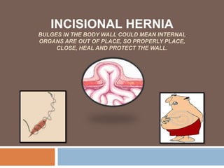
- 1. INCISIONAL HERNIA BULGES IN THE BODY WALL COULD MEAN INTERNAL ORGANS ARE OUT OF PLACE, SO PROPERLY PLACE, CLOSE, HEAL AND PROTECT THE WALL.
- 2. Presenting complaint(s) SM [NHN : 52020 2662] Admission Date : 15/02/16 Discharged Date : 19/02/16 SM, 40 y/o i-T/F admitted with Right Iliac Fossa Mass/Collection. She had RIF pain, vomiting, fever and diarrhea. 3rd admission for the same complaint(s). Planned open appendecectomy for clinical appendicitis 5/12 ptca but NO appe. done yet operative diagnosis was RIF abscess thus <50ml of pus drained from the RIF under general anesthesia.
- 3. Pathological Sieve – RIF MASS±PAIN Vascular-Aneurysms Inflammatory-Crohn’s disease, Appendicitis, Diverticulitis, Mesenteric adenitis, PID, Typhilitis Trauma-Hematoma Acquired- Incisional Hernia, Colocolic Interssusception (AIDS), Ectopic kidney, Ectopic pregnancy Metabolic- Hyperlipidemia, Hypercortisolemia Infection- Appendicular/Iliopsoas/Tubo-ovarian abscess, Ileocecal TB Neoplasm- Appendicular Carcinoid Tumor, Cecum Tumor, Ovarian Tumor, CRC and non Hodgkin Lymphoma
- 4. History of Presenting Complaint(s) Pt. says firstly she had “sharp-poky-persistent” pain localized in the RIF region then developed fever 3 days p.t.adm. 1 day prior she had (×2) of vomiting and diarrhea (“clear watery” Ø Bile/Blood/Mucus). ROS +(s): ↓ Appetite, ↓ Bowel output and Nausea (×1/7). PV Bleeding (×2/7). -(s): Generalized weakness, constipation, weight loss, pus vaginal discharge, hematuria, urgency, frequency, hesitancy, dysuria, menorrhagia, dysmenorrhea, amenorrhea
- 5. History… PMH 23/07/15 – 1st Admission Referral – Navua Hospital – Dx. Clinical Appendicitis Consented for Open Appendecectomy +/- Exploratory Laparotomy Surgical Notes: Under General Anesthesia, Pt. in supine position, Betadine prepared and draped, Lanz was done. Entered peritoneum safely and <50ml of pus drained from RIF. Mass noted (non- differentiable) appendix was plastered to cecum. Drain left in-situ. Closure of fascia with Safil 1/˚ and skin closure with Nylon 2/˚. Post-op care [PARU] and transferred to ASW. [Discharge Date: 28/07/15] 20/12/15 – 2nd Admission Presented with similar symptoms to the 3rd admission. Dx. Appendicular mass. Abdominal CT showed phlegmon with
- 6. History… Allergies – Nil known DH [as charted] – Cloxacillin 2g IV q6h Gentamycin 200mg IV OD Flagyl 400mg PO q8h Panadol 1g PO q6h Brufen 400mg PO q8h OB/GYN – P6G6, LNMP: started 2/7 p.t.adm. and currently was having her menses. Regular 2pds/d for 4 days. (-) Contraception SH – Married, Mother of 6, SDA Lives in Tokatoka Highway, Navua, does D.D and does gardening at home. Husband works at the local supermarket.
- 7. Physical Findings O/E: Middle aged woman, lying in a right lateral position. Pt. appears to be in pain (pt. rates it 6/10) Vitals: Temp: 37.9 BP: 112/82 PR: 89 RR: 20 HEENT: Nil(s)- pallor, jaundice, cyanosis. ABDOMEN: Lanz incision scar, Soft tissue mass on RIF ~6×5cm,~5cm Below Umbilicus protrusion, (++) Tender (+) Guarding (-) Rovsing’s sign, (-)distension Resonant percussion, Bowel sounds heard. Cough impulse-pain aggravated CHEST: Dual HS. Normal S1 and S2. (-)s murmurs, thrills, heaves Bilateral BS clear. (-)s creps, wheezes, stridor EXTREMITIES: Well perfused and warm. CR <2 secs. (-) Edema
- 8. Investigative Findings Bloods Done – WCC: 9600 Hgb: 12.6 MCV/PCV: 81/38 Platelet: 234,000, ESR: 30, Creatinine: 59 Albumin: 38 Ultrasonography Done – Mixed echoic mass at RIF over the surgical site [6.3×3.3×5.0cm], AV Uterus measures 9.7×3.6cm, regular outlines and echoes. Endometrium measures 1.1mm.
- 9. Assessment Lanz Incisional Hernia secondary to: • 5 months post planned appendecectomy + Exploratory Laparotomy incision • Suture technique • ≥ 40 yo • Poor healing
- 10. Treatment Plan Non Surgical Pain Relief: IV Morphine 4g PO q4h and Panadol 1g PO q6h Fluids: IV Normal Saline 1L q6h, Antibiotics: IV Antibiotics as charted: Cloxacillin 2g q6h, Gentamycin 200mg OD q8h, Flagyl 400mg PO q8h Surgical Hernia Repair: Seek consent if agree prep. Pt. NBM for >6hrs before OT. Proper pre-op, intra-op and post-op care. (Monitors: Vitals, O2 sat., Hgb levels, A/B, IDC, pain free)
- 11. Operative Assessment Surgical Operation – Incisional Hernia Repair Procedure – Under Spinal Anesthesia, Pt. vitals stabilized, Pt. in supine position, Betadine prepared and draped over abdomen, Incision through old scar, entered peritoneum safely, identified opened neck of sac, examined contents of sac (ORMENTUM AND CECUM) and was REDUCED. Appendectomy done also. Repaired by mattress stitches of non-absorbable (0/˚ Monofilament Premilene) suture for wound fascia closure. Complete skin closure with absorbable (4/˚ Monofilament Monocryl). Sterile dressing and admitted to PARU. Operative Diagnosis – Cecum Herniation (Cecum-
- 12. HERNIA Hernia: Abnormal protrusion of a viscus or part of a viscus through an abnormal or weak opening out of the confines of its normal original extremities. Classification(s): [Anatomic Location] – Inguinal, Femoral, Umbilical, Hiatus, Epigastric, Spigellian, Incisional, Obturator, Littre’s, Lumbar [Cause and Severity] – Congenital, Intra-parietal, Internal, Reducible, Irreducible, Incarcerated, Strangulated, Ischemic. Common Classification Used – Reducible or Irreducible with either Incarcerated, Strangulated, Ischemic with respect to its anatomic location.
- 13. Pathophysiology – Incisional Hernia • Incisional hernia (EHS)-any abdominal gap with or without a bulge in the area of postop. scar perceptible or palpable by clinical examination or imaging. 12-15% of abdominal surgeries may lead to IH.
- 14. Pathophysiology – Incisional Hernia Risk Factors: Surgical Technique Type of incision, Suture Material, Suture Technique Patient Related Poor wound healing Local infection and seroma formation >45 yo and M Concomitant disease(s)-Obesity, Anaemia, Immunosuppression, COPD, Malignancy, DM, AAA Exogenous toxins-Smoking Hereditary connective tissue disorder-type III pro-collagen disorder, Ehlers Danlos syndrome Evidence Based Medicine: IH is most likely associated with - • Vertical/midline incisions Non- synthetic suture e.g. catgut Multifilament sutures Absorbable fascia closure/sutures Non-Tricsolan coated sutures Incorrect Needle and Insecure knot Layered closure 1st post operative week-<5% tensile strength unwounded sutures <4 suture length/wound length ratio >10mm or <5mm stitch width No prophylactic mesh Patient related factors
- 15. Incisional Hernia Repair Simple Suture Hernia diameter is <3-4cm Open approach Incision through previous scar Hernia sac dissected sharply from surrounding tissue of abdominal wall until fascia identified circumferentially. Debrided fascial edges sutured together with mass closure technique Non-absorbable monofilament continuous sutures placed ~1cm from fascial edge and 1cm adjacent to the prior suture to avoid tight closure. Absorbable skin closure with monofilament sutures or staples or adhesive glue (Dermabond) Advantages Cost effective Less OT time Low rate of infection Disadvantages Recurrence rate >50% Tension sutures High post operative pain More seroma formation
- 16. Incisional Hernia Repair Mesh Placement Hernia diameter is >4cm Open/Laparoscopic approach Synthetic mesh e.g. polypropylene, ePTFE Mesh can be placed above fascia (onlay), below (sublay) or in between fascial edges (inlay). SUBLAY-GOLD STANDARD. Advantages Low recurrence rate 2-12% Less seroma formation Low post operative pain Tension free Reinforcement and reconstruction Disadvantages High rate of infection Costly More OT time
- 17. Summary and Conclusion Summary IH typically develops after abdominal incisions Risk factors of IH maybe due to surgical techniques and patient related factors Treatment of IH can be by open simple suture technique or open/laparoscopic mesh repair Conclusion Highest Incidence rate of IH are due to midline incisions Poor suture technique and wound healing are the major risk factors of IH Simple suture repair is for <3-4cm hernia diameter and has a higher recurrence rate but a lower risk of infection Mesh repair is for >4cm hernia diameter and has a lower recurrence rate but a higher risk of infection
- 18. THM and Recommendation Take Home Message IH is best assessed by thorough clinical history, examination and radiological investigation esp. USS and CTS Synthetic non absorbable, monofilament, continuous fascia closure sutures in simple suture technique is more effective Sublay (Gold Standard) method in mesh repair is more effective Laparoscopic approach has minimal complications Patient related factors such as BMI and smoking is modifiable Recommendation Decide on giving the most proper, less invasive and cost effective surgical technique: Make incisions as short as possible unless long incisions needed otherwise Close fascia with synthetic, non absorbable, monofilament, continuous suture Ensure Jenkins SL:WL ratio of 4:1 and <10mm->5mm stitch width More supply of mesh and should be made affordable Make the least invasive approach as you can laparoscopically unless open approach is needed otherwise Close F/U and R/V of pt. on post op Educate patient on Modifiable
- 19. References David C Brooks, MD and John Cone, MD-UpToDate- Incisional Hernia-Feb 2016 Jason S Mizell, MD FACS-UpToDate-Principles of Abdominal wound closure-Feb 2016 British Hernia Centre. 1990. British Hernia Centre. [ONLINE] Available at: https://www.hernia.org/. [Accessed 22 March 16]. European Hernia Society. 1979. European Hernia Society. [ONLINE] Available at:https://www.europeanherniasociety.eu/hernia.html. [Accessed 22 March 16].
- 20. SURGEONS WHO HAVE MADE ABDOMINAL WALL SURGERY THEIR SPECIAL FIELD OF INTEREST. GROUPE DE RECHERCHE ET D'ETUDE DE LA PAROI ABDOMINALE (GREPA) 1979, AVICENNE HOSPITAL IN BOBIGNY, PARIS, FRANCE. PROFS: CHEVERAL, RIVES, STOPPA, HUREAU, PERISSAT, ALEXANDRE ~Burotukula ALWAYS THINK FULL HOUSE!
Hinweis der Redaktion
- Read everything.
- Here I have my pathological sieve or DDs mnemonic as VITAMIN for RIF mass plus minus pain. So under vascular are aneurysms, under inflammatory are Crohn’s, Appendicitis, Diverticulitis, Mesenteric Adenitis, Pelvic Inflammatory Diseases and Typhilitis. Under trauma is hematoma. Acquired are incisional hernia, colocolic interssusception associated with AIDS, ectopic kidney and ectopic pregnancy. Under metabolic are hyperlipidemia and hypercortisolemia. Infection are Appendicular,Ileopsoas or tubo-ovarian abscess and Ileocecal TB and under neoplasm are Appendicular, Cecal, Colorectal, ovarian tumor and non Hodgkin lymphoma.
- So on history my patient said she initially had sharp poky persistent pain localized to the RIF then had fever 3 days prior to admission and 2 episodes of clear watery vomitus and diarrhea 1 day prior. She also had associated symptoms such as decreased appetite and bowel output and nausea 1 day prior and on VE she showed vaginal bleeding and she was on her 2nd day. So I also tried to rule out early or ectopic pregnancy, PID, and lower urinary tract symptoms in which she did not have yet confirmatory on investigations.
- On past medical history her first admission as a referral from Navua she was diagnosed with clinical appendicits and consented for Open appendecectomy plus minus exploratory laparotomy. Surgical notes stated that she was under GA and sterile condition. A lanz incision was made less than 50 mils of puss drained from RIF and appendix was fixed to the cecum and not removed. Fascia was closed with safil 1 and skin with nylon 2. On her 2nd admission she presented with similar symptoms, diagnosed with Appendicular mass in which her ACT showed phlegmon with adhesions to the abdominal wall. However she was given Antibiotics and discharged 6 days later.
- She has nil allergies known. Was given antibiotics and analgesics. She is a prima2 gravida2. Her last normal menstrual period was 2 days prior to admission and she was currently having menses which usually goes for 4 days. She is not on contraceptives. She’s married with 2 children, seven day adventist, lives with family in tokatoka highway navua and does domestic duties and casual gardening while husband works at the local supermarket. She’s a smoker for 5 years and drinks kava occasionally.
- On examination my patient was a middle aged women, seemed overweight, lying in a right lateral position and she rated her pain as 6 over 10. Her temperature was high. Heent, chest and extremities were remarkable while her abdomen showed a lanz incision scar and a protrusion about 5cm below umbilicus on inspection. She had soft tissue mass on right iliac fossa about 6 by 5 cm, plus plus tender and plus guarding on palpation. Most areas percussion was resonant, bowel sounds heard pain was aggravated when she coughed.
- On investigations bloods were done and only ESR was high. USS showed mixed echoic mass at right iliac fossa over the surgical site about 6.3 by 3.3 by 5cm. Her AV uterus measurements falls within normal since in a nulliparous woman the normal anteroposterior (AP) diameter is around 3-5cm with a normal uterine length of about 6-10cm these figures are increased in women who have had children and decreased in postmenopausal women. Since she was having her menses at that time her endometrial thickness was normal since 1-4mm is the range during menstrual phase. She had an ACT done but I could not retrieve the scan. However this is an USS and ACT of a 48y/o female post 4 months laparotomy with suspected IH taken from radiopedia.
- Therefore my assessment is that my patient had Lanz incisional hernia secondary to post planned appendecectomy and exploratory laparotomy 5 months prior. Associated with probably poor suture technique and the risk factors she has such as overweight, age, smoking and probably poor wound healing.
- Treatment plan includes both non surgical and surgical if needed be. My patient required both so she was given instant pain relief, fluids and antibiotics. Then she was requested to consent for hernia repair and she did thus she was properly preped for OT.
- Surgical notes stated that in OT she was under SA and sterile condition. Incision was made through old scar, neck of sac was identified, ormentum and cecum was reduced, appendecectomy was done, then fascia was closed with 0 monofilament premilene mattress stitches and the skin was completely closed with 4 monofilament monocryl. She was given pain relief and oxygen in PARU.
- Read everything.
- Read definition first….On the right are the common sites of incisions made on the abdomen. IH usually develop from sites such as medline. Paramedian, transverse, pfannestiel, lanz and mcburney’s incisions. Incisional with umbilical and epigastric hernia are types of ventral hernia which is hernia caused by the gut bulging through a muscular opening.
- Risk factors that lead to the development of incisional hernia can be either surgical technique or patient related factors. Surgical techniques depends on the type of incision made, the suture materials and the suture technique used. Then patient related factors such as read all that………..
- Hernia repair can be either done by simple suture technique or open slash laparoscopic mesh repair. First is simple suture technique. Indication is <3-4cm hernia diameter. Read…….
- 2nd is mesh placement. Indication is >4cm hernia diameter. Either by open or laparoscopic approach. Laparoscopic approach has a lower incidence of surgical site and mesh infection, repair is less painful and patient recovers more quickly. Synthetic meshes like polypropylene, polytetrafluoroethylene are used instead of biologic mesh such as porcine or bovine. The mesh can be placed above the fascia or ONLAY so with this technique the fascial edges are approximated and sutured together similar to a similar suture repair. Mesh is placed overlying the repair and affixed to the anterior abdominal wall fascia using sutures. Technique was refined and popularized by Pof. Chevrel. The recurrence rate at 5 yr follow up is 15%. Sublay mesh can be performed with an open or laparoscopic approach by placing the mesh in the retromuscular space posterior to the rectus abdominis. Recurrence rate is between 2-12% and is presently the GOLD STANDARD. Technique was popularized by Prof. Rives. Inlay mesh used only when onlay and sublay mesh cannot be performed because fascial defect is too large to primarily close. So mesh is sewn into the fascial defect and recurrence rate is >41%.