SlideShare ist ein Scribd-Unternehmen logo
1 von 122
Essentials of Radiation Therapy
Basil Tumaini MD, MMED Resident
03 August 2018
Muhimbili University
Outline
Introduction
Sources of ionizing radiation
Basis for radiotherapy
Clinical uses of RT
Radiation oncology team
Treatment process
Delivery of radiotherapy
Side effects and complications of RT
Summary
Introduction
 Radiation therapy sometimes called:
 radiotherapy
 irradiation
 Radiotherapy is the treatment of disease using
penetrating beams of high energy waves or streams
of particles called radiation (x-rays, γ-rays, electrons,
protons, or neutrons)
Sources of Ionizing Radiation
 Photons
 Gamma Rays
Emitted from a nucleus of a radioactive atom
 Cobalt treatment machine
 Radioisotopes used in brachytherapy
 X-rays
 Generated by a linear accelerator when
accelerated electrons hit a target
 Particle Beams
 Protons
 Neutrons
 Electrons
Most external beam radiation
treatments use photons generated
by a linear accelerator.
Source: Varian Medical Systems
Inc.
Cherry P, Duxbury A, editors. Practical radiotherapy: physics and equipment.
John Wiley & Sons; 2009 Sep 8.
Biologic Basis for Radiotherapy
 Radiation therapy works by damaging the
DNA of cells and destroys their ability to
reproduce
 Both normal and cancer cells can be
affected by radiation, but cancer cells
have generally impaired ability to repair
this damage, leading to cell death
 All tissues have a tolerance level, or
maximum dose, beyond which irreparable
damage may occur
Steel GG, Adams GE, Horwich A. The biological basis of radiotherapy.1989
 Radiation kills cells that are actively dividing.
 It also causes damage to the surrounding
tissue.
 Radiation doesn’t kill cells instantly, it may
take day to weeks depending on the cell
 Skin, bone marrow, lining of intestines affected
quickly
 Nerve, breast, brain, and bone tissue show
affected later
Fractionation
Fractionation, or dividing the total dose into small
daily fractions over several weeks, takes advantage of
differential repair abilities of normal and malignant
tissues
Fractionation spares normal tissue through repair
and repopulation while increasing damage to tumor
cells through redistribution and reoxygenation
Williams MV, James ND, Summers ET, Barrett A, Ash DV, Audit Sub-Committee.
National survey of radiotherapy fractionation practice in 2003. Clinical Oncology.
2006 Feb 1;18(1):3-14.
Radiocurability and radiosensitivity
 RADIOCURABILITY – refers to the eradication of
tumour at the primary or regional site and reflects a
direct effect of the irradiation - but this does not
equate with patients cure from cancer
 RADIOSENSITIVITY – is the measure of tumour
radiation response, thus describing the degree and
speed of regression during and immediately after
radiotherapy
Fertil B, Malaise EP. Inherent cellular radiosensitivity as a basic concept for
human tumor radiotherapy. International Journal of Radiation Oncology•
Biology• Physics. 1981 May 1;7(5):621-9.
Gerweck LE, Zaidi ST, Zietman A. Multivariate determinants of radiocurability
I: prediction of single fraction tumor control doses. International Journal of
Radiation Oncology* Biology* Physics. 1994 Apr 30;29(1):57-66.
Factors affecting radiosensitivity
 Histologic type
 High sensitivity: Malignant lymphoma, Seminoma
 Moderate sensitivity: Epithelial tumour (Carcinoma)
 Low sensitivity: Osteosarcoma, Malignant melanoma
 Oxygen concentration in tumour tissue:
 Radiosensitivity is low in the hypoxic state.
 Cell cycle: Radiosensitivity is high in M phase and low
in S phase
 Cancer-related genes: p53, Bel-2, Fas, VEGF
The Four R’s of Radiobiology
Four major factors are believed to affect tissue’s
response to fractionated radiation:
 Repair of sublethal damage to cells caused by radiation
between fractions
 Repopulation or regrowth of cells between fractions
 Redistribution of cells into radiosensitive phases of cell
cycle
 Reoxygenation of hypoxic cells to make them more
sensitive to radiation
Withers HR. The four R's of radiotherapy. InAdvances in radiation biology 1975
Jan 1 (Vol. 5, pp. 241-271). Elsevier.
Clinical Uses for RT
Therapeutic radiation serves two
major functions
 To cure cancer
 Destroy tumors that have not spread
 Kill residual microscopic disease left after
surgery or chemotherapy
 To reduce or palliate symptoms
 Shrink tumors affecting quality of life,
e.g., a lung tumor causing shortness of
breath
 Alleviate pain or neurologic symptoms by
reducing the size of a tumor
External beam radiation
treatments are usually
scheduled five days a week
and continue for one to ten
weeks
RT in Multidisciplinary Care
 Radiation therapy plays a major role in
the management of many common
cancers either alone or as an adjuvant
therapy with surgery and chemotherapy
 Sites commonly treated include breast,
prostate, lung, colorectal, pancreas,
esophagus, head and neck, brain, skin,
gynecologic, lymphomas, bladder cancers
and sarcomas
 Radiation is also frequently used to treat
brain and bone metastases as well as
cord compression
RT
 PRIMARY
 ADJUVANT
 NEO-ADJUVANT
 CONCURRENT
 PALLIATIVE
Palliative RT
 Commonly used to relieve pain from bone cancers
 ~ 50 percent of patients receive total relief from
their pain
 80 to 90 percent of patients will derive some relief
 Other palliative uses:
 Spinal cord compression
 Vascular compression, e.g., superior vena cava
syndrome
 Bronchial obstruction
 Bleeding from gastrointestinal or gynecologic
tumors
 Esophageal obstruction
Radiation is effective therapy for relief
of bone pain from cancer
The Radiation Oncology Team
 Radiation Oncologist
 The doctor who prescribes and oversees the radiation therapy treatments
 Medical Physicist
 Ensures that treatment plans are properly tailored for each patient, and is
responsible for the calibration and accuracy of treatment equipment
 Dosimetrist
 Works with the radiation oncologist and medical physicist to calculate the
proper dose of radiation given to the tumor
 Radiation Therapist
 Administers the daily radiation under the doctor’s prescription and supervision
 Radiation Oncology Nurse
 Interacts with the patient and family at the time of consultation, throughout the
treatment process and during follow-up care
The Treatment Process
Referral
Consultation
Simulation
Treatment Planning
Quality Assurance
Referral
Tissue diagnosis has been
established
Referring physician reviews
potential treatment options
with patient
Treatment options may include
radiation therapy, surgery,
chemotherapy or a
combination
It is important for a referring physician to
discuss all possible treatment options available
to the patient
Consultation
Radiation oncologist
determines whether radiation
therapy is appropriate
A treatment plan is developed
Care is coordinated with other
members of patient’s oncology
team
The radiation oncologist will discuss with the
patient which type of radiation therapy
treatment is best for their type of cancer
Simulation
Patient is set up in treatment
position on a dedicated CT scanner
 Immobilization devices may be
created to assure patient comfort and
daily reproducibility
 Reference marks or “tattoos” may be
placed on patient
CT simulation images are often
fused with PET or MRI scans for
treatment planning
Aird EG, Conway J. CT simulation for radiotherapy treatment planning. The British
journal of radiology. 2002 Dec;75(900):937-49.
Treatment Planning
 Physician outlines the target
and organs at risk
 Sophisticated software is used to
carefully derive an appropriate
treatment plan
• Computerized algorithms enable the
treatment plan to spare as much
healthy tissue as possible
 Medical physicist checks the chart
and dose calculations
 Radiation oncologist reviews and
approves final plan
Radiation oncologists work with medical
physicists and dosimetrists to create the
optimal treatment plan for each individualized
patient
Treatment Planning
• Indication for radiotherapy
• Goal of radiation therapy
• Planned treatment volume
• Planned treatment technique
• Planned treatment dose
Radiation oncologists work with medical
physicists and dosimetrists to create the
optimal treatment plan for each individualized
patient
Safety and Quality Assurance
Each radiation therapy treatment plan goes through
many safety checks
 The medical physicist checks the calibration of the linear
accelerator on a regular basis to assure the correct dose is
being delivered
 The radiation oncologist, along with the dosimetrist and
medical physicist go through a rigorous multi-step QA
process to be sure the plan can be safely delivered
 QA checks are done by the radiation therapist daily to ensure
that each patient is receiving the treatment that was
prescribed for them
Delivery of RT
External beam radiation therapy
typically delivers radiation using a linear
accelerator
Internal radiation therapy, called
brachytherapy, involves placing
radioactive sources into or near the
tumor
The modern unit of radiation is the
Gray (Gy), traditionally called the rad
1Gy = 100 centigray (cGy)
1cGy = 1 rad
The type of treatment used will depend on
the location, size and type of cancer.
RT
Types of External Beam RT
Two-dimensional radiation therapy
Three-dimensional conformal radiation therapy
(3-D CRT)
Intensity modulated radiation therapy (IMRT)
Image Guided Radiation Therapy (IGRT)
Intraoperative Radiation Therapy (IORT)
Stereotactic Radiotherapy (SRS/SBRT)
Particle Beam Therapy
Clinical radiation generators
 Kilovoltage Units
 Van de Graaff Generator
 Linear Accelerator (linacs)
 Betatron
 Microtron
 Cyclotron
 Machine Using Radionuclides (Radium-226, Cesium-137,
Cobalt-60)
 Heavy Particle Beams
Three-Dimensional Conformal
Radiation Therapy (3-D CRT)
Uses CT, PET or MRI scans to
create a 3-D picture of the tumor
and surrounding anatomy
Improved precision,
decreased normal tissue
damage
Huq S, Mayles P, Besa C. Transition from 2-D radiotherapy to 3-D conformal and
intensity modulated radiotherapy. IAEA-TECDOC; 2008.
Intensity Modulated Radiation
Therapy (IMRT)
A highly sophisticated form of 3-D
CRT allowing radiation to be shaped
more exactly to fit the tumor
 Radiation is broken into many
“beamlets,” the intensity of each can
be adjusted individually
IMRT allows higher doses of radition
to be delivered to the tumor while
sparing more healthy surrounding
tissue
Huq S, Mayles P, Besa C. Transition from 2-D radiotherapy to 3-D conformal and
intensity modulated radiotherapy. IAEA-TECDOC; 2008.
Image Guidance
For patients treated with 3-D or
IMRT
Physicians use frequent imaging
of the tumor, bony anatomy or
implanted fiducial markers for daily
set-up accuracy
Imaging performed using CT scans,
high quality X-rays, MRI or ultrasound
Motion of tumors can be tracked to
maximize tumor coverage and
minimize dose to normal tissues
Fiducial markers in prostate
visualized and aligned
Stereotactic Radiosurgery (SRS)
SRS is a specialized type of external
beam radiation that uses focused
radiation beams targeting a well-
defined tumor
o SRS relies on detailed imaging, 3-D
treatment planning and complex
immobilization for precise treatment
set-up to deliver the dose with
extreme accuracy
o Used on the brain or spine
o Typically delivered in a single
treatment or fraction
Stereotactic Body Radiotherapy
(SBRT)
SBRT refers to stereotactic radiation
treatments in 1-5 fractions on
specialized linear accelerators
o Uses sophisticated imaging, treatment
planning and immobilization
techniques
 Respiratory gating may be necessary for
motions management, e.g., lung tumors
o SBRT is used for a number of sites:
spine, lung, liver, brain, adrenals,
pancreas
o Data maturing for sites such as
prostate
Proton Beam Therapy
 Protons are charged particles that
deposit most of their energy at a
given depth, minimizing risk to
tissues beyond that point
 Allows for highly specific targeting of
tumors located near critical
structures
 Increasingly available in the U.S.
 Most commonly used in treatment of
pediatric, CNS and intraocular
malignancies
 Data maturing for use in other tumor
sites
Proton Gantry
Source: Mevion
Types of Internal RT
Intracavitary implants
 Radioactive sources are placed in a cavity near
the tumor (vagina, cervix, uterine)
Interstitial implants
 Sources placed directly into the tissue (breast,
prostate)
Intra-operative implants
 Surface applicator is in direct contact with the
surgical tumor bed
Brachytherapy
• Radioactive sources are implanted
into the tumor or surrounding
tissue
125I, 103Pd, 192Ir, 137Cs
• Purpose is to deliver high doses of
radiation to the desired target
while minimizing the dose to
surrounding normal tissues
Radioactive seeds for a
permanent prostate implant,
an example of low-dose-rate
brachytherapy.
Brachytherapy Dose Rate
Low-Dose-Rate (LDR)
Radiation delivered over days and
months
Prostate, breast, head and neck, and
gynecologic cancers may be treated
with LDR brachytherapy
High-Dose-Rate (HDR)
High energy source delivers the dose in
a matter of minutes rather than days
Gynecologic, breast, head and neck,
lung, skin and some prostate implants
may use HDR brachytherapy
LDR prostate implant
Permanent vs. Temporary Implants
 Permanent implants release small amounts of radiation
over a period of several months
 Examples include low-dose-rate prostate implants (“seeds”)
 Patients receiving permanent implants may be minimally radioactive and
should avoid close contact with children or pregnant women
 Temporary implants are left in the body for several hours
to several days
 Patient may require hospitalization during the implant depending on the
treatment site
 Examples include low-dose-rate GYN implants and high-dose-rate prostate
or breast implants
Intraoperative Radiation Therapy
(IORT)
IORT delivers a concentrated
dose of radiation therapy to a
tumor bed during surgery
Advantages
Decrease volume of tissue in boost
field
Ability to exclude part or all of dose-
limiting normal structures
Increase the effective dose
Multiple sites
Pancreas, stomach, lung, esophagus,
colorectal, sarcomas, pediatric
tumors, bladder, kidney, gyn
Several recent trials have shown
efficacy for breast cancer
Systemic RT
Radiation can also be delivered by an injection.
 Metastron (89Strontium), Quadramet(153Samarium) and
Xofigo (223Radium) are radioactive isotopes absorbed
primarily by cancer cells
 Used for treating bone metastases
 Radioactive isotopes may be attached to an antibody
targeted at tumor cells
 Zevalin, Bexxar for Lymphomas
 Radioactive “beads” may be used to treat primary or
metastatic liver cancer
 Y90-Microspheres
RT Side effects and complications
 The delivery of external beam
radiation treatments is painless and
usually scheduled five days a week for
one to ten weeks
 The effects of radiation therapy are
cumulative with most significant side
effects occurring near the end of the
treatment course.
 Side effects usually resolve over the course
of a few weeks
 There is a slight risk that radiation may
cause a secondary cancer many years after
treatment, but the risk is outweighed by
the potential for curative treatment with
radiation therapy
{Sabin Motwani will
send us image of mild
skin redness after RT
in a treatment field}.
Example of erythroderma after
several weeks of radiotherapy with
moist desquamation
Source:
sarahscancerjourney.blogspot.com
Common Radiation SE
Side effects during the treatment vary depending on
site of the treatment and affect the tissues in radiation
field:
 Breast – swelling, skin redness
 Abdomen – nausea, vomiting, diarrhea
 Chest – cough, shortness of breath, esophogeal
irritation
 Head and neck – taste alterations, dry mouth,
mucositis, skin redness
 Brain – hair loss, scalp redness
 Pelvis – diarrhea, cramping, urinary frequency, vaginal
irritation
 Prostate – impotence, urinary symptoms, diarrhea
 Fatigue is often seen when large areas are irradiated
Modern radiation therapy techniques have decreased
these side effects significantly
Unlike the systemic side effects
from chemotherapy, radiation
therapy usually only impacts the
area that received radiation
AE, risk factors, treatment
Berkey FJ. Managing the adverse effects of radiation
therapy. Am Fam Physician. 2010 Aug 15;82(4):381-8.
AE, risk factors, treatment …
Summary
Radiation therapy is a well established
modality for the treatment of numerous
malignancies
Radiation oncologists are specialists trained to
treat cancer with a variety of forms of
radiation
Treatment delivery is safe, quick and painless
Cancer immunotherapy
Facilitator:
Dr. Christina Malichewe
Presenter:
Basil Tumaini
Introduction
• Tumor immunotherapy requires the understanding of
tumor immunology to facilitate:
 understanding of the immunologic relationship between
the host and the tumor
 utilization of the immune response to tumors for
purpose of diagnosis, prophylaxis and therapy
Raval RR, Sharabi AB, Walker AJ, Drake CG, Sharma P. Tumor immunology and cancer immunotherapy:
summary of the 2013 SITC primer. Journal for immunotherapy of cancer. 2014 Dec;2(1):14.
How do cancer cells differ from normal?
 Clonal in origin
 Deregulated growth and lifespan
 Altered tissue affinity
 Resistance to control via apoptotic
signals
 Change in surface phenotype and markers
 Structural and biochemical changes
 Presence of tumour-specific antigens
48
Tumor antigen
Classification of tumor antigen
– Old classification;-classified into two
categories based on their patterns of
expression:
– Tumor-specific antigens (TSA’s)
– Tumor-associated antigens (TAA’s)
Modern classification -based on their molecular
structure and source
Renkvist N, Castelli C, Robbins PF, Parmiani G. A listing of human tumor antigens recognized by T cells.
Cancer Immunology, Immunotherapy. 2001 Mar 1;50(1):3-15.
Old classification
 Tumor-specific antigens (TSA’s)
Antigens unique to cancerous cells and are not present on their
normal counterpar
 Bcr-abl (e.g. in CML)
 CDK-4 / β-catenin (melanoma)
 Tumor-associated antigens (TAA’s)
Antigens on tumor cells that
– are qualitatively not different from those found on normal
cells
– but are over expressed i.e present at significantly increased
numbers on the cancer cell as products of cellular
oncogenes
– e.g. high levels of a growth factor receptor due to
increased expression of neu oncogene products
found in a number of human breast cells
– And ras oncogene products is present on some
human prostatic cancer cells
Tumour associated antigens
•MUC-1 (myeloma etc)
•α-fetoprotein (many)
•Her-2/neu (breast)
•WT-1 (many)
•myeloblastin (leukaemias)
•Survivin (many)
• old classification, is imperfect, because many
antigens thought to be tumor specific turned
out to be expressed by some normal cells as
well.
Modern classification of tumor antigens
7 categories
• Differ in both factors that induce malignancy
and immunochemical properties of tumor
antigens
• Important development in tumor immunology
is identifying tumor antigens that were
recognized by cytotoxic T lymphocytes
(CTLs), because CTLs are the major immune
defense mechanism against tumors.
1.Tumor Antigens Produced by Oncogenic Viruses
• there are viruses associated with cancers.
• these viruses produce proteins that are recognized as foreign
by the immune system.
• The most potent of these antigens are proteins produced by
latent DNA viruses; examples in humans include HPV and
EBV.
• There is abundant evidence that CTLs recognize antigens
of these viruses and that a competent immune system plays
a role in surveillance against virus-induced tumors because
of its ability to recognize and kill virus-infected cells.
- vaccines against HPV antigens have been found
effective in prevention of cervical cancers in
young females
-these antigens exhibit extensive immunologic cross
reactivity
– particular oncogenic virus induces expression of same
antigens in a tumor regardless of tissue origin or animal
species
2. Oncofetal Antigens.
• Antigens present during embryonic and fetal
development
–but they are either absent or present at very
low levels in normal adult tissue
–Become expressed in tumors
–Not immunogenic in the host
–can be detected by antisera prepared
against them
• E.g. Carcinoembryonic antigen (CEA) found
primarily in serum of patients with cancer of
GIT esp cancer of colon
–But non specific
–Elevated levels also been in the patients with
some types of lung cancer, pancreatic cancer
breast and stomach cancer
–Also elevated in patients with emphysema,
ulcerative colitis and pancreatitis,alcoholics
and heavy smokers
• α fetoprotein (AFP), which is normally present
in high concentrations in fetal and maternal
serum but absent from normal individuals
–Secreted by cells of a variety of cancers and
is found particularly in patients with
hepatomas and testicular teratocarcinomas
• Occasionally, virally induced tumors may
express oncofetal antigens, encoded by the
host genome
• they are not entirely tumor specific, they can
serve as serum markers for cancer.
• Association of oncodevelopmental antigens
with a wide variety of tumor types strongly
suggests that derepression of normal genes that
are usually repressed in the normal adult
individual in a concomitant malignancy
3.Products of other mutated genes
• Because of the genetic instability of tumor cells,
many different genes may be mutated in these
cells, including genes whose products are not
related to the transformed phenotype and have
no known function
• Products of these mutated genes are potential
tumor antigens
3.Products of other mutated genes …
• These antigens are extremely diverse because
the carcinogens that induce the tumors may
randomly mutagenize virtually any host gene
and the class I MHC antigen-presenting pathway
can display peptides from any mutated cytosolic
protein in each tumor.
• Mutated cellular proteins are found more
frequently in chemical carcinogen- or radiation-
induced tumors because chemical carcinogens
and radiation mutagenize many cellular genes.
4.Products of Mutated Oncogenes and Tumor Suppressor Genes.
• The products of altered(mutated) protooncogenes and tumor
suppressor genes are synthesized in the cytoplasm of the tumor
cells, and like any cytosolic protein, they may enter the class I
MHC antigen processing pathway and be recognized by CD8+ T
cells
• In addition, these proteins may enter the class II MHC antigen-
processing pathway in antigen-presenting cells that have
phagocytized dead tumor cells, and thus be recognized by CD4+
T cells also
4. Products of Mutated Oncogenes and Tumor Suppressor
Genes …
• Because these altered proteins are not present in normal
cells, they do not induce self-tolerance. Some cancer
patients have circulating CD4+ and CD8+ T cells that
can respond to the products of mutated oncogenes such
as RAS, p53, and BCR-ABL proteins.
• Because the mutant proteins are present only in tumors,
their peptides are expressed only in tumor cells.
• Since many tumors may carry the same mutation, such
antigens are shared by different tumors.
• Hence exhibit extensive immunologic cross reactivity
5.Over expressed or Aberrantly Expressed Cellular Proteins
• Tumor antigens may be normal cellular proteins that are
abnormally expressed in tumor cells and elicit immune
responses.
• some tumor antigens are structurally normal proteins that are
produced at low levels in normal cells and overexpressed in
tumor cells.
• This proteins in normal cell are produced in such small
amounts and in so few cells that it is not recognized by the
immune system and fails to induce tolerance.
• E.g One such antigen is tyrosinase, an enzyme involved in
melanin biosynthesis that is expressed only in normal
melanocytes and melanomas.
5.Over expressed or Aberrantly Expressed Cellular Proteins …
• T cells from melanoma patients recognize peptides
derived from tyrosinase, raising the possibility that
tyrosinase vaccines may stimulate such responses to
melanomas; clinical trials with these vaccines are
ongoing. On face value it is surprising that these
patients are able to respond to a normal self-antigen.
6. Altered Cell-Surface Glycolipids and Glycoproteins
• tumors express higher than normal levels
and/or abnormal forms of surface glycoproteins
and glycolipids, which may be diagnostic
markers and targets for therapy
• These altered molecules include gangliosides,
blood group antigens, and mucins
6. Altered Cell-Surface Glycolipids and Glycoproteins …
• Many antibodies have been raised in animals
that recognize the carbohydrate groups or
peptide cores of these molecules. Although
most of the epitopes recognized by these
antibodies are not specifically expressed on
tumors, they are present at higher levels on
cancer cells than on normal cells.
• This class of antigens is a target for cancer therapy
with specific antibodies.
• Eg. Among the glycolipids expressed at high
levels in melanomas are the gangliosides GM2,
GD2, and GD3.
• E.g. of mucin expressed in carcinoma are CA-125
and CA-19-9, expressed on ovarian carcinomas,
and MUC-1, expressed on breast carcinomas.
7. Cell Type-Specific Differentiation Antigens
• Tumors express molecules that are normally present on
the cells of origin. These antigens are called
differentiation antigens because they are specific for
particular lineages or differentiation stages of various
cell types.
• Their importance is as potential targets for
immunotherapy and for identifying the tissue of origin of
tumors
7. Cell Type-Specific Differentiation Antigens …
• These differentiation antigens are typically normal self-
antigens, and therefore they do not induce immune
responses in tumor-bearing hosts.
• For example, lymphomas may be diagnosed as B cell-
derived tumors by the detection of surface markers
characteristic of this lineage, such as CD10 and CD20.
• Antibodies against these molecules are also used for
tumor immunotherapy.
Antitumor Effector Mechanisms
• Cell-mediated immunity is the dominant anti-
tumor mechanism in vivo
• Although antibodies can be made against
tumors, there is no evidence that they play a
protective role under physiologic conditions
Cellular mechanisms
• Destruction by cytotoxic cells
• Antibody dependent cell mediated cytotoxicity
(ADCC)
• Destruction by activated macrophages
• Destruction by natural killer (NK) cells
T Lymphocytes
The T cell is the primary cell responsible for direct
recognition and killing of tumor cells. T cells carry out
immunologic surveillance, then proliferate and destroy
newly transformed tumor cells after recognizing TAAs.
Cytotoxic T cells
• Cytotoxic T cells (CTLs) CD8+ T cells:
attaching to class I MHC -peptide complex,
they destroy cancer cells by perforating the
membrane with enzymes or by triggering an
apoptotic pathway.
73
MAC or
B cell
(APC)
MHC 1
T
cytotoxic
cell
Perforins, apoptotic signals
Exogenous
antigen
T
cytotoxi
c
memory
cells
T
cytotoxic
effector
cells
T Cytotoxic
Cell Activity
in Tumor
Surveillance
Cancer
Cell
T
cytotoxic
cell
Endogenous
antigen
DIRECT PATHWAY
INDIRECT PATHWAY
Helper T cells
• CD4+ T cells: reacting to class II MHC peptide
complex, they secret cytokines.
• cytotoxic T cell response (Th1 helper T cells)
• antibody response (Th2 helper T cells)
Natural killer cells (NK)
• NK cells are lymphocytes that are capable of destroying
tumor cells without prior sensitization;
• they may provide the first line of defense against tumor
cells. After activation with IL-2, NK cells can lyse a wide
range of human tumors, including many that seem to be
nonimmunogenic for T cells.
• T cells and NK cells seem to provide complementary
antitumor mechanisms.
.
• Tumors that fail to express MHC class I antigens cannot be
recognized by T cells, but these tumors may trigger NK cells
because the latter are inhibited by recognition of normal
autologous class I MHC molecules
• The triggering receptors on NK cells are extremely diverse and
belong to several gene families.
• NKG2D proteins expressed on NK cells and some T cells are
important activating receptors.
• They recognize stress-induced antigens that are expressed on
tumor cells and cells that have incurred DNA damage and are at
risk for neoplastic transformation.
NATURAL KILLER CELL
NK
Target cell (infected or
cancerous)
Perforin and enzymes
killer activating receptor
Do not recognize tumor cell via antigen specific cell
surface receptor, but rather through receptors that
recognize loss of expression of MHC I molecules,
therefore detect “missing self” common in cancer.
78
Tumor surveillance by NK Cells
Tumor cells produce reactive oxygen species and stress induced ligands that
can be recognized by NK cells
Macrophages
• Macrophages can kill specific tumor cells when activated
by a combination of factors, including lymphokines
(soluble factors produced by T cells) and interferon.
• Activated macrophages exhibit cytotoxicity against tumor
cells in vitro.
• T cells, NK cells, and macrophages may collaborate in
antitumor reactivity, because interferon-γ, a cytokine
secreted by T cells and NK cells, is a potent activator of
macrophages.
• Activated macrophages may kill tumors by
mechanisms similar to those used to kill microbes
(e.g., production of reactive oxygen metabolites;) or
by secretion of tumor necrosis factor (TNF).
• tumor necrosis factor (TNF-) that has potent
antitumor activity.
• When TNF- is injected into tumor bearing animals,
it has been found to induce hemorrhage and necrosis
of the tumor
Dendritic cells
• Dendritic cells are dedicated antigen-
presenting cells present in barrier tissues (eg,
skin, lymph nodes). They play a central role in
initiation of tumor-specific immune response.
Palucka K, Banchereau J. Cancer immunotherapy via dendritic cells. Nature Reviews Cancer.
2012 Apr;12(4):265.
Humoral Mechanisms
• Although there is no evidence for the protective
effects of anti-tumor antibodies against
spontaneous tumors, administration of
monoclonal antibodies against tumor cells can be
therapeutically effective.
• A monoclonal antibody against CD20, a B cell
surface antigen, is widely used for treatment of
certain non-Hodgkin lymphomas.
Antibody - produced by B cells
Direct attack: blocking growth factor receptors, arresting
proliferation of tumor cells, or inducing apoptosis
-- is not usually sufficient to completely protect the body
Indirect attack: -- major protective efforts
(1)ADCC(antibody-dependent cell mediated cytotoxicity )
-- recruiting cells that have cytotoxicity, such as monocytes and
macrophages
(2) CDC (complement dependent cytotoxicity)
-- binding to receptor, initiating the complement system,
'complement cascade’, resulting in a membrane attack complex,
causing cell lysis and death
84
MAC
MHC II
MHC I
APC
T
helper
cell
T
helper
2 cell
IL-2
B Cell Eosinophil
IL-4 IL-5
T
helper
Memor
y cell
T
helper
Effector
cell
IL-1
T
cytotoxic
cell
T
cytotoxi
c
memory
cells
T
cytotoxic
effector
cells
Perforins, apoptotic signals
Interferon
1
Cancer
Cell
T
cytotoxic
cell
Endogenous
antigen
Perforins, apoptotic signals
Generally
ineffective
tumor
surveillance,
but some
ADCC
Tumor
antigen or
tumor cell
SUMMARY
Immunosurveillance
• Theory formulated in 1957 by Burnet and
Thomas, who proposed that lymphocytes act
as sentinels in recognizing and eliminating
continuously arising, nascent transformed
cells.
• An important host protection process that
inhibits carcinogenesis and maintains regular
cellular homeostasis
Immunoediting
• Immunoediting is a process by which a person is protected from
cancer growth and the development of tumour immunogenicity by
their immune system.
• It has three main phases:
– elimination
– equilibrium and
– escape
• The elimination phase consists of the following four phases
Dunn GP, Bruce AT, Ikeda H, Old LJ, Schreiber RD. Cancer immunoediting: from
immunosurveillance to tumor escape. Nature immunology. 2002 Nov;3(11):991.
Mittal D, Gubin MM, Schreiber RD, Smyth MJ. New insights into cancer immunoediting and its three
component phases—elimination, equilibrium and escape. Current opinion in immunology. 2014 Apr 1;27:16-
25.
Elimination: Phase 1
• The first phase of elimination involves the initiation of
antitumor immune response.
• Cells of the innate immune system recognise the presence
of a growing tumor which has undergone stromal
remodeling, causing local tissue damage
This is followed by the induction of inflammatory signals
which is essential for recruiting cells of the innate immune
system. (eg. natural killer cells, natural killer T cells,
macrophages and dendritic cells) to the tumor site.
production of IFN-gamma
Elimination: Phase 2
• In the second phase of elimination, newly
synthesised IFN-gamma induces tumor death (to a
limited amount) as well as promoting the
production of chemokines CXCL10, CXCL9 and
CXCL11.
• These chemokines play an important role in promoting
tumor death by blocking the formation of new blood
vessels.
• Tumor cell debris produced as a result of tumor death is
then ingested by dendritic cells, followed by the migration
of these dendritic cells to the draining lymph nodes.
• The recruitment of more immune cells also occurs and is
mediated by the chemokines .
Elimination: Phase 3
• In the third phase, natural killer cells and
macrophages transactivate one another via the
reciprocal production of IFN-gamma and IL-12.
• This again promotes more tumor killing by these cells
via apoptosis and the production of reactive oxygen
and nitrogen intermediates
• In the draining lymph nodes, tumor-specific dendritic
cells trigger the differentiation of Th1 cells which in
turn facilitates the development of CD8+ T cells
Elimination: Phase 4
• In the final phase of elimination, tumor-
specific CD4+ and CD8+ T cells home to
the tumor site and the cytolytic T
lymphocytes then destroy the antigen-
bearing tumor cells which remain at the
site.
Equilibrium and Escape
• Tumor cell variants which have survived the elimination
phase enter the equilibrium phase.
• lymphocytes and IFN-gamma exert a selection pressure on
tumor cells which are genetically unstable and rapidly
mutating.
• Tumor cells continue to grow and expand in an uncontrolled
manner and may eventually lead to malignancies.
• In the study of cancer immunoeditting, knockout mice have
been used for experimentation since human testing is not
possible.
92
Basic Tumor Immunosurveillance
Smyth, M. J. et al. Nature Immunology 2, 293 - 299 (2001)
1) The presence of tumor
cells and tumor antigens
initiates the release of
“danger” cytokines such
as IFN and heat shock
proteins (HSP).
2) These cause the
activation and maturation
of dendritic cells such
that they present tumor
antigens to CD8 and CD4
cells
3) subsequent T cytotoxic
destruction of the tumor
cells the occurs
93
Tumor Escape Mechanisms
• Low immunogenicity
• Antigen modulation
• Lack of costimulation
• Immune suppression by tumor cells or T
regulatory cells
• Antige n masking
• Apoptosis of cytotoxic T cells
Low immunogenicity
• Selective outgrowth of antigen-negative
variants: During tumor progression, strongly
immunogenic subclones may be eliminated
Antigen modulation
• Loss or reduced expression of MHC molecules:
Tumor cells may fail to express normal levels
of HLA class I molecules, thereby escaping
attack by cytotoxic T cells. Such cells, however,
may trigger NK cells.
Lack of costimulation
• Lack of costimulation: It may be recalled that
sensitization of T cells requires two signals, one by
foreign peptide presented by MHC molecules and the
other by costimulatory molecules although tumor cells
may express peptide antigens with class I molecules,
they often do not express costimulatory molecules. This
not only prevents sensitization, but also may render T
cells anergic or, worse, cause them to undergo
apoptosis.
•
Immunosuppression
• Many oncogenic agents (e.g., chemicals and
ionizing radiation) suppress host immune
responses. Tumors or tumor products may also be
immunosuppressive. For example, TGF-β,
secreted by many tumors, is a potent
immunosuppressant. In some cases, the immune
response induced by the tumor (e.g., activation of
regulatory T cells) may itself inhibit tumor
immunity.
98
1) Tumor cell production of immune suppressants such as
TGF-,
2) T regulatory cell stimulation with production of
immune suppressants such as TGF- 
1 2
Avoidance of tumor surveillance through
release of immune suppressants
Mapara Journal of Clinical Oncology. 22(6):1136-51, 2004
Antigen masking:
• Antigen masking: The cell-surface antigens of
tumors may be hidden, or masked, from the
immune system by glycocalyx molecules, such
as sialic acid-containing mucopolysaccharides.
This may be a consequence of the fact that
tumor cells often express more of these
glycocalyx molecules than normal cells do.
Apoptosis of cytotoxic T cells
• Apoptosis of cytotoxic T cells: Some
melanomas and hepatocellular carcinomas
express Fas ligand. It has been postulated that
these tumors kill Fas-expressing T
lymphocytes that come in contact with them,
thus eliminating tumor-specific T cells
101
Tumor cells induce apoptosis in T lymphocytes
via FAS activation
1) Cancer cells express FAS
ligand
2) Bind to FAS receptor on T
lymphocytes leading to
apoptosis
102
TUMOR ESCAPE MECHANISMS
T regulatory cells
Or kill them
Or T regulatory cells
Clinical Tumor Immunology
• Immunodiagnosis
• Immunoprophylaxis
• Immunotherapy
Immunodiagnosis
May be performed to achieve two separate goals
– Immunological detection of antigens specific to
tumor cells
– Assessment of hosts immune response to tumor
Immunodiagnosis …
Detection of tumor cells and their products by
immunological means
– Myeloma and Bence-Jones proteins e.g. plasma cell tumor
– AFP e.g. Liver cancer
– CEA e.g. GIT cancers
– PSA
– Immunological detection of other tumor cell markers e.g.
enzymes and hormones
– Detection of tumor specific antigens in the circulation or by
immunoimaging
Detection of anti tumor immune response
• Anti tumor antibodies
• Anti tumor cell mediated immunity
Cancer Immunotherapy
• Manipulation of Co-Stimulatory Signal
• Enhancement of APC Activity
• Cytokine Therapy
• Monoclonal Antibodies
• Cancer Vaccines
1863 1898 1957 1983 1985 1991, 4 2002 2009 2010 2011 2014
Description of
immune
infiltrates in
tumors by
Virchow
Treatment of
cancer with
bacterial
products
(“Coley’s toxin”)
Cancer
immuno-
surveillance
hypothesis
(Burnet,
Thomas)
1976
Treatment
of bladder
cancer with
BCG
IL-2
therapy for
cancer
Adoptive
cell therapy
Discovery of
human
tumor
antigens
(Boon,
others)
Adoptive T
cell therapy
HPV
vaccination
in VIN
FDA approval of
sipuleucel-T (DC
vaccine) in
prostate cancer
FDA approval of
anti-CTLA4
(ipilumimab) for
melanoma
FDA approval of
anti-PD1 for
melanoma
The history of cancer immunotherapy: from empirical
approaches to rational, science-based therapies
Mellman I, Coukos G, Dranoff G. Cancer immunotherapy comes of age. Nature; 2011 Dec
21;480:480. Available from: http://dx.doi.org/10.1038/nature10673
Manipulation of Co-Stimulatory Signal
• Tumor immunity can be enhanced by providing the co-
stimulatory signal necessary for activation of CTL
precursors (CTL-Ps)
• Fig. 22.11a
Manipulation of Co-Stimulatory Signal
Cont.
• Basis for Vaccine
– Prevent metastasis after surgical removal or
primary melanoma in human patients
Enhancement of APC Activity
• GM-CSF (Granulocyte-macrophage colony-
stimulating factor)
remember: CSFs are cytokines that induce the formation
of distinct hematopoietic cell lines
• Fig 22.11b
Cytokine Therapy
• Use of recombinant cytokines (singly or in
combination) to augment an immune
response against cancer
– Via isolation and cloning of various cytokine genes
such as:
– IFN-α, β, and γ
– Interleukin 1, 2, 4, 5, and 12
– GM-CSF and Tumor necrosis factor (TNF)
Cytokine Therapy Cont.
I. Interferons
• Most clinical trials involve IFN-α
• Has been shown to induce tumor regression in
hematologic malignancies i.e. leukemias,
lymphomas, melanomas and breast cancer
• All types of IFN increase MHC I expression
• IFN-γ also has also been shown to increase MHC
II expressionon macrophages and increase
activity of Tc cells, macrophages, and NKs
Cytokine Therapy Cont.
II. Tumor Necrosis Factors
• Kills some tumor cells
• Reduces proliferation of tumor cells without
affecting normal cells
How?
• Hemorrhagic necrosis and regression, inhibits
tumor induced vascularization (angio-genesis)
by damaging vascular endothelium
Cytokine Therapy Cont.
III. In Vitro-Activited LAK & TIL cells
A. Lymphocytes are activated against tumor
antigens in vitro
• Cultured with x-irradiated tumor cells in
presence of IL-2
• Generated lymphokine activated killer
cells (LAKs), which kill tumor cells
without affecting normal cells
In Vitro-Activated LAK and TIF cells Cont.
B. Tumors contain lymphocytes that have
infiltrated tumor and act in anti-tumor
response
• via biopsy, obtained cells and
expanded population in vitro with
• generated tumor-infiltrating lympho-
cytes (TILs)
Tumor immunoprophylaxis
• Immunization against tumor itself requires
that the tumor possess specific antigens and
that these antigens cross react
immunologically with any prepared vaccine
• Efficacy of immunoprophylaxis for protection
of humans and animals against spontaneous
tumors has not been sufficiently evaluated
118
Tumor Vaccines
• Killed tumor cells
• Purified tumor antigens
• DNA vaccines
• Dendritic cell vaccines
Pretherapy Week 4 Week 6
Injected
lesion
Clinical response of anti-PD1 refractory melanoma patient
treated with anti-PD1 plus in situ CMP-001
Photos courtesy of M. Milhem
Other references
• Levinson WE. Review of Medical Microbiology and
Immunology 15E. McGraw Hill Professional; 2018 Jun 22
• Kumar V, Cortran RS, Robbins SL. Robins basic Pathology. 7th
Edison. Saunders. Philadelphia. 2003:960-9
• Raval RR, Sharabi AB, Walker AJ, Drake CG, Sharma P. Tumor
immunology and cancer immunotherapy: summary of the
2013 SITC primer. Journal for immunotherapy of cancer. 2014
Dec;2(1):14

Weitere ähnliche Inhalte

Was ist angesagt?

Clinical response to normal tissue with radiation
Clinical response to normal tissue with radiationClinical response to normal tissue with radiation
Clinical response to normal tissue with radiationParag Roy
 
Palliation brain, spinal and bone mets
Palliation brain, spinal and bone metsPalliation brain, spinal and bone mets
Palliation brain, spinal and bone metsDrAyush Garg
 
Time, dose & fractionation,
Time, dose & fractionation,Time, dose & fractionation,
Time, dose & fractionation,Swarnita Sahu
 
TIME DOSE & FRACTIONATION
TIME DOSE & FRACTIONATIONTIME DOSE & FRACTIONATION
TIME DOSE & FRACTIONATIONIsha Jaiswal
 
Principles of radiation oncology
Principles of radiation oncologyPrinciples of radiation oncology
Principles of radiation oncologyRad Tech
 
Radiotherapy plan evaluation in brain tumours
Radiotherapy plan evaluation in brain tumoursRadiotherapy plan evaluation in brain tumours
Radiotherapy plan evaluation in brain tumoursAshutosh Mukherji
 
Raditherapy4idiots
Raditherapy4idiotsRaditherapy4idiots
Raditherapy4idiotsNHS
 
Principles of beam direction and use of simulators
Principles of beam direction and use of simulators Principles of beam direction and use of simulators
Principles of beam direction and use of simulators Anil Gupta
 
COMPLETE OVERVIEW ON ADAPTIVE RADIOTHERAPY OVER DAILY IMAGE GUIDED RADIOTHERAPY
COMPLETE OVERVIEW ON ADAPTIVE RADIOTHERAPY OVER DAILY IMAGE GUIDED RADIOTHERAPYCOMPLETE OVERVIEW ON ADAPTIVE RADIOTHERAPY OVER DAILY IMAGE GUIDED RADIOTHERAPY
COMPLETE OVERVIEW ON ADAPTIVE RADIOTHERAPY OVER DAILY IMAGE GUIDED RADIOTHERAPYSubrata Roy
 
Hemi body irradiation
Hemi body irradiationHemi body irradiation
Hemi body irradiationNilesh Kucha
 
Principals of brachytherapy shk
Principals of brachytherapy shk Principals of brachytherapy shk
Principals of brachytherapy shk Shashank Bansal
 
Robust Challenges of Bladder Protocol management ,Knowledge & Understanding
Robust Challenges of Bladder Protocol management ,Knowledge & UnderstandingRobust Challenges of Bladder Protocol management ,Knowledge & Understanding
Robust Challenges of Bladder Protocol management ,Knowledge & UnderstandingSubrata Roy
 
MOTION MANAGEMENT IN RADIOTHERAPY
MOTION MANAGEMENT IN RADIOTHERAPYMOTION MANAGEMENT IN RADIOTHERAPY
MOTION MANAGEMENT IN RADIOTHERAPYKanhu Charan
 
Contouring guidelines Cervix IMRT
Contouring guidelines Cervix IMRTContouring guidelines Cervix IMRT
Contouring guidelines Cervix IMRTDebarshi Lahiri
 

Was ist angesagt? (20)

Clinical response to normal tissue with radiation
Clinical response to normal tissue with radiationClinical response to normal tissue with radiation
Clinical response to normal tissue with radiation
 
Palliation brain, spinal and bone mets
Palliation brain, spinal and bone metsPalliation brain, spinal and bone mets
Palliation brain, spinal and bone mets
 
Time, dose & fractionation,
Time, dose & fractionation,Time, dose & fractionation,
Time, dose & fractionation,
 
TIME DOSE & FRACTIONATION
TIME DOSE & FRACTIONATIONTIME DOSE & FRACTIONATION
TIME DOSE & FRACTIONATION
 
Principles of radiation oncology
Principles of radiation oncologyPrinciples of radiation oncology
Principles of radiation oncology
 
Radiotherapy plan evaluation in brain tumours
Radiotherapy plan evaluation in brain tumoursRadiotherapy plan evaluation in brain tumours
Radiotherapy plan evaluation in brain tumours
 
accidents with radiotherapy
accidents with radiotherapyaccidents with radiotherapy
accidents with radiotherapy
 
Raditherapy4idiots
Raditherapy4idiotsRaditherapy4idiots
Raditherapy4idiots
 
Principles of beam direction and use of simulators
Principles of beam direction and use of simulators Principles of beam direction and use of simulators
Principles of beam direction and use of simulators
 
Tomotherapy
TomotherapyTomotherapy
Tomotherapy
 
Cell survival curve
Cell survival curveCell survival curve
Cell survival curve
 
5 rs radiotherapy
5 rs radiotherapy5 rs radiotherapy
5 rs radiotherapy
 
COMPLETE OVERVIEW ON ADAPTIVE RADIOTHERAPY OVER DAILY IMAGE GUIDED RADIOTHERAPY
COMPLETE OVERVIEW ON ADAPTIVE RADIOTHERAPY OVER DAILY IMAGE GUIDED RADIOTHERAPYCOMPLETE OVERVIEW ON ADAPTIVE RADIOTHERAPY OVER DAILY IMAGE GUIDED RADIOTHERAPY
COMPLETE OVERVIEW ON ADAPTIVE RADIOTHERAPY OVER DAILY IMAGE GUIDED RADIOTHERAPY
 
Hemi body irradiation
Hemi body irradiationHemi body irradiation
Hemi body irradiation
 
Principals of brachytherapy shk
Principals of brachytherapy shk Principals of brachytherapy shk
Principals of brachytherapy shk
 
Robust Challenges of Bladder Protocol management ,Knowledge & Understanding
Robust Challenges of Bladder Protocol management ,Knowledge & UnderstandingRobust Challenges of Bladder Protocol management ,Knowledge & Understanding
Robust Challenges of Bladder Protocol management ,Knowledge & Understanding
 
Radiotherapy
RadiotherapyRadiotherapy
Radiotherapy
 
Radiation planning steps
Radiation planning stepsRadiation planning steps
Radiation planning steps
 
MOTION MANAGEMENT IN RADIOTHERAPY
MOTION MANAGEMENT IN RADIOTHERAPYMOTION MANAGEMENT IN RADIOTHERAPY
MOTION MANAGEMENT IN RADIOTHERAPY
 
Contouring guidelines Cervix IMRT
Contouring guidelines Cervix IMRTContouring guidelines Cervix IMRT
Contouring guidelines Cervix IMRT
 

Ähnlich wie Essentials of radiation therapy and cancer immunotherapy by Dr. Basil Tumaini

Recent advances in radiation oncology final (1)
Recent advances in radiation oncology  final (1)Recent advances in radiation oncology  final (1)
Recent advances in radiation oncology final (1)prasanthkandra
 
RTforHealthCareProfessionals.ppt
RTforHealthCareProfessionals.pptRTforHealthCareProfessionals.ppt
RTforHealthCareProfessionals.pptShorenaGedenidze
 
RTforHealthCareProfessionals.ppt
RTforHealthCareProfessionals.pptRTforHealthCareProfessionals.ppt
RTforHealthCareProfessionals.pptKeynnaKem
 
RTforHealthCareProfessionals.pptx
RTforHealthCareProfessionals.pptxRTforHealthCareProfessionals.pptx
RTforHealthCareProfessionals.pptxNehaPandey199
 
Radiation therapy
Radiation therapyRadiation therapy
Radiation therapyRad Tech
 
AN ANALYSIS OF THE BENEFITS AND ADVANTAGES TO SRT
AN ANALYSIS OF THE BENEFITS AND ADVANTAGES TO SRTAN ANALYSIS OF THE BENEFITS AND ADVANTAGES TO SRT
AN ANALYSIS OF THE BENEFITS AND ADVANTAGES TO SRTMelissa McClement
 
Radiation Therapy_2013.ppt
Radiation Therapy_2013.pptRadiation Therapy_2013.ppt
Radiation Therapy_2013.pptFrancisKazoba
 
Dr.Shizan Pervez Radiation Therapy_2019.ppt
Dr.Shizan Pervez Radiation Therapy_2019.pptDr.Shizan Pervez Radiation Therapy_2019.ppt
Dr.Shizan Pervez Radiation Therapy_2019.pptdrshizanpervez786
 
Radiation Therapy of cancer patients _2013.ppt
Radiation Therapy of cancer patients _2013.pptRadiation Therapy of cancer patients _2013.ppt
Radiation Therapy of cancer patients _2013.pptBaljeet Kaur
 
Radiation therapy final copy (1)
Radiation therapy final copy (1)Radiation therapy final copy (1)
Radiation therapy final copy (1)OdeyGodwin1
 
Role of Radiotherapy in HCC
Role of Radiotherapy in HCCRole of Radiotherapy in HCC
Role of Radiotherapy in HCCPratap Tiwari
 
Radiation oncology
Radiation oncologyRadiation oncology
Radiation oncologyRad Tech
 
BOOK ON REIRRADIATION
BOOK ON REIRRADIATIONBOOK ON REIRRADIATION
BOOK ON REIRRADIATIONKanhu Charan
 
Radiation Therapy in Palliative Care Spring 2012
Radiation Therapy in Palliative Care Spring 2012Radiation Therapy in Palliative Care Spring 2012
Radiation Therapy in Palliative Care Spring 2012Christian Sinclair
 
R osborn rad-onc-101.2013
R osborn rad-onc-101.2013R osborn rad-onc-101.2013
R osborn rad-onc-101.2013Rex Osborn
 
Evolving Role of Radiation Therapists & Concernment of Risk Management in Mod...
Evolving Role of Radiation Therapists & Concernment of Risk Management in Mod...Evolving Role of Radiation Therapists & Concernment of Risk Management in Mod...
Evolving Role of Radiation Therapists & Concernment of Risk Management in Mod...Subrata Roy
 
Principles of Radiotherapy1 Darren Fray DM 1 mj.pptx
Principles of Radiotherapy1 Darren Fray DM 1 mj.pptxPrinciples of Radiotherapy1 Darren Fray DM 1 mj.pptx
Principles of Radiotherapy1 Darren Fray DM 1 mj.pptxMiguelJohnson8
 

Ähnlich wie Essentials of radiation therapy and cancer immunotherapy by Dr. Basil Tumaini (20)

Recent advances in radiation oncology final (1)
Recent advances in radiation oncology  final (1)Recent advances in radiation oncology  final (1)
Recent advances in radiation oncology final (1)
 
RTforHealthCareProfessionals.ppt
RTforHealthCareProfessionals.pptRTforHealthCareProfessionals.ppt
RTforHealthCareProfessionals.ppt
 
RTforHealthCareProfessionals.ppt
RTforHealthCareProfessionals.pptRTforHealthCareProfessionals.ppt
RTforHealthCareProfessionals.ppt
 
RTforHealthCareProfessionals.pptx
RTforHealthCareProfessionals.pptxRTforHealthCareProfessionals.pptx
RTforHealthCareProfessionals.pptx
 
Radiation therapy
Radiation therapyRadiation therapy
Radiation therapy
 
AN ANALYSIS OF THE BENEFITS AND ADVANTAGES TO SRT
AN ANALYSIS OF THE BENEFITS AND ADVANTAGES TO SRTAN ANALYSIS OF THE BENEFITS AND ADVANTAGES TO SRT
AN ANALYSIS OF THE BENEFITS AND ADVANTAGES TO SRT
 
Radiation Therapy_2013.ppt
Radiation Therapy_2013.pptRadiation Therapy_2013.ppt
Radiation Therapy_2013.ppt
 
Dr.Shizan Pervez Radiation Therapy_2019.ppt
Dr.Shizan Pervez Radiation Therapy_2019.pptDr.Shizan Pervez Radiation Therapy_2019.ppt
Dr.Shizan Pervez Radiation Therapy_2019.ppt
 
Radiation Therapy of cancer patients _2013.ppt
Radiation Therapy of cancer patients _2013.pptRadiation Therapy of cancer patients _2013.ppt
Radiation Therapy of cancer patients _2013.ppt
 
Radiation therapy final copy (1)
Radiation therapy final copy (1)Radiation therapy final copy (1)
Radiation therapy final copy (1)
 
Role of Radiotherapy in HCC
Role of Radiotherapy in HCCRole of Radiotherapy in HCC
Role of Radiotherapy in HCC
 
Radiation oncology
Radiation oncologyRadiation oncology
Radiation oncology
 
BOOK ON REIRRADIATION
BOOK ON REIRRADIATIONBOOK ON REIRRADIATION
BOOK ON REIRRADIATION
 
Novel rt in sarc2017
Novel rt in sarc2017Novel rt in sarc2017
Novel rt in sarc2017
 
Radiation Therapy in Palliative Care Spring 2012
Radiation Therapy in Palliative Care Spring 2012Radiation Therapy in Palliative Care Spring 2012
Radiation Therapy in Palliative Care Spring 2012
 
Mesotheliomahelp org treatment_radiation
Mesotheliomahelp org treatment_radiationMesotheliomahelp org treatment_radiation
Mesotheliomahelp org treatment_radiation
 
R osborn rad-onc-101.2013
R osborn rad-onc-101.2013R osborn rad-onc-101.2013
R osborn rad-onc-101.2013
 
Evolving Role of Radiation Therapists & Concernment of Risk Management in Mod...
Evolving Role of Radiation Therapists & Concernment of Risk Management in Mod...Evolving Role of Radiation Therapists & Concernment of Risk Management in Mod...
Evolving Role of Radiation Therapists & Concernment of Risk Management in Mod...
 
Principles of Radiotherapy1 Darren Fray DM 1 mj.pptx
Principles of Radiotherapy1 Darren Fray DM 1 mj.pptxPrinciples of Radiotherapy1 Darren Fray DM 1 mj.pptx
Principles of Radiotherapy1 Darren Fray DM 1 mj.pptx
 
What You Should Know about Radiation Therapy
What You Should Know about Radiation TherapyWhat You Should Know about Radiation Therapy
What You Should Know about Radiation Therapy
 

Mehr von Basil Tumaini

Antidepressants and anxiolytics by Dr. Basil Tumaini
Antidepressants and anxiolytics by Dr. Basil TumainiAntidepressants and anxiolytics by Dr. Basil Tumaini
Antidepressants and anxiolytics by Dr. Basil TumainiBasil Tumaini
 
Acute inflammatory arthropathies by Dr. Basil Tumaini
Acute inflammatory arthropathies by Dr. Basil TumainiAcute inflammatory arthropathies by Dr. Basil Tumaini
Acute inflammatory arthropathies by Dr. Basil TumainiBasil Tumaini
 
A pregnant woman by Dr. Basil Tumaini
A pregnant woman by Dr. Basil TumainiA pregnant woman by Dr. Basil Tumaini
A pregnant woman by Dr. Basil TumainiBasil Tumaini
 
Standardization of rates by Dr. Basil Tumaini
Standardization of rates by Dr. Basil TumainiStandardization of rates by Dr. Basil Tumaini
Standardization of rates by Dr. Basil TumainiBasil Tumaini
 
Rational use of antibiotics by Dr. Basil Tumaini
Rational use of antibiotics by Dr. Basil TumainiRational use of antibiotics by Dr. Basil Tumaini
Rational use of antibiotics by Dr. Basil TumainiBasil Tumaini
 
Porphyria by Dr. Basil Tumaini
Porphyria by Dr. Basil TumainiPorphyria by Dr. Basil Tumaini
Porphyria by Dr. Basil TumainiBasil Tumaini
 
Physical examination: nervous system and cardiovascular system
Physical examination: nervous system and cardiovascular systemPhysical examination: nervous system and cardiovascular system
Physical examination: nervous system and cardiovascular systemBasil Tumaini
 
Peritoneal diseases and ascites
Peritoneal diseases and ascitesPeritoneal diseases and ascites
Peritoneal diseases and ascitesBasil Tumaini
 
Peritoneal dialysis by Dr. Basil Tumaini
Peritoneal dialysis by Dr. Basil TumainiPeritoneal dialysis by Dr. Basil Tumaini
Peritoneal dialysis by Dr. Basil TumainiBasil Tumaini
 
Neurofibromatosis by Dr. Basil Tumaini
Neurofibromatosis by Dr. Basil TumainiNeurofibromatosis by Dr. Basil Tumaini
Neurofibromatosis by Dr. Basil TumainiBasil Tumaini
 
Multiple sclerosis by Dr. Basil B. Tumaini
Multiple sclerosis by Dr. Basil B. TumainiMultiple sclerosis by Dr. Basil B. Tumaini
Multiple sclerosis by Dr. Basil B. TumainiBasil Tumaini
 
Meiosis by Dr. Basil Tumaini
Meiosis by Dr. Basil TumainiMeiosis by Dr. Basil Tumaini
Meiosis by Dr. Basil TumainiBasil Tumaini
 
Management of hypertension by Dr. Basil Tumaini
Management of hypertension by Dr. Basil TumainiManagement of hypertension by Dr. Basil Tumaini
Management of hypertension by Dr. Basil TumainiBasil Tumaini
 
Liver cancer by Dr. Basil Tumaini
Liver cancer by Dr. Basil TumainiLiver cancer by Dr. Basil Tumaini
Liver cancer by Dr. Basil TumainiBasil Tumaini
 
Intestinal obstruction caused by volvulus by dr basil
Intestinal obstruction caused by volvulus by dr basilIntestinal obstruction caused by volvulus by dr basil
Intestinal obstruction caused by volvulus by dr basilBasil Tumaini
 
Infective endocarditis by Dr. Basil Tumaini and Dr. Shamsherali Ebrahim
Infective endocarditis by Dr. Basil Tumaini and Dr. Shamsherali EbrahimInfective endocarditis by Dr. Basil Tumaini and Dr. Shamsherali Ebrahim
Infective endocarditis by Dr. Basil Tumaini and Dr. Shamsherali EbrahimBasil Tumaini
 
Hyponatremia by Dr. Basil Tumaini
Hyponatremia by Dr. Basil TumainiHyponatremia by Dr. Basil Tumaini
Hyponatremia by Dr. Basil TumainiBasil Tumaini
 
Diabetic foot by Dr. Basil Tumaini and Dr. May Shoo
Diabetic foot by Dr. Basil Tumaini and Dr. May ShooDiabetic foot by Dr. Basil Tumaini and Dr. May Shoo
Diabetic foot by Dr. Basil Tumaini and Dr. May ShooBasil Tumaini
 
Diabetes mellitus management presentation by Dr. Basil Tumaini
Diabetes mellitus management presentation by Dr. Basil TumainiDiabetes mellitus management presentation by Dr. Basil Tumaini
Diabetes mellitus management presentation by Dr. Basil TumainiBasil Tumaini
 
Dengue fever by Dr. Basil Tumaini
Dengue fever by Dr. Basil TumainiDengue fever by Dr. Basil Tumaini
Dengue fever by Dr. Basil TumainiBasil Tumaini
 

Mehr von Basil Tumaini (20)

Antidepressants and anxiolytics by Dr. Basil Tumaini
Antidepressants and anxiolytics by Dr. Basil TumainiAntidepressants and anxiolytics by Dr. Basil Tumaini
Antidepressants and anxiolytics by Dr. Basil Tumaini
 
Acute inflammatory arthropathies by Dr. Basil Tumaini
Acute inflammatory arthropathies by Dr. Basil TumainiAcute inflammatory arthropathies by Dr. Basil Tumaini
Acute inflammatory arthropathies by Dr. Basil Tumaini
 
A pregnant woman by Dr. Basil Tumaini
A pregnant woman by Dr. Basil TumainiA pregnant woman by Dr. Basil Tumaini
A pregnant woman by Dr. Basil Tumaini
 
Standardization of rates by Dr. Basil Tumaini
Standardization of rates by Dr. Basil TumainiStandardization of rates by Dr. Basil Tumaini
Standardization of rates by Dr. Basil Tumaini
 
Rational use of antibiotics by Dr. Basil Tumaini
Rational use of antibiotics by Dr. Basil TumainiRational use of antibiotics by Dr. Basil Tumaini
Rational use of antibiotics by Dr. Basil Tumaini
 
Porphyria by Dr. Basil Tumaini
Porphyria by Dr. Basil TumainiPorphyria by Dr. Basil Tumaini
Porphyria by Dr. Basil Tumaini
 
Physical examination: nervous system and cardiovascular system
Physical examination: nervous system and cardiovascular systemPhysical examination: nervous system and cardiovascular system
Physical examination: nervous system and cardiovascular system
 
Peritoneal diseases and ascites
Peritoneal diseases and ascitesPeritoneal diseases and ascites
Peritoneal diseases and ascites
 
Peritoneal dialysis by Dr. Basil Tumaini
Peritoneal dialysis by Dr. Basil TumainiPeritoneal dialysis by Dr. Basil Tumaini
Peritoneal dialysis by Dr. Basil Tumaini
 
Neurofibromatosis by Dr. Basil Tumaini
Neurofibromatosis by Dr. Basil TumainiNeurofibromatosis by Dr. Basil Tumaini
Neurofibromatosis by Dr. Basil Tumaini
 
Multiple sclerosis by Dr. Basil B. Tumaini
Multiple sclerosis by Dr. Basil B. TumainiMultiple sclerosis by Dr. Basil B. Tumaini
Multiple sclerosis by Dr. Basil B. Tumaini
 
Meiosis by Dr. Basil Tumaini
Meiosis by Dr. Basil TumainiMeiosis by Dr. Basil Tumaini
Meiosis by Dr. Basil Tumaini
 
Management of hypertension by Dr. Basil Tumaini
Management of hypertension by Dr. Basil TumainiManagement of hypertension by Dr. Basil Tumaini
Management of hypertension by Dr. Basil Tumaini
 
Liver cancer by Dr. Basil Tumaini
Liver cancer by Dr. Basil TumainiLiver cancer by Dr. Basil Tumaini
Liver cancer by Dr. Basil Tumaini
 
Intestinal obstruction caused by volvulus by dr basil
Intestinal obstruction caused by volvulus by dr basilIntestinal obstruction caused by volvulus by dr basil
Intestinal obstruction caused by volvulus by dr basil
 
Infective endocarditis by Dr. Basil Tumaini and Dr. Shamsherali Ebrahim
Infective endocarditis by Dr. Basil Tumaini and Dr. Shamsherali EbrahimInfective endocarditis by Dr. Basil Tumaini and Dr. Shamsherali Ebrahim
Infective endocarditis by Dr. Basil Tumaini and Dr. Shamsherali Ebrahim
 
Hyponatremia by Dr. Basil Tumaini
Hyponatremia by Dr. Basil TumainiHyponatremia by Dr. Basil Tumaini
Hyponatremia by Dr. Basil Tumaini
 
Diabetic foot by Dr. Basil Tumaini and Dr. May Shoo
Diabetic foot by Dr. Basil Tumaini and Dr. May ShooDiabetic foot by Dr. Basil Tumaini and Dr. May Shoo
Diabetic foot by Dr. Basil Tumaini and Dr. May Shoo
 
Diabetes mellitus management presentation by Dr. Basil Tumaini
Diabetes mellitus management presentation by Dr. Basil TumainiDiabetes mellitus management presentation by Dr. Basil Tumaini
Diabetes mellitus management presentation by Dr. Basil Tumaini
 
Dengue fever by Dr. Basil Tumaini
Dengue fever by Dr. Basil TumainiDengue fever by Dr. Basil Tumaini
Dengue fever by Dr. Basil Tumaini
 

Kürzlich hochgeladen

Russian Escorts Girls Nehru Place ZINATHI 🔝9711199012 ☪ 24/7 Call Girls Delhi
Russian Escorts Girls  Nehru Place ZINATHI 🔝9711199012 ☪ 24/7 Call Girls DelhiRussian Escorts Girls  Nehru Place ZINATHI 🔝9711199012 ☪ 24/7 Call Girls Delhi
Russian Escorts Girls Nehru Place ZINATHI 🔝9711199012 ☪ 24/7 Call Girls DelhiAlinaDevecerski
 
Call Girls Gwalior Just Call 8617370543 Top Class Call Girl Service Available
Call Girls Gwalior Just Call 8617370543 Top Class Call Girl Service AvailableCall Girls Gwalior Just Call 8617370543 Top Class Call Girl Service Available
Call Girls Gwalior Just Call 8617370543 Top Class Call Girl Service AvailableDipal Arora
 
Call Girls Visakhapatnam Just Call 9907093804 Top Class Call Girl Service Ava...
Call Girls Visakhapatnam Just Call 9907093804 Top Class Call Girl Service Ava...Call Girls Visakhapatnam Just Call 9907093804 Top Class Call Girl Service Ava...
Call Girls Visakhapatnam Just Call 9907093804 Top Class Call Girl Service Ava...Dipal Arora
 
Bangalore Call Girl Whatsapp Number 100% Complete Your Sexual Needs
Bangalore Call Girl Whatsapp Number 100% Complete Your Sexual NeedsBangalore Call Girl Whatsapp Number 100% Complete Your Sexual Needs
Bangalore Call Girl Whatsapp Number 100% Complete Your Sexual NeedsGfnyt
 
Call Girls Ooty Just Call 9907093804 Top Class Call Girl Service Available
Call Girls Ooty Just Call 9907093804 Top Class Call Girl Service AvailableCall Girls Ooty Just Call 9907093804 Top Class Call Girl Service Available
Call Girls Ooty Just Call 9907093804 Top Class Call Girl Service AvailableDipal Arora
 
Lucknow Call girls - 8800925952 - 24x7 service with hotel room
Lucknow Call girls - 8800925952 - 24x7 service with hotel roomLucknow Call girls - 8800925952 - 24x7 service with hotel room
Lucknow Call girls - 8800925952 - 24x7 service with hotel roomdiscovermytutordmt
 
Call Girls Siliguri Just Call 9907093804 Top Class Call Girl Service Available
Call Girls Siliguri Just Call 9907093804 Top Class Call Girl Service AvailableCall Girls Siliguri Just Call 9907093804 Top Class Call Girl Service Available
Call Girls Siliguri Just Call 9907093804 Top Class Call Girl Service AvailableDipal Arora
 
Call Girls Horamavu WhatsApp Number 7001035870 Meeting With Bangalore Escorts
Call Girls Horamavu WhatsApp Number 7001035870 Meeting With Bangalore EscortsCall Girls Horamavu WhatsApp Number 7001035870 Meeting With Bangalore Escorts
Call Girls Horamavu WhatsApp Number 7001035870 Meeting With Bangalore Escortsvidya singh
 
Chandrapur Call girls 8617370543 Provides all area service COD available
Chandrapur Call girls 8617370543 Provides all area service COD availableChandrapur Call girls 8617370543 Provides all area service COD available
Chandrapur Call girls 8617370543 Provides all area service COD availableDipal Arora
 
Top Rated Bangalore Call Girls Richmond Circle ⟟ 8250192130 ⟟ Call Me For Gen...
Top Rated Bangalore Call Girls Richmond Circle ⟟ 8250192130 ⟟ Call Me For Gen...Top Rated Bangalore Call Girls Richmond Circle ⟟ 8250192130 ⟟ Call Me For Gen...
Top Rated Bangalore Call Girls Richmond Circle ⟟ 8250192130 ⟟ Call Me For Gen...narwatsonia7
 
Call Girls Faridabad Just Call 9907093804 Top Class Call Girl Service Available
Call Girls Faridabad Just Call 9907093804 Top Class Call Girl Service AvailableCall Girls Faridabad Just Call 9907093804 Top Class Call Girl Service Available
Call Girls Faridabad Just Call 9907093804 Top Class Call Girl Service AvailableDipal Arora
 
Call Girls Cuttack Just Call 9907093804 Top Class Call Girl Service Available
Call Girls Cuttack Just Call 9907093804 Top Class Call Girl Service AvailableCall Girls Cuttack Just Call 9907093804 Top Class Call Girl Service Available
Call Girls Cuttack Just Call 9907093804 Top Class Call Girl Service AvailableDipal Arora
 
VIP Mumbai Call Girls Hiranandani Gardens Just Call 9920874524 with A/C Room ...
VIP Mumbai Call Girls Hiranandani Gardens Just Call 9920874524 with A/C Room ...VIP Mumbai Call Girls Hiranandani Gardens Just Call 9920874524 with A/C Room ...
VIP Mumbai Call Girls Hiranandani Gardens Just Call 9920874524 with A/C Room ...Garima Khatri
 
Call Girls Service Jaipur Grishma WhatsApp ❤8445551418 VIP Call Girls Jaipur
Call Girls Service Jaipur Grishma WhatsApp ❤8445551418 VIP Call Girls JaipurCall Girls Service Jaipur Grishma WhatsApp ❤8445551418 VIP Call Girls Jaipur
Call Girls Service Jaipur Grishma WhatsApp ❤8445551418 VIP Call Girls Jaipurparulsinha
 
♛VVIP Hyderabad Call Girls Chintalkunta🖕7001035870🖕Riya Kappor Top Call Girl ...
♛VVIP Hyderabad Call Girls Chintalkunta🖕7001035870🖕Riya Kappor Top Call Girl ...♛VVIP Hyderabad Call Girls Chintalkunta🖕7001035870🖕Riya Kappor Top Call Girl ...
♛VVIP Hyderabad Call Girls Chintalkunta🖕7001035870🖕Riya Kappor Top Call Girl ...astropune
 
Call Girls Kochi Just Call 9907093804 Top Class Call Girl Service Available
Call Girls Kochi Just Call 9907093804 Top Class Call Girl Service AvailableCall Girls Kochi Just Call 9907093804 Top Class Call Girl Service Available
Call Girls Kochi Just Call 9907093804 Top Class Call Girl Service AvailableDipal Arora
 
The Most Attractive Hyderabad Call Girls Kothapet 𖠋 6297143586 𖠋 Will You Mis...
The Most Attractive Hyderabad Call Girls Kothapet 𖠋 6297143586 𖠋 Will You Mis...The Most Attractive Hyderabad Call Girls Kothapet 𖠋 6297143586 𖠋 Will You Mis...
The Most Attractive Hyderabad Call Girls Kothapet 𖠋 6297143586 𖠋 Will You Mis...chandars293
 
Call Girls Nagpur Just Call 9907093804 Top Class Call Girl Service Available
Call Girls Nagpur Just Call 9907093804 Top Class Call Girl Service AvailableCall Girls Nagpur Just Call 9907093804 Top Class Call Girl Service Available
Call Girls Nagpur Just Call 9907093804 Top Class Call Girl Service AvailableDipal Arora
 
Russian Call Girls in Jaipur Riya WhatsApp ❤8445551418 VIP Call Girls Jaipur
Russian Call Girls in Jaipur Riya WhatsApp ❤8445551418 VIP Call Girls JaipurRussian Call Girls in Jaipur Riya WhatsApp ❤8445551418 VIP Call Girls Jaipur
Russian Call Girls in Jaipur Riya WhatsApp ❤8445551418 VIP Call Girls Jaipurparulsinha
 
Best Rate (Guwahati ) Call Girls Guwahati ⟟ 8617370543 ⟟ High Class Call Girl...
Best Rate (Guwahati ) Call Girls Guwahati ⟟ 8617370543 ⟟ High Class Call Girl...Best Rate (Guwahati ) Call Girls Guwahati ⟟ 8617370543 ⟟ High Class Call Girl...
Best Rate (Guwahati ) Call Girls Guwahati ⟟ 8617370543 ⟟ High Class Call Girl...Dipal Arora
 

Kürzlich hochgeladen (20)

Russian Escorts Girls Nehru Place ZINATHI 🔝9711199012 ☪ 24/7 Call Girls Delhi
Russian Escorts Girls  Nehru Place ZINATHI 🔝9711199012 ☪ 24/7 Call Girls DelhiRussian Escorts Girls  Nehru Place ZINATHI 🔝9711199012 ☪ 24/7 Call Girls Delhi
Russian Escorts Girls Nehru Place ZINATHI 🔝9711199012 ☪ 24/7 Call Girls Delhi
 
Call Girls Gwalior Just Call 8617370543 Top Class Call Girl Service Available
Call Girls Gwalior Just Call 8617370543 Top Class Call Girl Service AvailableCall Girls Gwalior Just Call 8617370543 Top Class Call Girl Service Available
Call Girls Gwalior Just Call 8617370543 Top Class Call Girl Service Available
 
Call Girls Visakhapatnam Just Call 9907093804 Top Class Call Girl Service Ava...
Call Girls Visakhapatnam Just Call 9907093804 Top Class Call Girl Service Ava...Call Girls Visakhapatnam Just Call 9907093804 Top Class Call Girl Service Ava...
Call Girls Visakhapatnam Just Call 9907093804 Top Class Call Girl Service Ava...
 
Bangalore Call Girl Whatsapp Number 100% Complete Your Sexual Needs
Bangalore Call Girl Whatsapp Number 100% Complete Your Sexual NeedsBangalore Call Girl Whatsapp Number 100% Complete Your Sexual Needs
Bangalore Call Girl Whatsapp Number 100% Complete Your Sexual Needs
 
Call Girls Ooty Just Call 9907093804 Top Class Call Girl Service Available
Call Girls Ooty Just Call 9907093804 Top Class Call Girl Service AvailableCall Girls Ooty Just Call 9907093804 Top Class Call Girl Service Available
Call Girls Ooty Just Call 9907093804 Top Class Call Girl Service Available
 
Lucknow Call girls - 8800925952 - 24x7 service with hotel room
Lucknow Call girls - 8800925952 - 24x7 service with hotel roomLucknow Call girls - 8800925952 - 24x7 service with hotel room
Lucknow Call girls - 8800925952 - 24x7 service with hotel room
 
Call Girls Siliguri Just Call 9907093804 Top Class Call Girl Service Available
Call Girls Siliguri Just Call 9907093804 Top Class Call Girl Service AvailableCall Girls Siliguri Just Call 9907093804 Top Class Call Girl Service Available
Call Girls Siliguri Just Call 9907093804 Top Class Call Girl Service Available
 
Call Girls Horamavu WhatsApp Number 7001035870 Meeting With Bangalore Escorts
Call Girls Horamavu WhatsApp Number 7001035870 Meeting With Bangalore EscortsCall Girls Horamavu WhatsApp Number 7001035870 Meeting With Bangalore Escorts
Call Girls Horamavu WhatsApp Number 7001035870 Meeting With Bangalore Escorts
 
Chandrapur Call girls 8617370543 Provides all area service COD available
Chandrapur Call girls 8617370543 Provides all area service COD availableChandrapur Call girls 8617370543 Provides all area service COD available
Chandrapur Call girls 8617370543 Provides all area service COD available
 
Top Rated Bangalore Call Girls Richmond Circle ⟟ 8250192130 ⟟ Call Me For Gen...
Top Rated Bangalore Call Girls Richmond Circle ⟟ 8250192130 ⟟ Call Me For Gen...Top Rated Bangalore Call Girls Richmond Circle ⟟ 8250192130 ⟟ Call Me For Gen...
Top Rated Bangalore Call Girls Richmond Circle ⟟ 8250192130 ⟟ Call Me For Gen...
 
Call Girls Faridabad Just Call 9907093804 Top Class Call Girl Service Available
Call Girls Faridabad Just Call 9907093804 Top Class Call Girl Service AvailableCall Girls Faridabad Just Call 9907093804 Top Class Call Girl Service Available
Call Girls Faridabad Just Call 9907093804 Top Class Call Girl Service Available
 
Call Girls Cuttack Just Call 9907093804 Top Class Call Girl Service Available
Call Girls Cuttack Just Call 9907093804 Top Class Call Girl Service AvailableCall Girls Cuttack Just Call 9907093804 Top Class Call Girl Service Available
Call Girls Cuttack Just Call 9907093804 Top Class Call Girl Service Available
 
VIP Mumbai Call Girls Hiranandani Gardens Just Call 9920874524 with A/C Room ...
VIP Mumbai Call Girls Hiranandani Gardens Just Call 9920874524 with A/C Room ...VIP Mumbai Call Girls Hiranandani Gardens Just Call 9920874524 with A/C Room ...
VIP Mumbai Call Girls Hiranandani Gardens Just Call 9920874524 with A/C Room ...
 
Call Girls Service Jaipur Grishma WhatsApp ❤8445551418 VIP Call Girls Jaipur
Call Girls Service Jaipur Grishma WhatsApp ❤8445551418 VIP Call Girls JaipurCall Girls Service Jaipur Grishma WhatsApp ❤8445551418 VIP Call Girls Jaipur
Call Girls Service Jaipur Grishma WhatsApp ❤8445551418 VIP Call Girls Jaipur
 
♛VVIP Hyderabad Call Girls Chintalkunta🖕7001035870🖕Riya Kappor Top Call Girl ...
♛VVIP Hyderabad Call Girls Chintalkunta🖕7001035870🖕Riya Kappor Top Call Girl ...♛VVIP Hyderabad Call Girls Chintalkunta🖕7001035870🖕Riya Kappor Top Call Girl ...
♛VVIP Hyderabad Call Girls Chintalkunta🖕7001035870🖕Riya Kappor Top Call Girl ...
 
Call Girls Kochi Just Call 9907093804 Top Class Call Girl Service Available
Call Girls Kochi Just Call 9907093804 Top Class Call Girl Service AvailableCall Girls Kochi Just Call 9907093804 Top Class Call Girl Service Available
Call Girls Kochi Just Call 9907093804 Top Class Call Girl Service Available
 
The Most Attractive Hyderabad Call Girls Kothapet 𖠋 6297143586 𖠋 Will You Mis...
The Most Attractive Hyderabad Call Girls Kothapet 𖠋 6297143586 𖠋 Will You Mis...The Most Attractive Hyderabad Call Girls Kothapet 𖠋 6297143586 𖠋 Will You Mis...
The Most Attractive Hyderabad Call Girls Kothapet 𖠋 6297143586 𖠋 Will You Mis...
 
Call Girls Nagpur Just Call 9907093804 Top Class Call Girl Service Available
Call Girls Nagpur Just Call 9907093804 Top Class Call Girl Service AvailableCall Girls Nagpur Just Call 9907093804 Top Class Call Girl Service Available
Call Girls Nagpur Just Call 9907093804 Top Class Call Girl Service Available
 
Russian Call Girls in Jaipur Riya WhatsApp ❤8445551418 VIP Call Girls Jaipur
Russian Call Girls in Jaipur Riya WhatsApp ❤8445551418 VIP Call Girls JaipurRussian Call Girls in Jaipur Riya WhatsApp ❤8445551418 VIP Call Girls Jaipur
Russian Call Girls in Jaipur Riya WhatsApp ❤8445551418 VIP Call Girls Jaipur
 
Best Rate (Guwahati ) Call Girls Guwahati ⟟ 8617370543 ⟟ High Class Call Girl...
Best Rate (Guwahati ) Call Girls Guwahati ⟟ 8617370543 ⟟ High Class Call Girl...Best Rate (Guwahati ) Call Girls Guwahati ⟟ 8617370543 ⟟ High Class Call Girl...
Best Rate (Guwahati ) Call Girls Guwahati ⟟ 8617370543 ⟟ High Class Call Girl...
 

Essentials of radiation therapy and cancer immunotherapy by Dr. Basil Tumaini

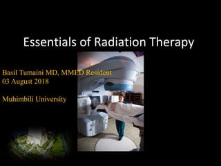
  • 1. Essentials of Radiation Therapy Basil Tumaini MD, MMED Resident 03 August 2018 Muhimbili University
  • 2. Outline Introduction Sources of ionizing radiation Basis for radiotherapy Clinical uses of RT Radiation oncology team Treatment process Delivery of radiotherapy Side effects and complications of RT Summary
  • 3. Introduction  Radiation therapy sometimes called:  radiotherapy  irradiation  Radiotherapy is the treatment of disease using penetrating beams of high energy waves or streams of particles called radiation (x-rays, γ-rays, electrons, protons, or neutrons)
  • 4. Sources of Ionizing Radiation  Photons  Gamma Rays Emitted from a nucleus of a radioactive atom  Cobalt treatment machine  Radioisotopes used in brachytherapy  X-rays  Generated by a linear accelerator when accelerated electrons hit a target  Particle Beams  Protons  Neutrons  Electrons Most external beam radiation treatments use photons generated by a linear accelerator. Source: Varian Medical Systems Inc. Cherry P, Duxbury A, editors. Practical radiotherapy: physics and equipment. John Wiley & Sons; 2009 Sep 8.
  • 5. Biologic Basis for Radiotherapy  Radiation therapy works by damaging the DNA of cells and destroys their ability to reproduce  Both normal and cancer cells can be affected by radiation, but cancer cells have generally impaired ability to repair this damage, leading to cell death  All tissues have a tolerance level, or maximum dose, beyond which irreparable damage may occur Steel GG, Adams GE, Horwich A. The biological basis of radiotherapy.1989
  • 6.  Radiation kills cells that are actively dividing.  It also causes damage to the surrounding tissue.  Radiation doesn’t kill cells instantly, it may take day to weeks depending on the cell  Skin, bone marrow, lining of intestines affected quickly  Nerve, breast, brain, and bone tissue show affected later
  • 7. Fractionation Fractionation, or dividing the total dose into small daily fractions over several weeks, takes advantage of differential repair abilities of normal and malignant tissues Fractionation spares normal tissue through repair and repopulation while increasing damage to tumor cells through redistribution and reoxygenation Williams MV, James ND, Summers ET, Barrett A, Ash DV, Audit Sub-Committee. National survey of radiotherapy fractionation practice in 2003. Clinical Oncology. 2006 Feb 1;18(1):3-14.
  • 8. Radiocurability and radiosensitivity  RADIOCURABILITY – refers to the eradication of tumour at the primary or regional site and reflects a direct effect of the irradiation - but this does not equate with patients cure from cancer  RADIOSENSITIVITY – is the measure of tumour radiation response, thus describing the degree and speed of regression during and immediately after radiotherapy Fertil B, Malaise EP. Inherent cellular radiosensitivity as a basic concept for human tumor radiotherapy. International Journal of Radiation Oncology• Biology• Physics. 1981 May 1;7(5):621-9. Gerweck LE, Zaidi ST, Zietman A. Multivariate determinants of radiocurability I: prediction of single fraction tumor control doses. International Journal of Radiation Oncology* Biology* Physics. 1994 Apr 30;29(1):57-66.
  • 9. Factors affecting radiosensitivity  Histologic type  High sensitivity: Malignant lymphoma, Seminoma  Moderate sensitivity: Epithelial tumour (Carcinoma)  Low sensitivity: Osteosarcoma, Malignant melanoma  Oxygen concentration in tumour tissue:  Radiosensitivity is low in the hypoxic state.  Cell cycle: Radiosensitivity is high in M phase and low in S phase  Cancer-related genes: p53, Bel-2, Fas, VEGF
  • 10. The Four R’s of Radiobiology Four major factors are believed to affect tissue’s response to fractionated radiation:  Repair of sublethal damage to cells caused by radiation between fractions  Repopulation or regrowth of cells between fractions  Redistribution of cells into radiosensitive phases of cell cycle  Reoxygenation of hypoxic cells to make them more sensitive to radiation Withers HR. The four R's of radiotherapy. InAdvances in radiation biology 1975 Jan 1 (Vol. 5, pp. 241-271). Elsevier.
  • 11. Clinical Uses for RT Therapeutic radiation serves two major functions  To cure cancer  Destroy tumors that have not spread  Kill residual microscopic disease left after surgery or chemotherapy  To reduce or palliate symptoms  Shrink tumors affecting quality of life, e.g., a lung tumor causing shortness of breath  Alleviate pain or neurologic symptoms by reducing the size of a tumor External beam radiation treatments are usually scheduled five days a week and continue for one to ten weeks
  • 12. RT in Multidisciplinary Care  Radiation therapy plays a major role in the management of many common cancers either alone or as an adjuvant therapy with surgery and chemotherapy  Sites commonly treated include breast, prostate, lung, colorectal, pancreas, esophagus, head and neck, brain, skin, gynecologic, lymphomas, bladder cancers and sarcomas  Radiation is also frequently used to treat brain and bone metastases as well as cord compression
  • 13. RT  PRIMARY  ADJUVANT  NEO-ADJUVANT  CONCURRENT  PALLIATIVE
  • 14. Palliative RT  Commonly used to relieve pain from bone cancers  ~ 50 percent of patients receive total relief from their pain  80 to 90 percent of patients will derive some relief  Other palliative uses:  Spinal cord compression  Vascular compression, e.g., superior vena cava syndrome  Bronchial obstruction  Bleeding from gastrointestinal or gynecologic tumors  Esophageal obstruction Radiation is effective therapy for relief of bone pain from cancer
  • 15. The Radiation Oncology Team  Radiation Oncologist  The doctor who prescribes and oversees the radiation therapy treatments  Medical Physicist  Ensures that treatment plans are properly tailored for each patient, and is responsible for the calibration and accuracy of treatment equipment  Dosimetrist  Works with the radiation oncologist and medical physicist to calculate the proper dose of radiation given to the tumor  Radiation Therapist  Administers the daily radiation under the doctor’s prescription and supervision  Radiation Oncology Nurse  Interacts with the patient and family at the time of consultation, throughout the treatment process and during follow-up care
  • 17. Referral Tissue diagnosis has been established Referring physician reviews potential treatment options with patient Treatment options may include radiation therapy, surgery, chemotherapy or a combination It is important for a referring physician to discuss all possible treatment options available to the patient
  • 18. Consultation Radiation oncologist determines whether radiation therapy is appropriate A treatment plan is developed Care is coordinated with other members of patient’s oncology team The radiation oncologist will discuss with the patient which type of radiation therapy treatment is best for their type of cancer
  • 19. Simulation Patient is set up in treatment position on a dedicated CT scanner  Immobilization devices may be created to assure patient comfort and daily reproducibility  Reference marks or “tattoos” may be placed on patient CT simulation images are often fused with PET or MRI scans for treatment planning Aird EG, Conway J. CT simulation for radiotherapy treatment planning. The British journal of radiology. 2002 Dec;75(900):937-49.
  • 20. Treatment Planning  Physician outlines the target and organs at risk  Sophisticated software is used to carefully derive an appropriate treatment plan • Computerized algorithms enable the treatment plan to spare as much healthy tissue as possible  Medical physicist checks the chart and dose calculations  Radiation oncologist reviews and approves final plan Radiation oncologists work with medical physicists and dosimetrists to create the optimal treatment plan for each individualized patient
  • 21. Treatment Planning • Indication for radiotherapy • Goal of radiation therapy • Planned treatment volume • Planned treatment technique • Planned treatment dose Radiation oncologists work with medical physicists and dosimetrists to create the optimal treatment plan for each individualized patient
  • 22. Safety and Quality Assurance Each radiation therapy treatment plan goes through many safety checks  The medical physicist checks the calibration of the linear accelerator on a regular basis to assure the correct dose is being delivered  The radiation oncologist, along with the dosimetrist and medical physicist go through a rigorous multi-step QA process to be sure the plan can be safely delivered  QA checks are done by the radiation therapist daily to ensure that each patient is receiving the treatment that was prescribed for them
  • 23. Delivery of RT External beam radiation therapy typically delivers radiation using a linear accelerator Internal radiation therapy, called brachytherapy, involves placing radioactive sources into or near the tumor The modern unit of radiation is the Gray (Gy), traditionally called the rad 1Gy = 100 centigray (cGy) 1cGy = 1 rad The type of treatment used will depend on the location, size and type of cancer.
  • 24. RT
  • 25. Types of External Beam RT Two-dimensional radiation therapy Three-dimensional conformal radiation therapy (3-D CRT) Intensity modulated radiation therapy (IMRT) Image Guided Radiation Therapy (IGRT) Intraoperative Radiation Therapy (IORT) Stereotactic Radiotherapy (SRS/SBRT) Particle Beam Therapy
  • 26. Clinical radiation generators  Kilovoltage Units  Van de Graaff Generator  Linear Accelerator (linacs)  Betatron  Microtron  Cyclotron  Machine Using Radionuclides (Radium-226, Cesium-137, Cobalt-60)  Heavy Particle Beams
  • 27. Three-Dimensional Conformal Radiation Therapy (3-D CRT) Uses CT, PET or MRI scans to create a 3-D picture of the tumor and surrounding anatomy Improved precision, decreased normal tissue damage Huq S, Mayles P, Besa C. Transition from 2-D radiotherapy to 3-D conformal and intensity modulated radiotherapy. IAEA-TECDOC; 2008.
  • 28. Intensity Modulated Radiation Therapy (IMRT) A highly sophisticated form of 3-D CRT allowing radiation to be shaped more exactly to fit the tumor  Radiation is broken into many “beamlets,” the intensity of each can be adjusted individually IMRT allows higher doses of radition to be delivered to the tumor while sparing more healthy surrounding tissue Huq S, Mayles P, Besa C. Transition from 2-D radiotherapy to 3-D conformal and intensity modulated radiotherapy. IAEA-TECDOC; 2008.
  • 29. Image Guidance For patients treated with 3-D or IMRT Physicians use frequent imaging of the tumor, bony anatomy or implanted fiducial markers for daily set-up accuracy Imaging performed using CT scans, high quality X-rays, MRI or ultrasound Motion of tumors can be tracked to maximize tumor coverage and minimize dose to normal tissues Fiducial markers in prostate visualized and aligned
  • 30. Stereotactic Radiosurgery (SRS) SRS is a specialized type of external beam radiation that uses focused radiation beams targeting a well- defined tumor o SRS relies on detailed imaging, 3-D treatment planning and complex immobilization for precise treatment set-up to deliver the dose with extreme accuracy o Used on the brain or spine o Typically delivered in a single treatment or fraction
  • 31. Stereotactic Body Radiotherapy (SBRT) SBRT refers to stereotactic radiation treatments in 1-5 fractions on specialized linear accelerators o Uses sophisticated imaging, treatment planning and immobilization techniques  Respiratory gating may be necessary for motions management, e.g., lung tumors o SBRT is used for a number of sites: spine, lung, liver, brain, adrenals, pancreas o Data maturing for sites such as prostate
  • 32. Proton Beam Therapy  Protons are charged particles that deposit most of their energy at a given depth, minimizing risk to tissues beyond that point  Allows for highly specific targeting of tumors located near critical structures  Increasingly available in the U.S.  Most commonly used in treatment of pediatric, CNS and intraocular malignancies  Data maturing for use in other tumor sites Proton Gantry Source: Mevion
  • 33. Types of Internal RT Intracavitary implants  Radioactive sources are placed in a cavity near the tumor (vagina, cervix, uterine) Interstitial implants  Sources placed directly into the tissue (breast, prostate) Intra-operative implants  Surface applicator is in direct contact with the surgical tumor bed
  • 34. Brachytherapy • Radioactive sources are implanted into the tumor or surrounding tissue 125I, 103Pd, 192Ir, 137Cs • Purpose is to deliver high doses of radiation to the desired target while minimizing the dose to surrounding normal tissues Radioactive seeds for a permanent prostate implant, an example of low-dose-rate brachytherapy.
  • 35. Brachytherapy Dose Rate Low-Dose-Rate (LDR) Radiation delivered over days and months Prostate, breast, head and neck, and gynecologic cancers may be treated with LDR brachytherapy High-Dose-Rate (HDR) High energy source delivers the dose in a matter of minutes rather than days Gynecologic, breast, head and neck, lung, skin and some prostate implants may use HDR brachytherapy LDR prostate implant
  • 36. Permanent vs. Temporary Implants  Permanent implants release small amounts of radiation over a period of several months  Examples include low-dose-rate prostate implants (“seeds”)  Patients receiving permanent implants may be minimally radioactive and should avoid close contact with children or pregnant women  Temporary implants are left in the body for several hours to several days  Patient may require hospitalization during the implant depending on the treatment site  Examples include low-dose-rate GYN implants and high-dose-rate prostate or breast implants
  • 37. Intraoperative Radiation Therapy (IORT) IORT delivers a concentrated dose of radiation therapy to a tumor bed during surgery Advantages Decrease volume of tissue in boost field Ability to exclude part or all of dose- limiting normal structures Increase the effective dose Multiple sites Pancreas, stomach, lung, esophagus, colorectal, sarcomas, pediatric tumors, bladder, kidney, gyn Several recent trials have shown efficacy for breast cancer
  • 38. Systemic RT Radiation can also be delivered by an injection.  Metastron (89Strontium), Quadramet(153Samarium) and Xofigo (223Radium) are radioactive isotopes absorbed primarily by cancer cells  Used for treating bone metastases  Radioactive isotopes may be attached to an antibody targeted at tumor cells  Zevalin, Bexxar for Lymphomas  Radioactive “beads” may be used to treat primary or metastatic liver cancer  Y90-Microspheres
  • 39. RT Side effects and complications  The delivery of external beam radiation treatments is painless and usually scheduled five days a week for one to ten weeks  The effects of radiation therapy are cumulative with most significant side effects occurring near the end of the treatment course.  Side effects usually resolve over the course of a few weeks  There is a slight risk that radiation may cause a secondary cancer many years after treatment, but the risk is outweighed by the potential for curative treatment with radiation therapy {Sabin Motwani will send us image of mild skin redness after RT in a treatment field}. Example of erythroderma after several weeks of radiotherapy with moist desquamation Source: sarahscancerjourney.blogspot.com
  • 40. Common Radiation SE Side effects during the treatment vary depending on site of the treatment and affect the tissues in radiation field:  Breast – swelling, skin redness  Abdomen – nausea, vomiting, diarrhea  Chest – cough, shortness of breath, esophogeal irritation  Head and neck – taste alterations, dry mouth, mucositis, skin redness  Brain – hair loss, scalp redness  Pelvis – diarrhea, cramping, urinary frequency, vaginal irritation  Prostate – impotence, urinary symptoms, diarrhea  Fatigue is often seen when large areas are irradiated Modern radiation therapy techniques have decreased these side effects significantly Unlike the systemic side effects from chemotherapy, radiation therapy usually only impacts the area that received radiation
  • 41. AE, risk factors, treatment Berkey FJ. Managing the adverse effects of radiation therapy. Am Fam Physician. 2010 Aug 15;82(4):381-8.
  • 42. AE, risk factors, treatment …
  • 43. AE, risk factors, treatment …
  • 44. Summary Radiation therapy is a well established modality for the treatment of numerous malignancies Radiation oncologists are specialists trained to treat cancer with a variety of forms of radiation Treatment delivery is safe, quick and painless
  • 45. Cancer immunotherapy Facilitator: Dr. Christina Malichewe Presenter: Basil Tumaini
  • 46. Introduction • Tumor immunotherapy requires the understanding of tumor immunology to facilitate:  understanding of the immunologic relationship between the host and the tumor  utilization of the immune response to tumors for purpose of diagnosis, prophylaxis and therapy Raval RR, Sharabi AB, Walker AJ, Drake CG, Sharma P. Tumor immunology and cancer immunotherapy: summary of the 2013 SITC primer. Journal for immunotherapy of cancer. 2014 Dec;2(1):14.
  • 47. How do cancer cells differ from normal?  Clonal in origin  Deregulated growth and lifespan  Altered tissue affinity  Resistance to control via apoptotic signals  Change in surface phenotype and markers  Structural and biochemical changes  Presence of tumour-specific antigens
  • 48. 48 Tumor antigen Classification of tumor antigen – Old classification;-classified into two categories based on their patterns of expression: – Tumor-specific antigens (TSA’s) – Tumor-associated antigens (TAA’s) Modern classification -based on their molecular structure and source Renkvist N, Castelli C, Robbins PF, Parmiani G. A listing of human tumor antigens recognized by T cells. Cancer Immunology, Immunotherapy. 2001 Mar 1;50(1):3-15.
  • 49. Old classification  Tumor-specific antigens (TSA’s) Antigens unique to cancerous cells and are not present on their normal counterpar  Bcr-abl (e.g. in CML)  CDK-4 / β-catenin (melanoma)  Tumor-associated antigens (TAA’s) Antigens on tumor cells that – are qualitatively not different from those found on normal cells – but are over expressed i.e present at significantly increased numbers on the cancer cell as products of cellular oncogenes
  • 50. – e.g. high levels of a growth factor receptor due to increased expression of neu oncogene products found in a number of human breast cells – And ras oncogene products is present on some human prostatic cancer cells Tumour associated antigens •MUC-1 (myeloma etc) •α-fetoprotein (many) •Her-2/neu (breast) •WT-1 (many) •myeloblastin (leukaemias) •Survivin (many)
  • 51. • old classification, is imperfect, because many antigens thought to be tumor specific turned out to be expressed by some normal cells as well.
  • 52. Modern classification of tumor antigens 7 categories • Differ in both factors that induce malignancy and immunochemical properties of tumor antigens • Important development in tumor immunology is identifying tumor antigens that were recognized by cytotoxic T lymphocytes (CTLs), because CTLs are the major immune defense mechanism against tumors.
  • 53. 1.Tumor Antigens Produced by Oncogenic Viruses • there are viruses associated with cancers. • these viruses produce proteins that are recognized as foreign by the immune system. • The most potent of these antigens are proteins produced by latent DNA viruses; examples in humans include HPV and EBV. • There is abundant evidence that CTLs recognize antigens of these viruses and that a competent immune system plays a role in surveillance against virus-induced tumors because of its ability to recognize and kill virus-infected cells.
  • 54. - vaccines against HPV antigens have been found effective in prevention of cervical cancers in young females -these antigens exhibit extensive immunologic cross reactivity – particular oncogenic virus induces expression of same antigens in a tumor regardless of tissue origin or animal species
  • 55. 2. Oncofetal Antigens. • Antigens present during embryonic and fetal development –but they are either absent or present at very low levels in normal adult tissue –Become expressed in tumors –Not immunogenic in the host –can be detected by antisera prepared against them
  • 56. • E.g. Carcinoembryonic antigen (CEA) found primarily in serum of patients with cancer of GIT esp cancer of colon –But non specific –Elevated levels also been in the patients with some types of lung cancer, pancreatic cancer breast and stomach cancer –Also elevated in patients with emphysema, ulcerative colitis and pancreatitis,alcoholics and heavy smokers
  • 57. • α fetoprotein (AFP), which is normally present in high concentrations in fetal and maternal serum but absent from normal individuals –Secreted by cells of a variety of cancers and is found particularly in patients with hepatomas and testicular teratocarcinomas • Occasionally, virally induced tumors may express oncofetal antigens, encoded by the host genome • they are not entirely tumor specific, they can serve as serum markers for cancer.
  • 58. • Association of oncodevelopmental antigens with a wide variety of tumor types strongly suggests that derepression of normal genes that are usually repressed in the normal adult individual in a concomitant malignancy
  • 59. 3.Products of other mutated genes • Because of the genetic instability of tumor cells, many different genes may be mutated in these cells, including genes whose products are not related to the transformed phenotype and have no known function • Products of these mutated genes are potential tumor antigens
  • 60. 3.Products of other mutated genes … • These antigens are extremely diverse because the carcinogens that induce the tumors may randomly mutagenize virtually any host gene and the class I MHC antigen-presenting pathway can display peptides from any mutated cytosolic protein in each tumor. • Mutated cellular proteins are found more frequently in chemical carcinogen- or radiation- induced tumors because chemical carcinogens and radiation mutagenize many cellular genes.
  • 61. 4.Products of Mutated Oncogenes and Tumor Suppressor Genes. • The products of altered(mutated) protooncogenes and tumor suppressor genes are synthesized in the cytoplasm of the tumor cells, and like any cytosolic protein, they may enter the class I MHC antigen processing pathway and be recognized by CD8+ T cells • In addition, these proteins may enter the class II MHC antigen- processing pathway in antigen-presenting cells that have phagocytized dead tumor cells, and thus be recognized by CD4+ T cells also
  • 62. 4. Products of Mutated Oncogenes and Tumor Suppressor Genes … • Because these altered proteins are not present in normal cells, they do not induce self-tolerance. Some cancer patients have circulating CD4+ and CD8+ T cells that can respond to the products of mutated oncogenes such as RAS, p53, and BCR-ABL proteins. • Because the mutant proteins are present only in tumors, their peptides are expressed only in tumor cells. • Since many tumors may carry the same mutation, such antigens are shared by different tumors. • Hence exhibit extensive immunologic cross reactivity
  • 63. 5.Over expressed or Aberrantly Expressed Cellular Proteins • Tumor antigens may be normal cellular proteins that are abnormally expressed in tumor cells and elicit immune responses. • some tumor antigens are structurally normal proteins that are produced at low levels in normal cells and overexpressed in tumor cells. • This proteins in normal cell are produced in such small amounts and in so few cells that it is not recognized by the immune system and fails to induce tolerance. • E.g One such antigen is tyrosinase, an enzyme involved in melanin biosynthesis that is expressed only in normal melanocytes and melanomas.
  • 64. 5.Over expressed or Aberrantly Expressed Cellular Proteins … • T cells from melanoma patients recognize peptides derived from tyrosinase, raising the possibility that tyrosinase vaccines may stimulate such responses to melanomas; clinical trials with these vaccines are ongoing. On face value it is surprising that these patients are able to respond to a normal self-antigen.
  • 65. 6. Altered Cell-Surface Glycolipids and Glycoproteins • tumors express higher than normal levels and/or abnormal forms of surface glycoproteins and glycolipids, which may be diagnostic markers and targets for therapy • These altered molecules include gangliosides, blood group antigens, and mucins
  • 66. 6. Altered Cell-Surface Glycolipids and Glycoproteins … • Many antibodies have been raised in animals that recognize the carbohydrate groups or peptide cores of these molecules. Although most of the epitopes recognized by these antibodies are not specifically expressed on tumors, they are present at higher levels on cancer cells than on normal cells.
  • 67. • This class of antigens is a target for cancer therapy with specific antibodies. • Eg. Among the glycolipids expressed at high levels in melanomas are the gangliosides GM2, GD2, and GD3. • E.g. of mucin expressed in carcinoma are CA-125 and CA-19-9, expressed on ovarian carcinomas, and MUC-1, expressed on breast carcinomas.
  • 68. 7. Cell Type-Specific Differentiation Antigens • Tumors express molecules that are normally present on the cells of origin. These antigens are called differentiation antigens because they are specific for particular lineages or differentiation stages of various cell types. • Their importance is as potential targets for immunotherapy and for identifying the tissue of origin of tumors
  • 69. 7. Cell Type-Specific Differentiation Antigens … • These differentiation antigens are typically normal self- antigens, and therefore they do not induce immune responses in tumor-bearing hosts. • For example, lymphomas may be diagnosed as B cell- derived tumors by the detection of surface markers characteristic of this lineage, such as CD10 and CD20. • Antibodies against these molecules are also used for tumor immunotherapy.
  • 70. Antitumor Effector Mechanisms • Cell-mediated immunity is the dominant anti- tumor mechanism in vivo • Although antibodies can be made against tumors, there is no evidence that they play a protective role under physiologic conditions
  • 71. Cellular mechanisms • Destruction by cytotoxic cells • Antibody dependent cell mediated cytotoxicity (ADCC) • Destruction by activated macrophages • Destruction by natural killer (NK) cells T Lymphocytes The T cell is the primary cell responsible for direct recognition and killing of tumor cells. T cells carry out immunologic surveillance, then proliferate and destroy newly transformed tumor cells after recognizing TAAs.
  • 72. Cytotoxic T cells • Cytotoxic T cells (CTLs) CD8+ T cells: attaching to class I MHC -peptide complex, they destroy cancer cells by perforating the membrane with enzymes or by triggering an apoptotic pathway.
  • 73. 73 MAC or B cell (APC) MHC 1 T cytotoxic cell Perforins, apoptotic signals Exogenous antigen T cytotoxi c memory cells T cytotoxic effector cells T Cytotoxic Cell Activity in Tumor Surveillance Cancer Cell T cytotoxic cell Endogenous antigen DIRECT PATHWAY INDIRECT PATHWAY
  • 74. Helper T cells • CD4+ T cells: reacting to class II MHC peptide complex, they secret cytokines. • cytotoxic T cell response (Th1 helper T cells) • antibody response (Th2 helper T cells)
  • 75. Natural killer cells (NK) • NK cells are lymphocytes that are capable of destroying tumor cells without prior sensitization; • they may provide the first line of defense against tumor cells. After activation with IL-2, NK cells can lyse a wide range of human tumors, including many that seem to be nonimmunogenic for T cells. • T cells and NK cells seem to provide complementary antitumor mechanisms. .
  • 76. • Tumors that fail to express MHC class I antigens cannot be recognized by T cells, but these tumors may trigger NK cells because the latter are inhibited by recognition of normal autologous class I MHC molecules • The triggering receptors on NK cells are extremely diverse and belong to several gene families. • NKG2D proteins expressed on NK cells and some T cells are important activating receptors. • They recognize stress-induced antigens that are expressed on tumor cells and cells that have incurred DNA damage and are at risk for neoplastic transformation.
  • 77. NATURAL KILLER CELL NK Target cell (infected or cancerous) Perforin and enzymes killer activating receptor Do not recognize tumor cell via antigen specific cell surface receptor, but rather through receptors that recognize loss of expression of MHC I molecules, therefore detect “missing self” common in cancer.
  • 78. 78 Tumor surveillance by NK Cells Tumor cells produce reactive oxygen species and stress induced ligands that can be recognized by NK cells
  • 79. Macrophages • Macrophages can kill specific tumor cells when activated by a combination of factors, including lymphokines (soluble factors produced by T cells) and interferon. • Activated macrophages exhibit cytotoxicity against tumor cells in vitro. • T cells, NK cells, and macrophages may collaborate in antitumor reactivity, because interferon-γ, a cytokine secreted by T cells and NK cells, is a potent activator of macrophages.
  • 80. • Activated macrophages may kill tumors by mechanisms similar to those used to kill microbes (e.g., production of reactive oxygen metabolites;) or by secretion of tumor necrosis factor (TNF). • tumor necrosis factor (TNF-) that has potent antitumor activity. • When TNF- is injected into tumor bearing animals, it has been found to induce hemorrhage and necrosis of the tumor
  • 81. Dendritic cells • Dendritic cells are dedicated antigen- presenting cells present in barrier tissues (eg, skin, lymph nodes). They play a central role in initiation of tumor-specific immune response. Palucka K, Banchereau J. Cancer immunotherapy via dendritic cells. Nature Reviews Cancer. 2012 Apr;12(4):265.
  • 82. Humoral Mechanisms • Although there is no evidence for the protective effects of anti-tumor antibodies against spontaneous tumors, administration of monoclonal antibodies against tumor cells can be therapeutically effective. • A monoclonal antibody against CD20, a B cell surface antigen, is widely used for treatment of certain non-Hodgkin lymphomas.
  • 83. Antibody - produced by B cells Direct attack: blocking growth factor receptors, arresting proliferation of tumor cells, or inducing apoptosis -- is not usually sufficient to completely protect the body Indirect attack: -- major protective efforts (1)ADCC(antibody-dependent cell mediated cytotoxicity ) -- recruiting cells that have cytotoxicity, such as monocytes and macrophages (2) CDC (complement dependent cytotoxicity) -- binding to receptor, initiating the complement system, 'complement cascade’, resulting in a membrane attack complex, causing cell lysis and death
  • 84. 84 MAC MHC II MHC I APC T helper cell T helper 2 cell IL-2 B Cell Eosinophil IL-4 IL-5 T helper Memor y cell T helper Effector cell IL-1 T cytotoxic cell T cytotoxi c memory cells T cytotoxic effector cells Perforins, apoptotic signals Interferon 1 Cancer Cell T cytotoxic cell Endogenous antigen Perforins, apoptotic signals Generally ineffective tumor surveillance, but some ADCC Tumor antigen or tumor cell SUMMARY
  • 85. Immunosurveillance • Theory formulated in 1957 by Burnet and Thomas, who proposed that lymphocytes act as sentinels in recognizing and eliminating continuously arising, nascent transformed cells. • An important host protection process that inhibits carcinogenesis and maintains regular cellular homeostasis
  • 86. Immunoediting • Immunoediting is a process by which a person is protected from cancer growth and the development of tumour immunogenicity by their immune system. • It has three main phases: – elimination – equilibrium and – escape • The elimination phase consists of the following four phases Dunn GP, Bruce AT, Ikeda H, Old LJ, Schreiber RD. Cancer immunoediting: from immunosurveillance to tumor escape. Nature immunology. 2002 Nov;3(11):991. Mittal D, Gubin MM, Schreiber RD, Smyth MJ. New insights into cancer immunoediting and its three component phases—elimination, equilibrium and escape. Current opinion in immunology. 2014 Apr 1;27:16- 25.
  • 87. Elimination: Phase 1 • The first phase of elimination involves the initiation of antitumor immune response. • Cells of the innate immune system recognise the presence of a growing tumor which has undergone stromal remodeling, causing local tissue damage This is followed by the induction of inflammatory signals which is essential for recruiting cells of the innate immune system. (eg. natural killer cells, natural killer T cells, macrophages and dendritic cells) to the tumor site. production of IFN-gamma
  • 88. Elimination: Phase 2 • In the second phase of elimination, newly synthesised IFN-gamma induces tumor death (to a limited amount) as well as promoting the production of chemokines CXCL10, CXCL9 and CXCL11. • These chemokines play an important role in promoting tumor death by blocking the formation of new blood vessels. • Tumor cell debris produced as a result of tumor death is then ingested by dendritic cells, followed by the migration of these dendritic cells to the draining lymph nodes. • The recruitment of more immune cells also occurs and is mediated by the chemokines .
  • 89. Elimination: Phase 3 • In the third phase, natural killer cells and macrophages transactivate one another via the reciprocal production of IFN-gamma and IL-12. • This again promotes more tumor killing by these cells via apoptosis and the production of reactive oxygen and nitrogen intermediates • In the draining lymph nodes, tumor-specific dendritic cells trigger the differentiation of Th1 cells which in turn facilitates the development of CD8+ T cells
  • 90. Elimination: Phase 4 • In the final phase of elimination, tumor- specific CD4+ and CD8+ T cells home to the tumor site and the cytolytic T lymphocytes then destroy the antigen- bearing tumor cells which remain at the site.
  • 91. Equilibrium and Escape • Tumor cell variants which have survived the elimination phase enter the equilibrium phase. • lymphocytes and IFN-gamma exert a selection pressure on tumor cells which are genetically unstable and rapidly mutating. • Tumor cells continue to grow and expand in an uncontrolled manner and may eventually lead to malignancies. • In the study of cancer immunoeditting, knockout mice have been used for experimentation since human testing is not possible.
  • 92. 92 Basic Tumor Immunosurveillance Smyth, M. J. et al. Nature Immunology 2, 293 - 299 (2001) 1) The presence of tumor cells and tumor antigens initiates the release of “danger” cytokines such as IFN and heat shock proteins (HSP). 2) These cause the activation and maturation of dendritic cells such that they present tumor antigens to CD8 and CD4 cells 3) subsequent T cytotoxic destruction of the tumor cells the occurs
  • 93. 93 Tumor Escape Mechanisms • Low immunogenicity • Antigen modulation • Lack of costimulation • Immune suppression by tumor cells or T regulatory cells • Antige n masking • Apoptosis of cytotoxic T cells
  • 94. Low immunogenicity • Selective outgrowth of antigen-negative variants: During tumor progression, strongly immunogenic subclones may be eliminated
  • 95. Antigen modulation • Loss or reduced expression of MHC molecules: Tumor cells may fail to express normal levels of HLA class I molecules, thereby escaping attack by cytotoxic T cells. Such cells, however, may trigger NK cells.
  • 96. Lack of costimulation • Lack of costimulation: It may be recalled that sensitization of T cells requires two signals, one by foreign peptide presented by MHC molecules and the other by costimulatory molecules although tumor cells may express peptide antigens with class I molecules, they often do not express costimulatory molecules. This not only prevents sensitization, but also may render T cells anergic or, worse, cause them to undergo apoptosis. •
  • 97. Immunosuppression • Many oncogenic agents (e.g., chemicals and ionizing radiation) suppress host immune responses. Tumors or tumor products may also be immunosuppressive. For example, TGF-β, secreted by many tumors, is a potent immunosuppressant. In some cases, the immune response induced by the tumor (e.g., activation of regulatory T cells) may itself inhibit tumor immunity.
  • 98. 98 1) Tumor cell production of immune suppressants such as TGF-, 2) T regulatory cell stimulation with production of immune suppressants such as TGF-  1 2 Avoidance of tumor surveillance through release of immune suppressants Mapara Journal of Clinical Oncology. 22(6):1136-51, 2004
  • 99. Antigen masking: • Antigen masking: The cell-surface antigens of tumors may be hidden, or masked, from the immune system by glycocalyx molecules, such as sialic acid-containing mucopolysaccharides. This may be a consequence of the fact that tumor cells often express more of these glycocalyx molecules than normal cells do.
  • 100. Apoptosis of cytotoxic T cells • Apoptosis of cytotoxic T cells: Some melanomas and hepatocellular carcinomas express Fas ligand. It has been postulated that these tumors kill Fas-expressing T lymphocytes that come in contact with them, thus eliminating tumor-specific T cells
  • 101. 101 Tumor cells induce apoptosis in T lymphocytes via FAS activation 1) Cancer cells express FAS ligand 2) Bind to FAS receptor on T lymphocytes leading to apoptosis
  • 102. 102 TUMOR ESCAPE MECHANISMS T regulatory cells Or kill them Or T regulatory cells
  • 103. Clinical Tumor Immunology • Immunodiagnosis • Immunoprophylaxis • Immunotherapy
  • 104. Immunodiagnosis May be performed to achieve two separate goals – Immunological detection of antigens specific to tumor cells – Assessment of hosts immune response to tumor
  • 105. Immunodiagnosis … Detection of tumor cells and their products by immunological means – Myeloma and Bence-Jones proteins e.g. plasma cell tumor – AFP e.g. Liver cancer – CEA e.g. GIT cancers – PSA – Immunological detection of other tumor cell markers e.g. enzymes and hormones – Detection of tumor specific antigens in the circulation or by immunoimaging
  • 106. Detection of anti tumor immune response • Anti tumor antibodies • Anti tumor cell mediated immunity
  • 107. Cancer Immunotherapy • Manipulation of Co-Stimulatory Signal • Enhancement of APC Activity • Cytokine Therapy • Monoclonal Antibodies • Cancer Vaccines
  • 108. 1863 1898 1957 1983 1985 1991, 4 2002 2009 2010 2011 2014 Description of immune infiltrates in tumors by Virchow Treatment of cancer with bacterial products (“Coley’s toxin”) Cancer immuno- surveillance hypothesis (Burnet, Thomas) 1976 Treatment of bladder cancer with BCG IL-2 therapy for cancer Adoptive cell therapy Discovery of human tumor antigens (Boon, others) Adoptive T cell therapy HPV vaccination in VIN FDA approval of sipuleucel-T (DC vaccine) in prostate cancer FDA approval of anti-CTLA4 (ipilumimab) for melanoma FDA approval of anti-PD1 for melanoma The history of cancer immunotherapy: from empirical approaches to rational, science-based therapies Mellman I, Coukos G, Dranoff G. Cancer immunotherapy comes of age. Nature; 2011 Dec 21;480:480. Available from: http://dx.doi.org/10.1038/nature10673
  • 109. Manipulation of Co-Stimulatory Signal • Tumor immunity can be enhanced by providing the co- stimulatory signal necessary for activation of CTL precursors (CTL-Ps) • Fig. 22.11a
  • 110. Manipulation of Co-Stimulatory Signal Cont. • Basis for Vaccine – Prevent metastasis after surgical removal or primary melanoma in human patients
  • 111. Enhancement of APC Activity • GM-CSF (Granulocyte-macrophage colony- stimulating factor) remember: CSFs are cytokines that induce the formation of distinct hematopoietic cell lines • Fig 22.11b
  • 112. Cytokine Therapy • Use of recombinant cytokines (singly or in combination) to augment an immune response against cancer – Via isolation and cloning of various cytokine genes such as: – IFN-α, β, and γ – Interleukin 1, 2, 4, 5, and 12 – GM-CSF and Tumor necrosis factor (TNF)
  • 113. Cytokine Therapy Cont. I. Interferons • Most clinical trials involve IFN-α • Has been shown to induce tumor regression in hematologic malignancies i.e. leukemias, lymphomas, melanomas and breast cancer • All types of IFN increase MHC I expression • IFN-γ also has also been shown to increase MHC II expressionon macrophages and increase activity of Tc cells, macrophages, and NKs
  • 114. Cytokine Therapy Cont. II. Tumor Necrosis Factors • Kills some tumor cells • Reduces proliferation of tumor cells without affecting normal cells How? • Hemorrhagic necrosis and regression, inhibits tumor induced vascularization (angio-genesis) by damaging vascular endothelium
  • 115. Cytokine Therapy Cont. III. In Vitro-Activited LAK & TIL cells A. Lymphocytes are activated against tumor antigens in vitro • Cultured with x-irradiated tumor cells in presence of IL-2 • Generated lymphokine activated killer cells (LAKs), which kill tumor cells without affecting normal cells
  • 116. In Vitro-Activated LAK and TIF cells Cont. B. Tumors contain lymphocytes that have infiltrated tumor and act in anti-tumor response • via biopsy, obtained cells and expanded population in vitro with • generated tumor-infiltrating lympho- cytes (TILs)
  • 117. Tumor immunoprophylaxis • Immunization against tumor itself requires that the tumor possess specific antigens and that these antigens cross react immunologically with any prepared vaccine • Efficacy of immunoprophylaxis for protection of humans and animals against spontaneous tumors has not been sufficiently evaluated
  • 118. 118 Tumor Vaccines • Killed tumor cells • Purified tumor antigens • DNA vaccines • Dendritic cell vaccines
  • 119. Pretherapy Week 4 Week 6 Injected lesion Clinical response of anti-PD1 refractory melanoma patient treated with anti-PD1 plus in situ CMP-001 Photos courtesy of M. Milhem
  • 120.
  • 121.
  • 122. Other references • Levinson WE. Review of Medical Microbiology and Immunology 15E. McGraw Hill Professional; 2018 Jun 22 • Kumar V, Cortran RS, Robbins SL. Robins basic Pathology. 7th Edison. Saunders. Philadelphia. 2003:960-9 • Raval RR, Sharabi AB, Walker AJ, Drake CG, Sharma P. Tumor immunology and cancer immunotherapy: summary of the 2013 SITC primer. Journal for immunotherapy of cancer. 2014 Dec;2(1):14

Hinweis der Redaktion

  1. An overview of topics included in the presentation.
  2. Photon=in physics, a corpuscle of energy or particle of light; a quantum of light or other electromagnetic radiation
  3. Radiation therapy has multiple sources to be used for delivery. Most radiation therapy treatments are delivered using photons which are either delivered with Gamma Rays (such as radioisotopes used in brachytherapy) and X-rays (generated by a linear accelerator) Additional sources include particle beams such as protons, neutrons and electrons
  4. Radiation therapy damages the DNA of cells – normal cancer cells are able to repair themselves but cancer cells have an impaired ability to repair the damage, unlike healthy cells.
  5. Umbra and penumbra
  6. Between each treatment, healthy cells are able to repair themselves and cancer cells slowly break down. As the cell life continues, the repeated treatments continue to kill the cancer cells as they become sensitive to the radiation.
  7. Radiation therapy typically has two primary uses: To cure cancer by destroying tumors that have not spread or to kill residual disease left after chemotherapy or surgery To reduce or palliate symptoms by shrinking tumors that affect quality of life or alleviate pain or neurologic symptoms by reducing the tumor size
  8. Radiation therapy treatment plans often include adjuvant therapies, such as surgery and/or chemotherapy, in order to to enhance its effectiveness and reduce the chance of the tumor recurring.
  9. Palliative radiation therapy is commonly used to relieve pain from bone cancers. It has been shown that 80 to 90 percent of patients derive some relief from this. Additional palliative uses include spinal cord compression, vascular compression, bronchial or esophageal obstruction or bleeding from GI for gyn tumors.
  10. The radiation therapy treatment team works closely to ensure that patients are receiving safe, quality treatment.
  11. Because the referring physician kicks off the treatment process, it is extremely important for the referring physician to have a basic understanding of potential treatment options available.
  12. Once a patient has been diagnosed with a cancer, it is important for their referring physician to discuss all of the possible treatment options available to the patient.
  13. A radiation oncologist will discuss with the patient if radiation is the appropriate treatment for their cancer. Care is then coordinated with other members of the patient’s oncology team, which could include a medical oncologist or surgical oncologist. It is important for the referring physician to be kept in the loop about the treatment plan.
  14. Simulation is an important step in the beginning of the treatment process. The patient is set up for treatment so that immobilization devices can be created, reference marks may be placed on the patient. Measurements and additional scans may be taken at this time.
  15. The radiation oncologist works very closely with the medical physicist and dosimetrist to create the optimal individualized plan for each patient. The radiation oncologist uses consensus atlases to outline the target and avoidance structures and then uses sophisticated software to create an appropriate treatment plan. Using nationally approved constraints, the treatment team creates a plan that will spare as much healthy tissue as possible.
  16. The radiation oncologist works very closely with the medical physicist and dosimetrist to create the optimal individualized plan for each patient. The radiation oncologist uses consensus atlases to outline the target and avoidance structures and then uses sophisticated software to create an appropriate treatment plan. Using nationally approved constraints, the treatment team creates a plan that will spare as much healthy tissue as possible.
  17. Safety is very important in the delivery of radiation therapy and each radiation therapy treatment team goes through many checks and balances to ensure that patients are receiving the correct dose to the correct area. The radiation oncologist works with the dosimetrist and medial physicist to go through rigourous quality assurance processes to be sure that the radiation therapy plan can be safely delivered. In addition, each facility works follows state and federal regulations to help with their checks and balances.
  18. Radiation therapy is delivered in two ways, External Beam Radiation Therapy and Internal Radiation Therapy. The absorbed dose is the quantity of radiation absorbed per unit mass of absorbing material. The RAD, or Radiation absorbed dose, is the traditional basic unit. The modern unit is the Gray (Gy), and is defined as 1 joule absorbed/kg. A dose may be prescribed as a Gy or cGy (centigray).
  19. Two dimensional radiation therapy uses X-rays to localize tumor. The other types of External Beam Radiation Therapy are much more
  20. There are specially designed linear accelerators with built in imaging capabilities that allow for simultaneous imaging to assure precise radiation delivery. Motions of tumors can be tracked to ensure that the maximum dose is delivered to the tumor while minimizing dose to the normal tissues.
  21. Stereotactic Radiotherapy, commonly referred to as SRS, was developed in 1949 to treat small targets in the brain that could not be surgically removed. It uses a 3-D coordinate system device and fiducial markers, along with specialized immobilization to deliver very precise treatment and is typically delivered in a single fraction.
  22. SRS is now more commonly referred to as stereotactic body radiotherapy or SBRT and is used to treat a number of sites in addition to the brain including spine, lung, liver, adrenal, pancreas and prostate) There are several external beam machines used to deliver SBRT such as linear accelerators, Cyberknife and Gamma Knife. SBRT is typically delivered in a few high-dose fractions.
  23. Proton therapy is increasingly becoming available in the United States with more centers opening each year. Protons have historically been used to treat CNS tumors and pediatric cancers.
  24. Low-dose-rate brachytherapy is delivered over the course of 48 to 120 hours and is typically used to treat prostate, breast, head and neck and gyn cancers. High-dose-rate brachytherapy is delivered in minutes rather than days and is typically used to treat certain types of gyn, breast, head and neck, lung and skin cancers. Some prostate cancers may be treated with high-dose-rate brachytherapy.
  25. Brachytherapy is delivered through permanent or temporary implants depending on the cancer and location. Permanent implants remain in the patient and eventually lose their radioactivity. An example of this is prostate seed implants. Temporary implants may require hospitalization and are left in the body for several hours to days and are then removed. An example of this is high-dose-rate breast implants.
  26. Systemic radiation therapy uses radiopharmaceuticals, given by injection or intravenously, that collect where the cancer is and deliver their radiation to kill the cancer cells. Some of the radioactive isotopes used are listed on this slide.
  27. While the delivery of external beam radiation therapy is painless, patients may experience side effects throughout their treatments and at the end. Side effects typically resolve within a few weeks of treatment completion. Side effects can be anything from skin redness in the are treated to GI discomfort. There is a risk of secondary cancers from treatment, but the benefits from the potential cure usually far outweighs the risk.
  28. Modern radiation therapy techniques have decreased these side effects significantly
  29. Radiation therapy is safe and effective and should be considered by patients and referring physicians as treatment for numerous cancers.
  30. Ctl – cytotoxic t lymphocyrte