SlideShare ist ein Scribd-Unternehmen logo
1 von 19
Nuclear magnetic resonance (NMR)
• Nuclear magnetic resonance (NMR) spectroscopy is a
physicochemical technique used to obtain structural
information about molecules.
• It is based on the physical phenomenon of magnetic
resonance that was first demonstrated by Isidor I. Rabi
in 1938. In the 1940s, two research groups
independently obtained the first successful
measurements of NMR in condensed matter.
• The two principal investigators of these groups, Felix
Bloch from Stanford University and Edward M. Purcell
from Harvard University, were jointly awarded with the
Nobel Prize in Physics in 1952 for their contributions to
the field of magnetic resonance
Since those early days, NMR spectroscopy progressed
concurrently with advances in many other fields, such as
mathematics, physics and informatics.
In the 1960s, the implementation of superconducting magnets
and computers to NMR equipment opened the door to a great
improvement in sensitivity and the possibility to design new
types of NMR experiments.
As a consequence, scientists have developed a myriad of novel
methodologies to study complex systems, such as membrane
proteins, metabolically complex samples, or even biological
tissues.
What is NMR
NMR spectroscopy is a physicochemical analysis technique that is based on
the interaction of an externally applied radiofrequency radiation with atomic
nuclei.
During this interaction there is a net exchange of energy which leads to a
change in an intrinsic property of the atomic nuclei called nuclear spin.
The nuclear spin is defined by a quantic number (I), which varies depending
on the considered isotope.
Only atomic nuclei with I ≠ 0 are detectable by NMR spectroscopy (NMR-
active nuclei, such as 1H, 2H, 13C and 15N).
These NMR-active nuclei behave as tiny magnets (magnetic dipoles), capable
of aligning with external magnetic fields (a process called magnetization).
The force of those tiny magnets is defined by a constant known as the
magnetogyric ratio (γ), whose value depends on the isotope.
Nuclear spins of some NMR-active nuclei are able to adopt two different
orientations when they align to an external magnetic field (B0).
One orientation corresponds to the lowest energy level of the nucleus
(parallel to the external magnetic field), and the other one is associated to
the highest energy level of the nucleus (antiparallel to the external magnetic
field) (Figure 1, left panel).
The difference between energy levels (ΔE) depends on the magnetic field and
the magnetogyric ratio (Eq. 1) and affects the sensitivity of the technique
(Figure 1, right panel).
Figure 1: Nuclear spin orientations of a sample aligned (parallel and
antiparallel) with the direction of an external magnetic field B0 (left panel).
Distribution of nuclear spin populations in the two possible energy levels in
nuclei with I = ½ (right panel).
• Magnetic resonance is achieved when nuclei are irradiated
with radiofrequency.
• This causes transitions between energy levels, which involves
changes in the orientation of nuclear spins.
• When atomic nuclei are under the effect of a magnetic field,
nuclear magnetic dipoles are not statically aligned with the
magnetic field B0, but rather move like a spinning top
(precession movement) around an axis parallel to the
direction of the field (Figure 2, left panel).
• The frequency of this precession movement, called Larmor
frequency (νL), is defined by the magnetogyric ratio and the
magnetic field:
As a consequence of this precession movement, the magnetic vector
(μ) associated with the nuclear magnetic dipoles possesses a
component parallel to the magnetic field (μz) and another component
perpendicular to the magnetic field (μxy), with this last one having a
net value of zero in the absence of external perturbations.
In an NMR experiment, it is not possible to measure the signal in
the z direction, as the magnetic field is too intense in that direction.
Therefore, it is necessary to transfer the magnetization of the z
component to the xy plane.
For this purpose, a magnetic pulse containing frequencies close to the
Larmor frequency is applied perpendicular to B0 to reach the
resonance of nuclear spins, which generates a non-zero μxy
component.
• After this pulse, a relaxation process takes place and the μxy
component gradually recovers its net value of zero (Figure 2,
right panel).
• As a consequence of this relaxation, energy is emitted as
radiofrequency, producing a characteristic signal called free
induction decay (FID) which is registered by the detector.
• This FID is subsequently transformed into a plot of intensities
versus frequencies known as an NMR spectrum.
Figure 2: Nuclear spin behavior under the influence of an external magnetic field
(left panel). Scheme of a basic NMR experiment in which the magnetization is
transferred to the xy plane upon the application of a magnetic pulse (right panel).
Figure 3: General design of an NMR spectrometer with
its principal components.
• NMR spectrometers consist of three main components: a
superconducting magnet, a probe and a complex electronic system
(console) controlled by a workstation (Figure 3).
• The magnet is responsible for the generation of a strong magnetic field
that aligns the nuclear spins of the atoms present in the sample.
• Nowadays, the magnets used in NMR spectroscopy are based on
superconducting materials, and thus, they require very low
temperatures to work (around 4 K).
• For this reason, NMR spectrometers contain a cooling system
composed of an inner jacket filled with liquid helium which is
refrigerated by an additional jacket filled with liquid nitrogen, and
many layers of thermal isolating materials
• The superconducting magnet surrounds a cylindrical chamber known as
the “probe”, which is a crucial component of the instrument. The sample
is introduced into the probe and thus placed under the influence of the
magnetic field.
• Additionally, the probe contains a series of magnetic coils that are also
located around the sample (Figure 4). These coils have multiple purposes.
• On one hand, they are used to irradiate the radiofrequency pulses and to
detect and collect the NMR signal emitted by the sample.
• On the other hand, they also enable control of the magnetic field
homogeneity and the application of pulse gradients that are used in some
NMR experiments.
Figure 4: Internal components of an NMR spectrometer,
including a detailed view of the probe.
• Finally, the electronic system of the spectrometer controls all the
experimental conditions and enables the set up and modification of
every parameter of the NMR experiment through the workstation.
• This system is also responsible for data acquisition and subsequent
mathematical transformation into an NMR spectrum.
• The spectrum contains a series of peaks of different intensities as a
function of a magnitude known as the chemical shift that is derived
from the Larmor frequency of the different atomic nuclei present in
the sample.
How to read an NMR spectrum and what it tells you
• The signal detected by an NMR spectrometer (the FID) must be
transformed prior to analysis.
• As the Larmor frequency is dependent upon the intensity of the
magnetic field, it varies from instrument to instrument.
• For this reason, a mathematical transformation is performed to
provide a relative magnitude called chemical shift (δ) (see Eq. 3).
• Unlike the Larmor frequency, this magnitude is independent of the
magnetic field and the value can be compared across instruments.
• Where νL is the observed Larmor frequency of a nucleus and
νL0 is the Larmor frequency of a reference nucleus, both in
Hz.
• By convention, chemical shift is always expressed in parts per
million (ppm).
• The zero value of the chemical shift scale is set using a
reference compound (such as tetramethylsilane (TMS) or
sodium trimethylsilylpropanesulfonate (DSS) for 1H).
Figure 5: 1H solution NMR spectrum of acetic acid. The signals correspond to
the two different 1H nuclei present in the molecule and their areas are
proportional to the number of nuclei contributing to the signal
• An NMR spectrum provides a lot of information about the
molecules present in the sample.
• First, chemical groups within a molecule can be identified
from chemical shift values.
• In the example provided in Figure 5, acetic acid (H3C-COOH)
has four protons so you could be forgiven for expecting to see
four signals in the spectrum.
• However, the three protons of the methyl group (CH3) are
magnetically equivalent and therefore have the same
chemical shift. This means that one signal comes from the
CH3 group and the other one, from the proton in the
carboxylic acid group (COOH).
• Secondly, in 1H-NMR spectra, signal area is proportional to
the number of atomic nuclei producing that signal (this does
not apply to 13C-NMR spectra).
• In this example, if the areas of both signals were to be
calculated, the most intense signal will be three times larger
than the other.
• This is in accordance with the fact that one signal represents
the three protons from the CH3 group (signal at δ = 2.0 ppm)
and the other one the proton from the COOH group (signal at
δ = 11.5 ppm)

Weitere ähnliche Inhalte

Ähnlich wie NMR ssc.pptx (20)

Prabhakar singh ii sem-paper v-nmr and epr spectroscopy
Prabhakar singh  ii sem-paper v-nmr and epr spectroscopyPrabhakar singh  ii sem-paper v-nmr and epr spectroscopy
Prabhakar singh ii sem-paper v-nmr and epr spectroscopy
 
Proton nmr by Bhushan Chavan
Proton nmr by Bhushan ChavanProton nmr by Bhushan Chavan
Proton nmr by Bhushan Chavan
 
NMR, principle, chemical shift , valu,13 C, application
NMR, principle, chemical shift , valu,13 C, applicationNMR, principle, chemical shift , valu,13 C, application
NMR, principle, chemical shift , valu,13 C, application
 
spectroscopy nmr for basic principles nmr
spectroscopy nmr for basic principles nmrspectroscopy nmr for basic principles nmr
spectroscopy nmr for basic principles nmr
 
7pr-NMR spectroscopy.pdf
7pr-NMR spectroscopy.pdf7pr-NMR spectroscopy.pdf
7pr-NMR spectroscopy.pdf
 
Nmr
NmrNmr
Nmr
 
NMR.pptx
NMR.pptxNMR.pptx
NMR.pptx
 
NUCLEAR MAGNETIC RESONANCE (NMR) SPECTROSCOPY
NUCLEAR MAGNETIC RESONANCE (NMR) SPECTROSCOPYNUCLEAR MAGNETIC RESONANCE (NMR) SPECTROSCOPY
NUCLEAR MAGNETIC RESONANCE (NMR) SPECTROSCOPY
 
NMR
NMRNMR
NMR
 
Nmr spectroscopy
Nmr spectroscopyNmr spectroscopy
Nmr spectroscopy
 
Nuclear magnetic resonanace spectroscopy
Nuclear magnetic resonanace spectroscopyNuclear magnetic resonanace spectroscopy
Nuclear magnetic resonanace spectroscopy
 
Nmr spectroscopy
Nmr spectroscopyNmr spectroscopy
Nmr spectroscopy
 
NMR, principle and instrumentation by kk sahu sir
NMR, principle and instrumentation by kk sahu sirNMR, principle and instrumentation by kk sahu sir
NMR, principle and instrumentation by kk sahu sir
 
Nuclear magnetic resonance (NMR)
Nuclear magnetic resonance (NMR)Nuclear magnetic resonance (NMR)
Nuclear magnetic resonance (NMR)
 
Nmr
NmrNmr
Nmr
 
NMR
NMRNMR
NMR
 
NMR
NMRNMR
NMR
 
NMR spectroscopy
NMR spectroscopyNMR spectroscopy
NMR spectroscopy
 
nmr2_pl.ppt
nmr2_pl.pptnmr2_pl.ppt
nmr2_pl.ppt
 
Nmr theory
Nmr theoryNmr theory
Nmr theory
 

Kürzlich hochgeladen

Recombinant DNA technology (Immunological screening)
Recombinant DNA technology (Immunological screening)Recombinant DNA technology (Immunological screening)
Recombinant DNA technology (Immunological screening)PraveenaKalaiselvan1
 
Pests of cotton_Sucking_Pests_Dr.UPR.pdf
Pests of cotton_Sucking_Pests_Dr.UPR.pdfPests of cotton_Sucking_Pests_Dr.UPR.pdf
Pests of cotton_Sucking_Pests_Dr.UPR.pdfPirithiRaju
 
❤Jammu Kashmir Call Girls 8617697112 Personal Whatsapp Number 💦✅.
❤Jammu Kashmir Call Girls 8617697112 Personal Whatsapp Number 💦✅.❤Jammu Kashmir Call Girls 8617697112 Personal Whatsapp Number 💦✅.
❤Jammu Kashmir Call Girls 8617697112 Personal Whatsapp Number 💦✅.Nitya salvi
 
VIRUSES structure and classification ppt by Dr.Prince C P
VIRUSES structure and classification ppt by Dr.Prince C PVIRUSES structure and classification ppt by Dr.Prince C P
VIRUSES structure and classification ppt by Dr.Prince C PPRINCE C P
 
GUIDELINES ON SIMILAR BIOLOGICS Regulatory Requirements for Marketing Authori...
GUIDELINES ON SIMILAR BIOLOGICS Regulatory Requirements for Marketing Authori...GUIDELINES ON SIMILAR BIOLOGICS Regulatory Requirements for Marketing Authori...
GUIDELINES ON SIMILAR BIOLOGICS Regulatory Requirements for Marketing Authori...Lokesh Kothari
 
Hubble Asteroid Hunter III. Physical properties of newly found asteroids
Hubble Asteroid Hunter III. Physical properties of newly found asteroidsHubble Asteroid Hunter III. Physical properties of newly found asteroids
Hubble Asteroid Hunter III. Physical properties of newly found asteroidsSérgio Sacani
 
9654467111 Call Girls In Raj Nagar Delhi Short 1500 Night 6000
9654467111 Call Girls In Raj Nagar Delhi Short 1500 Night 60009654467111 Call Girls In Raj Nagar Delhi Short 1500 Night 6000
9654467111 Call Girls In Raj Nagar Delhi Short 1500 Night 6000Sapana Sha
 
Bacterial Identification and Classifications
Bacterial Identification and ClassificationsBacterial Identification and Classifications
Bacterial Identification and ClassificationsAreesha Ahmad
 
GBSN - Microbiology (Unit 1)
GBSN - Microbiology (Unit 1)GBSN - Microbiology (Unit 1)
GBSN - Microbiology (Unit 1)Areesha Ahmad
 
TEST BANK For Radiologic Science for Technologists, 12th Edition by Stewart C...
TEST BANK For Radiologic Science for Technologists, 12th Edition by Stewart C...TEST BANK For Radiologic Science for Technologists, 12th Edition by Stewart C...
TEST BANK For Radiologic Science for Technologists, 12th Edition by Stewart C...ssifa0344
 
High Class Escorts in Hyderabad ₹7.5k Pick Up & Drop With Cash Payment 969456...
High Class Escorts in Hyderabad ₹7.5k Pick Up & Drop With Cash Payment 969456...High Class Escorts in Hyderabad ₹7.5k Pick Up & Drop With Cash Payment 969456...
High Class Escorts in Hyderabad ₹7.5k Pick Up & Drop With Cash Payment 969456...chandars293
 
Nanoparticles synthesis and characterization​ ​
Nanoparticles synthesis and characterization​  ​Nanoparticles synthesis and characterization​  ​
Nanoparticles synthesis and characterization​ ​kaibalyasahoo82800
 
Presentation Vikram Lander by Vedansh Gupta.pptx
Presentation Vikram Lander by Vedansh Gupta.pptxPresentation Vikram Lander by Vedansh Gupta.pptx
Presentation Vikram Lander by Vedansh Gupta.pptxgindu3009
 
GBSN - Microbiology (Unit 2)
GBSN - Microbiology (Unit 2)GBSN - Microbiology (Unit 2)
GBSN - Microbiology (Unit 2)Areesha Ahmad
 
GBSN - Biochemistry (Unit 1)
GBSN - Biochemistry (Unit 1)GBSN - Biochemistry (Unit 1)
GBSN - Biochemistry (Unit 1)Areesha Ahmad
 
Biological Classification BioHack (3).pdf
Biological Classification BioHack (3).pdfBiological Classification BioHack (3).pdf
Biological Classification BioHack (3).pdfmuntazimhurra
 
Biogenic Sulfur Gases as Biosignatures on Temperate Sub-Neptune Waterworlds
Biogenic Sulfur Gases as Biosignatures on Temperate Sub-Neptune WaterworldsBiogenic Sulfur Gases as Biosignatures on Temperate Sub-Neptune Waterworlds
Biogenic Sulfur Gases as Biosignatures on Temperate Sub-Neptune WaterworldsSérgio Sacani
 
SCIENCE-4-QUARTER4-WEEK-4-PPT-1 (1).pptx
SCIENCE-4-QUARTER4-WEEK-4-PPT-1 (1).pptxSCIENCE-4-QUARTER4-WEEK-4-PPT-1 (1).pptx
SCIENCE-4-QUARTER4-WEEK-4-PPT-1 (1).pptxRizalinePalanog2
 
Pulmonary drug delivery system M.pharm -2nd sem P'ceutics
Pulmonary drug delivery system M.pharm -2nd sem P'ceuticsPulmonary drug delivery system M.pharm -2nd sem P'ceutics
Pulmonary drug delivery system M.pharm -2nd sem P'ceuticssakshisoni2385
 
COST ESTIMATION FOR A RESEARCH PROJECT.pptx
COST ESTIMATION FOR A RESEARCH PROJECT.pptxCOST ESTIMATION FOR A RESEARCH PROJECT.pptx
COST ESTIMATION FOR A RESEARCH PROJECT.pptxFarihaAbdulRasheed
 

Kürzlich hochgeladen (20)

Recombinant DNA technology (Immunological screening)
Recombinant DNA technology (Immunological screening)Recombinant DNA technology (Immunological screening)
Recombinant DNA technology (Immunological screening)
 
Pests of cotton_Sucking_Pests_Dr.UPR.pdf
Pests of cotton_Sucking_Pests_Dr.UPR.pdfPests of cotton_Sucking_Pests_Dr.UPR.pdf
Pests of cotton_Sucking_Pests_Dr.UPR.pdf
 
❤Jammu Kashmir Call Girls 8617697112 Personal Whatsapp Number 💦✅.
❤Jammu Kashmir Call Girls 8617697112 Personal Whatsapp Number 💦✅.❤Jammu Kashmir Call Girls 8617697112 Personal Whatsapp Number 💦✅.
❤Jammu Kashmir Call Girls 8617697112 Personal Whatsapp Number 💦✅.
 
VIRUSES structure and classification ppt by Dr.Prince C P
VIRUSES structure and classification ppt by Dr.Prince C PVIRUSES structure and classification ppt by Dr.Prince C P
VIRUSES structure and classification ppt by Dr.Prince C P
 
GUIDELINES ON SIMILAR BIOLOGICS Regulatory Requirements for Marketing Authori...
GUIDELINES ON SIMILAR BIOLOGICS Regulatory Requirements for Marketing Authori...GUIDELINES ON SIMILAR BIOLOGICS Regulatory Requirements for Marketing Authori...
GUIDELINES ON SIMILAR BIOLOGICS Regulatory Requirements for Marketing Authori...
 
Hubble Asteroid Hunter III. Physical properties of newly found asteroids
Hubble Asteroid Hunter III. Physical properties of newly found asteroidsHubble Asteroid Hunter III. Physical properties of newly found asteroids
Hubble Asteroid Hunter III. Physical properties of newly found asteroids
 
9654467111 Call Girls In Raj Nagar Delhi Short 1500 Night 6000
9654467111 Call Girls In Raj Nagar Delhi Short 1500 Night 60009654467111 Call Girls In Raj Nagar Delhi Short 1500 Night 6000
9654467111 Call Girls In Raj Nagar Delhi Short 1500 Night 6000
 
Bacterial Identification and Classifications
Bacterial Identification and ClassificationsBacterial Identification and Classifications
Bacterial Identification and Classifications
 
GBSN - Microbiology (Unit 1)
GBSN - Microbiology (Unit 1)GBSN - Microbiology (Unit 1)
GBSN - Microbiology (Unit 1)
 
TEST BANK For Radiologic Science for Technologists, 12th Edition by Stewart C...
TEST BANK For Radiologic Science for Technologists, 12th Edition by Stewart C...TEST BANK For Radiologic Science for Technologists, 12th Edition by Stewart C...
TEST BANK For Radiologic Science for Technologists, 12th Edition by Stewart C...
 
High Class Escorts in Hyderabad ₹7.5k Pick Up & Drop With Cash Payment 969456...
High Class Escorts in Hyderabad ₹7.5k Pick Up & Drop With Cash Payment 969456...High Class Escorts in Hyderabad ₹7.5k Pick Up & Drop With Cash Payment 969456...
High Class Escorts in Hyderabad ₹7.5k Pick Up & Drop With Cash Payment 969456...
 
Nanoparticles synthesis and characterization​ ​
Nanoparticles synthesis and characterization​  ​Nanoparticles synthesis and characterization​  ​
Nanoparticles synthesis and characterization​ ​
 
Presentation Vikram Lander by Vedansh Gupta.pptx
Presentation Vikram Lander by Vedansh Gupta.pptxPresentation Vikram Lander by Vedansh Gupta.pptx
Presentation Vikram Lander by Vedansh Gupta.pptx
 
GBSN - Microbiology (Unit 2)
GBSN - Microbiology (Unit 2)GBSN - Microbiology (Unit 2)
GBSN - Microbiology (Unit 2)
 
GBSN - Biochemistry (Unit 1)
GBSN - Biochemistry (Unit 1)GBSN - Biochemistry (Unit 1)
GBSN - Biochemistry (Unit 1)
 
Biological Classification BioHack (3).pdf
Biological Classification BioHack (3).pdfBiological Classification BioHack (3).pdf
Biological Classification BioHack (3).pdf
 
Biogenic Sulfur Gases as Biosignatures on Temperate Sub-Neptune Waterworlds
Biogenic Sulfur Gases as Biosignatures on Temperate Sub-Neptune WaterworldsBiogenic Sulfur Gases as Biosignatures on Temperate Sub-Neptune Waterworlds
Biogenic Sulfur Gases as Biosignatures on Temperate Sub-Neptune Waterworlds
 
SCIENCE-4-QUARTER4-WEEK-4-PPT-1 (1).pptx
SCIENCE-4-QUARTER4-WEEK-4-PPT-1 (1).pptxSCIENCE-4-QUARTER4-WEEK-4-PPT-1 (1).pptx
SCIENCE-4-QUARTER4-WEEK-4-PPT-1 (1).pptx
 
Pulmonary drug delivery system M.pharm -2nd sem P'ceutics
Pulmonary drug delivery system M.pharm -2nd sem P'ceuticsPulmonary drug delivery system M.pharm -2nd sem P'ceutics
Pulmonary drug delivery system M.pharm -2nd sem P'ceutics
 
COST ESTIMATION FOR A RESEARCH PROJECT.pptx
COST ESTIMATION FOR A RESEARCH PROJECT.pptxCOST ESTIMATION FOR A RESEARCH PROJECT.pptx
COST ESTIMATION FOR A RESEARCH PROJECT.pptx
 

NMR ssc.pptx

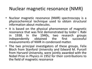
  • 1. Nuclear magnetic resonance (NMR) • Nuclear magnetic resonance (NMR) spectroscopy is a physicochemical technique used to obtain structural information about molecules. • It is based on the physical phenomenon of magnetic resonance that was first demonstrated by Isidor I. Rabi in 1938. In the 1940s, two research groups independently obtained the first successful measurements of NMR in condensed matter. • The two principal investigators of these groups, Felix Bloch from Stanford University and Edward M. Purcell from Harvard University, were jointly awarded with the Nobel Prize in Physics in 1952 for their contributions to the field of magnetic resonance
  • 2. Since those early days, NMR spectroscopy progressed concurrently with advances in many other fields, such as mathematics, physics and informatics. In the 1960s, the implementation of superconducting magnets and computers to NMR equipment opened the door to a great improvement in sensitivity and the possibility to design new types of NMR experiments. As a consequence, scientists have developed a myriad of novel methodologies to study complex systems, such as membrane proteins, metabolically complex samples, or even biological tissues.
  • 3. What is NMR NMR spectroscopy is a physicochemical analysis technique that is based on the interaction of an externally applied radiofrequency radiation with atomic nuclei. During this interaction there is a net exchange of energy which leads to a change in an intrinsic property of the atomic nuclei called nuclear spin. The nuclear spin is defined by a quantic number (I), which varies depending on the considered isotope. Only atomic nuclei with I ≠ 0 are detectable by NMR spectroscopy (NMR- active nuclei, such as 1H, 2H, 13C and 15N).
  • 4. These NMR-active nuclei behave as tiny magnets (magnetic dipoles), capable of aligning with external magnetic fields (a process called magnetization). The force of those tiny magnets is defined by a constant known as the magnetogyric ratio (γ), whose value depends on the isotope. Nuclear spins of some NMR-active nuclei are able to adopt two different orientations when they align to an external magnetic field (B0). One orientation corresponds to the lowest energy level of the nucleus (parallel to the external magnetic field), and the other one is associated to the highest energy level of the nucleus (antiparallel to the external magnetic field) (Figure 1, left panel). The difference between energy levels (ΔE) depends on the magnetic field and the magnetogyric ratio (Eq. 1) and affects the sensitivity of the technique (Figure 1, right panel).
  • 5. Figure 1: Nuclear spin orientations of a sample aligned (parallel and antiparallel) with the direction of an external magnetic field B0 (left panel). Distribution of nuclear spin populations in the two possible energy levels in nuclei with I = ½ (right panel).
  • 6. • Magnetic resonance is achieved when nuclei are irradiated with radiofrequency. • This causes transitions between energy levels, which involves changes in the orientation of nuclear spins. • When atomic nuclei are under the effect of a magnetic field, nuclear magnetic dipoles are not statically aligned with the magnetic field B0, but rather move like a spinning top (precession movement) around an axis parallel to the direction of the field (Figure 2, left panel). • The frequency of this precession movement, called Larmor frequency (νL), is defined by the magnetogyric ratio and the magnetic field:
  • 7. As a consequence of this precession movement, the magnetic vector (μ) associated with the nuclear magnetic dipoles possesses a component parallel to the magnetic field (μz) and another component perpendicular to the magnetic field (μxy), with this last one having a net value of zero in the absence of external perturbations. In an NMR experiment, it is not possible to measure the signal in the z direction, as the magnetic field is too intense in that direction. Therefore, it is necessary to transfer the magnetization of the z component to the xy plane. For this purpose, a magnetic pulse containing frequencies close to the Larmor frequency is applied perpendicular to B0 to reach the resonance of nuclear spins, which generates a non-zero μxy component.
  • 8. • After this pulse, a relaxation process takes place and the μxy component gradually recovers its net value of zero (Figure 2, right panel). • As a consequence of this relaxation, energy is emitted as radiofrequency, producing a characteristic signal called free induction decay (FID) which is registered by the detector. • This FID is subsequently transformed into a plot of intensities versus frequencies known as an NMR spectrum.
  • 9. Figure 2: Nuclear spin behavior under the influence of an external magnetic field (left panel). Scheme of a basic NMR experiment in which the magnetization is transferred to the xy plane upon the application of a magnetic pulse (right panel).
  • 10. Figure 3: General design of an NMR spectrometer with its principal components.
  • 11. • NMR spectrometers consist of three main components: a superconducting magnet, a probe and a complex electronic system (console) controlled by a workstation (Figure 3). • The magnet is responsible for the generation of a strong magnetic field that aligns the nuclear spins of the atoms present in the sample. • Nowadays, the magnets used in NMR spectroscopy are based on superconducting materials, and thus, they require very low temperatures to work (around 4 K). • For this reason, NMR spectrometers contain a cooling system composed of an inner jacket filled with liquid helium which is refrigerated by an additional jacket filled with liquid nitrogen, and many layers of thermal isolating materials
  • 12. • The superconducting magnet surrounds a cylindrical chamber known as the “probe”, which is a crucial component of the instrument. The sample is introduced into the probe and thus placed under the influence of the magnetic field. • Additionally, the probe contains a series of magnetic coils that are also located around the sample (Figure 4). These coils have multiple purposes. • On one hand, they are used to irradiate the radiofrequency pulses and to detect and collect the NMR signal emitted by the sample. • On the other hand, they also enable control of the magnetic field homogeneity and the application of pulse gradients that are used in some NMR experiments.
  • 13. Figure 4: Internal components of an NMR spectrometer, including a detailed view of the probe.
  • 14. • Finally, the electronic system of the spectrometer controls all the experimental conditions and enables the set up and modification of every parameter of the NMR experiment through the workstation. • This system is also responsible for data acquisition and subsequent mathematical transformation into an NMR spectrum. • The spectrum contains a series of peaks of different intensities as a function of a magnitude known as the chemical shift that is derived from the Larmor frequency of the different atomic nuclei present in the sample.
  • 15. How to read an NMR spectrum and what it tells you • The signal detected by an NMR spectrometer (the FID) must be transformed prior to analysis. • As the Larmor frequency is dependent upon the intensity of the magnetic field, it varies from instrument to instrument. • For this reason, a mathematical transformation is performed to provide a relative magnitude called chemical shift (δ) (see Eq. 3). • Unlike the Larmor frequency, this magnitude is independent of the magnetic field and the value can be compared across instruments.
  • 16. • Where νL is the observed Larmor frequency of a nucleus and νL0 is the Larmor frequency of a reference nucleus, both in Hz. • By convention, chemical shift is always expressed in parts per million (ppm). • The zero value of the chemical shift scale is set using a reference compound (such as tetramethylsilane (TMS) or sodium trimethylsilylpropanesulfonate (DSS) for 1H).
  • 17. Figure 5: 1H solution NMR spectrum of acetic acid. The signals correspond to the two different 1H nuclei present in the molecule and their areas are proportional to the number of nuclei contributing to the signal
  • 18. • An NMR spectrum provides a lot of information about the molecules present in the sample. • First, chemical groups within a molecule can be identified from chemical shift values. • In the example provided in Figure 5, acetic acid (H3C-COOH) has four protons so you could be forgiven for expecting to see four signals in the spectrum. • However, the three protons of the methyl group (CH3) are magnetically equivalent and therefore have the same chemical shift. This means that one signal comes from the CH3 group and the other one, from the proton in the carboxylic acid group (COOH).
  • 19. • Secondly, in 1H-NMR spectra, signal area is proportional to the number of atomic nuclei producing that signal (this does not apply to 13C-NMR spectra). • In this example, if the areas of both signals were to be calculated, the most intense signal will be three times larger than the other. • This is in accordance with the fact that one signal represents the three protons from the CH3 group (signal at δ = 2.0 ppm) and the other one the proton from the COOH group (signal at δ = 11.5 ppm)