2018 BDSRA Roine CLN3
•
1 gefällt mir•38 views

Decreased structural brain network integration in juvenile neuronal ceroid lipofuscinosis
Empfohlen

Empfohlen
Weitere ähnliche Inhalte
Was ist angesagt?
Was ist angesagt? (13)
Ähnlich wie 2018 BDSRA Roine CLN3
Ähnlich wie 2018 BDSRA Roine CLN3 (20)
Mehr von Batten Disease Support and Research Association
Mehr von Batten Disease Support and Research Association (20)
Kürzlich hochgeladen
Kürzlich hochgeladen (20)
2018 BDSRA Roine CLN3
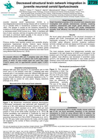
- 1. HELSINGIN YLIOPISTO HELSINGFORS UNIVERSITET UNIVERSITY OF HELSINKI LÄÄKETIETEELLINEN TIEDEKUNTA MEDICINSKA FAKULTETEN FACULTY OF MEDICINE Graph theoretical analyses Graph theoretical properties of the structural brain networks were investigated both globally (betweenness centrality, clustering coefficient, characteristic path length, global efficiency, small- worldness, degree, and strength) and locally (betweenness centrality, local efficiency, and strength) (Bullmore and Sporns, 2009) Results We found global abnormalities in the structural connectome of patients with CLN3. Characteristic path length (p=0.003) was significantly increased in CLN3. In addition, small-worldness (p=0.009), global efficiency (p=0.02), clustering coefficient (p=0.02), strength (p=0.02) and degree (p=0.02) were decreased, but did not endure a Bonferroni correction for multiple comparisons. The local analyses showed that betweenness centrality was decreased in the right thalamus (p=0.007) and increased in the posterior part of the right midcingulate cortex (p=0.003) in CLN3. In addition, local efficiency was decreased in seventeen nodes (p<0.01), and strength was decreased in nine nodes (p<0.01). However, none of the local results endured a Bonferroni correction for multiple comparisons. CLN3 Juvenile neuronal ceroid lipofuscinosis (CLN3) is a neurodegenerative lysosomal storage disease with an incidence of 7.0 in 100,000 births in Scandinavia (Uvebrant and Hagberg, 1997). The disease is caused by the recessive inheritance of mutations in CLN3 gene located on chromosome 16p12 encoding a membrane protein CLN3 (Lerner et al., 1995). It manifests with loss of vision, seizures, and loss of cognitive and motor functions, and leads to premature death typically in the second or third decade of life (Jalanko and Braulke, 2009). Previous MRI studies Previous studies have reported cerebral and cerebellar atrophy, progressive hippocampal atrophy, thalamic signal intensity alterations, and decreased white matter volume in the corona radiata (Autti et al., 2007; Tokola et al., 2014). In addition, global and widespread local microstructural alterations have been found using diffusion MRI (Roine et al., 2018). Subjects We acquired diffusion MRI and T1-weighted data with Philips Achieva 3T machine from 14 patients with CLN3 (aged 9.6±3.4 years), of which 12 were imaged again two years later (aged 11.4±3.2 years), and 14 age-matched controls (aged 11.2±2.3 years). Acquisition and preprocessing Diffusion MRI data were acquired in 32 orientations with b=1000 s/mm2 and a 2 mm isotropic voxel size. T1-weighted data were acquired with a 1 mm isotropic voxel size. Diffusion MRI data were corrected for subject motion (Leemans and Jones, 2009), eddy current induced distortions, and nonlinearly registered to the T1- weighted data to correct for echo planar imaging distortions (Irfanoglu et al., 2012). Conclusions Although conventional MRI is usually visually normal under the age of ten, we found decreased integration of the structural connectome in CLN3 by using diffusion MRI. In addition, the centrality of the thalamus was decreased, which is supported by the previous MRI findings related to thalamic signal intensity alterations. Decreased structural brain network integration in juvenile neuronal ceroid lipofuscinosis Roine U.1, Roine T.1,2, Tokola A.1, Balk M.1, Mannerkoski M.3, Åberg L.4, Lönnqvist T.5, Autti T.1 1HUS Medical Imaging Center, Radiology, University of Helsinki and Helsinki University Hospital, Finland, 2Turku Brain and Mind Center, University of Turku, Finland, 3Child Psychiatry, University of Helsinki and Helsinki University Hospital, Helsinki, Finland, 4Department of Psychiatry, University of Helsinki and Helsinki University Hospital, Finland, 5Department of Child Neurology, University of Helsinki and Helsinki University Hospital, Finland Figure 1. A) Whole-brain constrained spherical deconvolution (CSD)-based tractography was performed with ExploreDTI by using up to 4th order spherical harmonics (Tournier et al., 2007; Leemans et al., 2009; Jeurissen et al., 2011). CSD can track through regions with complex (e.g. crossing) fiber configurations, present in most of the white matter (Jeurissen et al., 2013). B) Cortical parcellation was performed in FreeSurfer by using the Destrieux atlas (Destrieux et al., 2009) combined with the subcortical structures from FSL’s FIRST (Patenaude et al., 2011). C) The 164 gray matter areas become the nodes, and the edges are weighted by the number of fibers connecting a pair of gray matter regions. 2739 A B C Reconstruction of structural brain networks Figure 2. The structural connectome in CLN3. The size of the nodes illustrates the size of the gray matter area, and the colour of the nodes shows whether there are differences in betweenness centrality. The size of the edges is proportional to the number of fiber tracts connecting the nodes. References Autti et al. (2007). Eur J Neurol, 14(4), 447-450.; Bullmore and Sporns (2009). Nat Rev Neurosci, 10(3), 186-198; Destrieux et al. (2009). NeuroImage, 47, S151; Irfanoglu et al. (2012). NeuroImage, 61(1), 275-288; Jalanko and Braulke (2009). Biochim Biophys Acta, 1793(4), 697-709; Jeurissen et al. (2011). Hum Brain Mapp, 32(3), 461-479; Jeurissen et al. (2013). Hum Brain Mapp, 34(11), 2747-2766; Leemans et al. (2009). In Proc Intl Soc Mag Reson Med 17, 3537; Leemans and Jones (2009). Magn Reson Med, 61(6), 1336-1349; Lerner et al. (1995). Cell, 82(6), 949-957; Patenaude et al. (2011). NeuroImage, 56(3), 907-922; Roine et al., (2018). Am J Neuroradiol, DOI:10.3174/ajnr.A5687; Tokola et al. (2014). Pediatr Neurol, 50(2), 158-163; Tournier et al. (2007). NeuroImage, 35(4), 1459–1472; Uvebrant and Hagberg (1997). Neuropediatrics, 28(1), 6-8. The structural connectome in CLN3 Acknowledgements This work was supported by the Noah’s Hope/Hope 4 Bridget (NH/H4B), Thisbe and Noah Scott Foundation, and the Batten Disease Support and Research Association, Emil Aaltonen Foundation, Pehr Oscar Klingendahl Fund, and Arvo and Lea Ylppö Foundation.