2018 BDSRA Hughes CLN6
•
1 gefällt mir•93 views

Secretion of proteins and extracellular vesicles in CLN6 Batten disease
Empfohlen

Empfohlen
Weitere ähnliche Inhalte
Was ist angesagt?
Was ist angesagt? (20)
Ähnlich wie 2018 BDSRA Hughes CLN6
Ähnlich wie 2018 BDSRA Hughes CLN6 (20)
Mehr von Batten Disease Support and Research Association
Mehr von Batten Disease Support and Research Association (20)
Kürzlich hochgeladen
Kürzlich hochgeladen (20)
2018 BDSRA Hughes CLN6
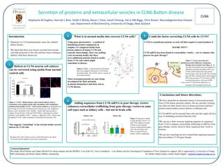
- 1. CLN6 Secretion of proteins and extracellular vesicles in CLN6 Batten disease Stephanie M Hughes, Hannah L Best, Hollie E Wicky, Alison J Clare, Isaiah Cheong, Harry AM Biggs, Chris Brown. Neurodegenerative Disease Lab, Department of Biochemistry, University of Otago, New Zealand Introduction • Mutations in CLN6 predominantly cause late infantile Batten disease. • We found that there were factors secreted from normal cells that protected CLN6 cells and have been working to identify what these factors are. Conclusions and future directions: •We have identified an abnormal release of lysosomal proteins from CLN6 mouse neuronal cultures. We are currently working out what role these factors have in disease processes and how normal control media corrects deficits in affected cells. •CLN6 mRNA can be secreted from cells and this might still be a way of mediating correction between cells. •We can use a short exosome targeting sequence or sequences from CLN6 to enhance extracellular trafficking of mRNAs from gene therapy vectors, however these sequences don’t work in neurons. •We are now searching for new extracellular targeting sequences that can enhance CLN6 gene therapy. Acknowledgements: We thank David Palmer and Nadia Mitchell for sheep samples and the BSDRA, Cure Kids NZ, Gray Foundation – Cure Batten and the Neurological Foundation of New Zealand for support. HB is supported by a University of Otago PhD scholarship and Roche Hanns Möhler scholarship. For further details please contact Steph Hughes stephanie.hughes@otago.ac.nz What is in normal media that corrects CLN6 cells? Using mass spectrometry – a method of identifying protein components in samples, we compared media from neuron cultures of mouse CLN6 and controls. Interestingly, there was nothing extra in normal media, but there were additional proteins found in media from CLN6 cells which might contribute to disease. These lysosomal proteins are now being investigated for their potential as disease biomarkers and their role in CLN6 disease. Could the factor correcting CLN6 cells be CLN6? CLN6 is a membrane protein, so each cell that requires it must produce it. MAYBE NOT?? CLN6 mRNA has been found in extracellular vesicles – can we enhance this process for gene therapy? Figure 3. Cross-correction of a membrane protein deficiency using gene therapy and exosomes (our idea). A. An exosome localisation tag (red) added to a membrane protein-encoding cDNA (e.g. CLN6) will be delivered to the mouse brain using a viral vector. B. The exosome tag will allow incorporation of the tagged RNA into exosomes. C. Exosomes will be secreted from donor cells and will D. cross-correct CLN6 protein deficiency in recipient cells throughout the brain. Defects in CLN6 neuron cell cultures can be corrected using media from normal control cells Figure 1. CLN6-/- Batten disease ovine neural cultures show a restoration in lysosomal activity after incubation with conditioned medium from healthy CLN6+/+ ovine neural cultures. a. Quantification of average LysoTracker Red fluorescent intensity per cell, in affected culture, after incubation with ‘healthy” media. b-d. Representative images of LysoTracker (red) and DAPI (blue), indicating lysosomal function, in b. healthy CLN6+/+ culture, c. affected CLN6-/- culture and d. affected CLN6-/- after incubation with media from healthy cells. ’ This suggests “something” in the normal media corrects defects in CLN6 cells. We thank Prof. Dave Palmer and Dr. Nadia Mitchell for the sheep CLN6 and control samples used in these experiments. Figure 2. Right: A representation of proteins that were uniquely detected in CLN6 mouse neuronal media. The red box shows a group of lysosomal proteins. Adding sequences from CLN6 mRNA to gene therapy vectors enhances extracellular trafficking from gene therapy vectors in some cell types such as kidney cells – but not in brain cells. Kidney cells Brain cells Figure 3. Sequences added to gene therapy vectors (.25 and UTR) enhance transfer of RNA to extracellular vesicles released from kidney cells, but not brain cells. Here we used GFP as a marker of RNA trafficking. 1 2 3 4