Haemolytic disease of new born
•Als PPTX, PDF herunterladen•
3 gefällt mir•854 views

Haemolytic disease of new born

Empfohlen
Weitere ähnliche Inhalte
Was ist angesagt?
Was ist angesagt? (20)
Ähnlich wie Haemolytic disease of new born
Ähnlich wie Haemolytic disease of new born (20)
Kürzlich hochgeladen
Kürzlich hochgeladen (20)
Haemolytic disease of new born
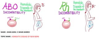
- 1. NAME : KHAN AZRA .Y. NISAR AHMED TOPIC NAME : HEMOLYTIC DISEASE OF NEW BORN
- 2. WHAT IS HEMOLYTIC DISEASE OF NEW BORN ? Hemolytic disease of the new born,also known as hemolytic disease of the fetus and new born ,HDN ,HDFN ,erythroblatosis foetalis. HDN usually occurs as a result of rhesus incompatibility. It develops when an Rh-negative mother carries an Rh-positive fetus. Normally, the fetal RBC are separated from the mother’s circulation by the layer of cells In the placenta called trophoblast .however ,during late pregnancy ,and especially during the process of child birth ,the fetal RBC may escape into mother’s circulation. Once these cells reach the mother’s circulation ,they are perceived as foreign and therefore provoke an antibody response. Maternal IgG antibodies provoked in this way can cross the placenta and reach the fetal circulation ,where they react with the fetal RBC and cause their destruction.
- 3. 0f HDN
- 4. What causes HDN in a newborn? All people have a blood type (A, B, AB, or O). Everyone also has an Rh factor (positive or negative). There can be a problem if a mother and baby have a different blood type and Rh factor. HDN happens most often when an Rh negative mother has a baby with an Rh positive father. If the baby's Rh factor is positive, like his or her father's, this can be an issue if the baby's red blood cells cross to the Rh negative mother. This often happens at birth when the placenta breaks away. But it may also happen any time the mother’s and baby's blood cells mix. This can occur during a miscarriage or fall. It may also happen during a prenatal test. These can include amniocentesis or chorionic villus sampling. These tests use a needle to take a sample of tissue. They may cause bleeding.
- 6. PATHOPHYSIOLOGY: • The three most common models in which a woman becomes sensitized toward (i.e., produces IgG antibodies against) a particular antigen are hemorrhage, blood transfusion, and ABO incompatibility. • Fetal-maternal hemorrhage, which is the movement of fetal blood cells across the placenta, can occur during abortion, ectopic pregnancy, childbirth, ruptures in the placenta during pregnancy (often caused by trauma), or medical procedures carried out during pregnancy that breach the uterine wall. In subsequent pregnancies, if there is a similar incompatibility in the fetus, thes antibodies are then able to cross the placenta into the fetal bloodstream to attach to the red blood cells and cause their destruction (hemolysis). This is a major cause of HDN, because 75% of pregnancies result in some contact between fetal and maternal blood, and 15-50% of pregnancies have hemorrhages with the potential for immune sensitization. • The woman may have received a therapeutic blood transfusion. ABO blood group system and the D antigen of the Rhesus (Rh) blood group system typing are routine prior to transfusion. • The third sensitization model can occur in women of blood type O. • Women of blood type O are more prone than women of types A and B to making IgG anti-A and anti-B antibodies, and these IgG antibodies are able to cross the placenta. For unknown reasons, the incidence of maternal antibodies against type A and B antigens of the IgG type that could potentially cause hemolytic disease of the newborn is greater than the observed incidence of "ABO disease." About 15% of pregnancies involve a type O mother and a type A or type B child; only 3% of these pregnancies result in hemolytic disease due to A/B/O incompatibility.
- 7. SIGNS AND SYMPTOMS: Signs of hemolytic disease of the newborn include a positive direct Coombs test (also called direct agglutination test), elevated cord bilirubin levels, and hemolytic anemia. It is possible for a newborn with this disease to have neutropenia and neonatal alloimmune thrombocytopenia as well. Hemolysis leads to elevated bilirubin levels. After delivery bilirubin is no longer cleared (via the placenta) from the neonate's blood and the symptoms of jaundice (yellowish skin and yellow discoloration of the whites of the eyes, or icterus) increase within 24 hours after birth. Like other forms of severe neonatal jaundice, there is the possibility of the neonate developing acute or chronic kernicterus, however the risk of kernicterus in HDN is higher because of the rapid and massive destruction of blood cells. Untreated profound anemia can cause high-output heart failure, with pallor, enlarged liver and/or spleen, generalized swelling,kernicterus, and respiratory distress. HDN can be the cause of hydrops fetalis, an often-severe form of prenatal heart failure that causes fetal edema.
- 8. DIAGNOSIS: • Blood test : Testing is done to look for for Rh positive antibodies Mother’s blood. • Ultrasound: This test can show enlarged organs or fluid buildup in baby. • Amniocentesis: This test is done to check the amount of bilirubin in the amniotic fluid. In this test, a needle is put into mother’s abdominal and uterine wall. It goes through to the amniotic sac. The needle takes a sample of amniotic fluid. Lily’s graph is use to evaluate the level of bile pigment in amniotic fluid to measure hemolysis in the fetous. • Percutaneous umbilical cord blood sampling: This test is also called fetal blood sampling. In this test, a blood sample is taken from baby’s umbilical cord. Blood is checked for antibodies, bilirubin, and anemia. This is done to check if baby needs an intrauterine blood transfusion. • Coombs test: The direct Coombs test detects antibodies that are stuck to the surface of the red blood cells. Since these antibodies sometimes destroy red blood cells, a person can be anemic and this test can help clarify the condition. The indirect Coombs detects antibodies that are floating freely in the blood. These antibiodies could act against certain red blood cells and the test can be done to diagnose reactions to a blood transfusion.
- 9. After birth testing. Hgb - the infant's hemoglobin should be tested from cord blood. Reticulocyte count - Reticulocytes are elevated when the infant is producing more red blood cells in response to anemia. A rise in the retic count can mean that an infant may not need additional transfusions. Low retic is observed in infants treated with IUT and in those with HDN from anti-Kell. Neutrophils - as neutropenia is one of the complications of HDN, the neutrophil count should be checked. Thrombocytes - as thrombocytopenia is one of the complications of HDN, the thrombocyte count should be checked. Bilirubin should be tested from cord blood. Ferritin - because most infants affected by HDN have iron overload, a ferritin must be run before giving the infant any additional iron. Newborn Screening Tests - Transfusion with donor blood during pregnancy or shortly after birth can affect the results of the Newborn Screening Tests. It is recommended to wait and retest 10–12 months after last transfusion. In some cases, DNA testing from saliva can be used to rule out certain conditions.
- 10. TYPES OF HDN: o Types of HDN are classified by the type of antigens involved. • The main types are: ABO, HDN, Rhesus HDN, Kell HDN, and other antibodies. • ABO hemolytic disease of the newborn can range from mild to severe, but generally it is a mild disease. It can be caused by anti-A and anti-B antibodies. • Rhesus D hemolytic disease of the newborn (often called Rh disease) is the most common form of severe HDN. Rhesus c hemolytic disease of the newborn can range from a mild to severe disease - is the third most common form of severe HDN. Rhesus e and rhesus C hemolytic disease of the newborn are rare. Combinations of antibodies, for example, anti-Rhc and anti-RhE occurring together can be especially severe. • Anti-Kell hemolytic disease of the newborn is most commonly caused by anti-K 1 antibodies, the second most common form of severe HDN. Over half of the cases of anti-K 1 related HDN are caused by multiple blood transfusions. Antibodies to the other Kell antigens are rare.
- 11. Spectrum of hemolytic disease of newborn Rh D- hemolytic disease, ABO- hemolytic disease and hemolytic disease due to alloantibodies other than anti D comprise the complete spectrum of HDFN. • Rh D hemolytic disease This condition is still commonly seen in many developing countries including India and it is likely that inadequate prenatal care or failure to administer Rh IG is responsible for this. Studies have shown that Rh (D) positive women are just as likely as D negative women to form alloantibodies.maternal anti D alloantibody and feto-maternal ABO incompatibility are the two major causes of HDFN. However, occasional cases of severe HDFN due to non ABO, non Rh D antigens do occur. There can be considerable variation in the clinical severity of Rh hemolytic disease though direct antiglobulin test (DAT) is usually positive. • ABO hemolytic disease With routine immunoprophylaxis with Rh IG, ABO incompatibility is now the single largest cause of HDFN in the western world. ABO hemolytic disease occurs almost exclusively in infants of blood group A or B born to O group mothers, because Ig G anti A, anti B, occur more commonly in group O than group A or B individuals. However, rare cases of ABO incompatibility have been reported in group A2 mothers with high titer anti B. Hemolytic disease of the newborn due to ABO incompatibility may also be seen in Rh (D) negative women.
- 12. • Hemolytic disease due to antibodies other than anti D, anti A and anti B The prevalence of non Rh D alloantibodies in pregnancy has been found to range from 0.15 to 1.1%.[14] In a study from Norway, about one third of the new cases of immunization to the Rh system were found in Rh (D) positive women and included anti C and anti E. scientist have described two cases of severe HDFN, born to two Rh D positive mothers, both detected postnatally. Though the mothers were under regular antenatal supervision, the maternal alloimmunization to Rh (c) antigen went undetected.At times, maternal alloimmunization could be to more than one antigen. scientists recently identified maternal anti C and anti E alloantibodies in a Rh (D) positive mother who delivered an infant with hemolytic disease of newborn (unpublished data). Failure of alloantibodies of some antigen systems, eg. Le, Lu, to cause HDFN may be due to paucity of antigen sites on fetal red cells or absorption of antibodies by fetal antigen in the placenta. When alloantibodies are detected in the antenatal antibody screening, the antibody must be identified, since the alloantibody specificity is a clue to its potential risk for hemolysis.
- 13. HOW HDN IS TREATED In NEW BORN: Treatment will depend on child’s symptoms, age, and general health. It will also depend on how severe the condition is. During pregnancy, treatment for HDN may include the following. . Interauterine blood transfusion:This test puts red blood cells into baby's circulation. In this test, a needle is placed through mother’s uterus. It goes into baby’s abdominal cavity to a vein in the umbilical cord. baby may need sedative medicine to keep him or her from moving. mother may need to have more than 1 transfusion. Intravenous immunoglobulin:IVIG is a solution made from blood plasma. It contains antibodies to help the baby's immune system. IVIG reduces your baby’s breakdown of red blood cells. It may also lower his or her bilirubin levels. Exchange transfusion:This test removes baby’s blood that has a high bilirubin level. It replaces it with fresh blood that has a normal bilirubin level. This raises baby’s red blood cell count. It also lowers his Or her bilirubin level.
- 14. What can be done to prevent hemolytic disease of the newborn? HDN can be prevented. Almost all women will have a blood test to learn their blood type early in pregnancy. If mother is Rh negative and have not been sensitized, mother get a medicine called Rh immunoglobulin (RhoGAM). This medicine can stop mother’s antibodies from reacting to baby’s Rh positive cells. Many women get RhoGAM around week 28 of pregnancy. If baby is Rh positive, mother get a second dose of medicine within 72 hours of giving birth. If baby is Rh negative,mother won’t need a second dose
- 16. Antibody specific information • Anti-D is the only preventable form of HDN. Since the 1968 introduction of Rho-D immunoglobulin, (Rhogam), which prevents the production of maternal Rho-D antibodies, the incidence of anti-D HDN has decreased dramatically. • Anti-C and anti-c can both show a negative DAT but still have a severely affected infant. An indirect Coombs must also be run. • Anti-M also recommends antigen testing to rule out the presence of HDN as the direct coombs can come back negative in a severely affected infant. • Anti-Kell can cause severe anemia regardless of titer. Anti-Kell suppresses the bone marrow,[18] by inhibiting the erythroid progenitor cells. • Kidd antigens are also present on the endothelial cells of the kidneys. • One study done by Moran et al., found that titers are not reliable for anti-E. Their most severe case of hemolytic disease of the newborn occurred with titers 1:2. Moran states that it would be unwise routinely to dismiss anti-E as being of little clinical consequence.
- 17. BIBLIOGRAPHY: Sameer Wagle,Muhammad Asalm(2018).Hemolytic disease of newborn.Medical Encyclopedia,[Accessed:Dec 28 2017] Hemolytic disease of new born[Internet].Available from://www.emedicine.Medscape.com Hemolytic disease of new born[Internet].Available from://www.medline.plus.gov{[Accessed:Dec 13 2017]