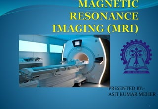
SlideShare ist ein Scribd-Unternehmen logo
1 von 41
PRESENTED BY:-
ASIT KUMAR MEHER
1
CONTENTS
 INTRODUCTION
 HISTORY
 MRI VS CT SCAN
 HOW MRI WORKS
 COMPONENTS OF MRI
 COMPUTER SYSTEM
 COOLING OF MAGNETS
 ADVANTAGES
 DISADVANTAGES
 SHAPE OF MRI MACHINE
2
Introduction
 MRI is a type of scan that uses strong magnetic fields
and radio waves to produce detailed images of inside of
the body.
 An MRI scanner is a large tube that contains powerful
magnets.You lie inside the tube during scan.
 MRI perhaps the best application of superconductivity
which directly affected the humanity across the globe.
3
Introduction
 Prof Peter Mansfield was awarded Nobel
prize in 2003 for his discovery in MRI with
Prof Paul C Lauterbur of USA.
 The concept of NMR imaging used in
present day MRI system was proposed
by Paul Lauterbur as early as 1973.
4
Notes
 MRI is perhaps the best application of superconductivity which directly affected the
humanity across the globe. It is a common tool with the radiologist in diagnostic
hospitals for imaging various soft tissue parts of the human body and for detecting
tumors. The concept of NMR imaging used in present day MRI systems was proposed
by Paul Lauterbur as early as 1973. MRI exploits the presence of vast amount of
hydrogen (protons) in a human body as the water content in a human body is said to
be about 80 %. When protons in the tissues of the body, aligned in a static magnetic
field (B0), are subjected to resonant RF excitation, they absorb energy. Proton relaxes
back and emits resonant signal which is a characteristic of the tissue. The signal is
picked-up by a receiver located inside the magnet bore and is used to construct the
image using Fourier transform. Since the NMR signal frequency is proportional to the
magnetic field the whole tissue can be mapped by assigning different values of the
proton frequency to different proton locations in the sample using well computed
field gradient. All MRIs use proton NMR for mapping proton density which is
different in different types of tissues. The images show contrast which helps in
identifying these tissues and the changes occurring in a sample tissue. MRI turns out
to be an ideal technique for soft tissue regions of the body such as brain, eyes and soft
tissue part of the head. Since bones have low density of protons they appear as dark
regions.
5
History
6
MRI VS CT SCAN
CT SCAN
• Uses X-rays for imaging.
• Exposure to ionizing radiation.
• Resolution problem.
• Injection of a contrast medium
(dye) can cause kidney.
• problems or result in allergic or
injection-site reactions in some
people.
• Less cost than MRI.
• Quick process and easily available.
ct-scan-vs-mri-scan-4-638.jpg
MRI
• Uses large external field, RF pulse
and 3 different gradient fields.
• MRI machines do not emit
ionizing radiation.
• Good resolution & 3-D
reconstruction.
• Gadolinium contrast is relatively
nontoxic.
• More cost.
• Lengthy process and non
availability.
ct-scan-vs-mri-scan-6-638.jpg
7
Notes
 Why we are using MRI instead of CT scan , here some
comparison between this two.
 CT scan uses x ray technology to produce image but MRI
uses large magnetic field to elicit image.
 Certain advantages of CT scan over MRI as it is less
expensive, easily available , quick process .
 But still we are going for MRI technology because it has no
ionization radiation so no harm to body, produce good
resolution 3D image and each n every inner injury can be
detected. So every one now preferring MRI although it has
high cost.
8
HOW MRI WORKS
 MRI exploits the presence of vast amount of hydrogen in a
human body as the water content in human body is said to
be about 80%.
 At the centre of each hydrogen atom is an even smaller
particle , called proton. Protons are like tiny magnets and
are very sensitive to magnetic fields and has magnetic spin.
 MRI utilizes this magnetic spin properties of protons of
hydrogen to elicit images.
 Then why our body can’t like magnets?
9
HOW MRI WORKS
10
•The protons i.e. hydrogen ions in a body
•are spinning in a haphazard fashion and
•cancel all the magnetism.
•That is our natural state.
•When there is large magnetic field acts
•on our body, protons in our body line up in
•same direction.
•In same way that magnet can pull the
needle of a compass.
Notes
 Human body is largely made of water molecules, which
consists of smaller particles i.e hydrogen and oxygen atoms.
 Protons lies at the centre of each atom, which is sensitive to
any magnetic fields and hence this proton serves as a
magnet. Normally water molecules in our body are
randomly arranged, but upon entering on the MRI scanner
first magnet causes body’s water molecules to align in one
direction and second magnet was then turned on and off in
a series of quick pulses, causing each hydrogen atom to
alter their alignment and quickly , switches back to their
original relaxed state, when switched off.
11
HOW MRI WORKS
12
COMPONETS OF MRI
13
1) Main magnet
(superconducting
magnet)
2)Gradient coils
3)RF coils(radiofrequency)
Schematic diagram of MRI scanner
Notes
 A superconducting magnet is the heart and most expensive part of an MRI
scanner.MRI magnets need high homogeneity and high temporal stability
similar to NMR spectrometers. However, the magnetic field requirement in
the present day MRI scanners for clinical use is limited to 3T only. Another
major difference with NMR magnet is that sample size is much larger.
 The main magnet is superconducting, cooled to LHe temperature and
mounted in an efficient cryostat with a horizontal bore to accommodate the
patient. Inside the main magnet is a set of gradient coils for changing the
field along the X, Y and Z directions required for imaging. Inside the gradient
coils are the RF coils producing the field B1 for rotating the spin by an angle
dictated by the pulse sequence. These coils also detect the signal emitted by
the spins inside the body. At the centre is a patient table which is computer
controlled.
 The magnet, the RF body coil and the gradient coil assembly represent the
three major subsystems that comprise the resonance module of the MR
scanner.
14
COMPONETS OF MRI
15
16
COMPONETS OF MRI
1.Superconducting magnet
 A superconducting magnet is the heart and most
expensive part of an MRI scanner.
 The magnetic field requirement in the present day MRI
scanners for clinical use is limited to 3T only.
 The main magnet is superconducting, cooled to LHe
temperature and mounted in an efficient cryostat with
a horizontal bore to accommodate the patient.
5195771_orig.gif
17
COMPONETS OF MRI
2.Gradient coils
 Gradient coils are used to produce deliberate variations
in the main magnetic field.
 There are usually three sets of gradient coils, one for
each direction.
 The variation in the magnetic field permits localization
of image slices as well as phase encoding and frequency
encoding.
 The set of gradient coils for the z axis are Helmholtz
pairs, and for the x and y axis paired saddle coils.
18
Notes
 It generates secondary magnetic field with in primary
magnetic field, they are located in bore of primary magnet.
They are arranged in opposition to each other to +ve and
–ve pulse.
 Gradient coils are set of magnetization coils, which cause of
variation in magnetic field. They must be able to cause
spatial variation along the direction of man magnetic field.
 They are along with RF pulse are responsible for slice and
voxel formation.
 Gradient is extra magnetic field which is added to the
magnetic field.
19
COMPONENTS OF MRI
2.Gradient coils
 X coil – create a varying
magnetic field from left to
right.
 Y coil- create a varying
magnetic field from top to
bottom.
 Z coil- create a varying
Magnetic field from head to toe.
20
21
COMPONETS OF MRI
3. RF Coils
 Same as Radio waves – high wavelength, low energy
electromagnetic waves.
 RF coils are the "antenna" of the MRI system
 That transmit the RF signal and receives the return signal.
 They are simply a loop of wire either circular or
rectangular.
 Inside the gradient coils are the RF coils producing the
field B for rotating the spin by an angle dictated by the
pulse sequence. These coils also detect the signal emitted
by the spins inside the body. At the centre is a patient table
which is computer controlled.
22
23
COMPONENTS OF MRI
3.RF Coils
Start RF pulses (Excitation- Protons jump to higher energy
state by absorbing radiation).
24
COMPONENTS OF MRI
3.RF coils
Stop RF pulses (Relaxation- Protons return to
their original state emitting radiation)
25
Notes
 RF used to transmit RF pulses receiving signals in MRI produce
best possible images. It can make magnetization of hydrogen
nuclei , turn it 90 degree away from magnetic field.
 Some low energy (parallel protons) flip to a high energy (anti
parallel) state decreasing longitudinal magnetization.
 Protons process in phase, at a result net magnetization vector
turns towards the transverse plane, i.e. right angles to the
primary magnetic field = transverse magnetization.
 Each proton is rotating around its axis 63,000,000 rotation per
second. The 63MHz rotation is in the frequency range called
Radio frequency.
 Rotation speed α magnetic field strength
26
COMPUTER SYSTEM
27
Notes
 Receives RF signal and performs analog to digital
conversion.
 Digital signal representing image of body part is stored in
temporary image space or case space. It store digital signal
during data acquisition, digital signal then sent to an image
processor were a mathematical formula called Fourier
transformation is applied to image of MRI scan is displayed
on a monitor.
28
COOLING OF MAGNETS
 MRI (magnetic resonant imaging) machines work by generating a very
large magnetic field using a super conducting magnet and many coils
of wires through which a current is passed. Maintaining a large
magnetic field needs a lot of energy, and this is accomplished using
superconductivity, which involves trying to reduce the resistance in
the wires to almost zero. This is done by bathing the wires in a
continuous supply of liquid helium at -269.1C.
 A typical MRI scanner uses 1,700 liters of liquid helium, which needs
to be topped up periodically.
 Recently small special purpose refrigerators have been proposed for
recondensation of evaporated helium, which together with a
cryocooler for the radiation shields give a complete closed refrigeration
system.
29
COOLING OF MAGNETS
309652071_orig.gif
Notes
 In this figure the cryostat has an outer vacuum case (OVC) made of metal, one thermal shield (usually at a
temperature of 40–50 K) and the helium vessel, housing the magnet assembly. Top left shows a typical
cryocooler in its vertical orientation, ready to fit into the cryocooler sleeve, as indicated.
 The liquid helium fill level to keep the magnet superconducting at 4 K is also shown. For a complete fill,
typically 1500–2000 l is used. Depending on the temperature gradient that may develop inside the magnet
(from bottom to top) and on the superconducting coil design, which defines coil stability, lower fill volumes
may be tolerable. The minimum allowable volume may also differ between the ramping process and the
subsequent persistent operation of the ramped magnet.
 Any advanced/alternative cryogenic concept for MRI applications needs to address all the following operating
modes:
 Energy saving pre-cooling of the magnet down to the
 operating temperature (usually done with liquid nitrogen or a recoverable liquid helium facility).
 Magnet ramp up to full field, preferably with captured boil-off helium gas during ramp.
 Normal operating condition (NOC) with extra heat loads (due to gradient heating) that reduce the cryogenic
margin, and ensure no helium loss (zero boil-off/recovery).
 Ramp down.
 Shipping ‘ride-through’ (from factory to MRI site), optimizing losses to minimize the cost.
 Cooldown to operating temperature or refill at the customer site, with high-efficiency transfer.
 Safe ramp up at the customer site.
Cryocooler technology is constantly progressing. Currently, the dual-stage cryocooler cools the thermal shield
thermally linked to its first stage. The second stage is connected to the recondenser which re-liquefies
escaping helium gas from the helium vessel.
31
COOLING OF MAGNETS
32
LASER COOLING SYSTEM(LCS)
• LCS is one of the recent technologies used to cool magnet in
MRI. The temperature of a laser system can determine its
lifetime, performance and safety.
• In laser cooling, atomic and molecular samples are cooled down
to nearly absolute zero through the interaction with one or more
laser fields.
• The basic principle of laser cooling is Doppler effect .
• The Doppler effect, or Doppler shift, is the change in wavelength
and frequency caused by the movement of an observer relative
to the source.
LCS
 In Doppler effect the frequency of light is tuned slightly below an
electronic transition in the atom. Because the light is detuned to lower
frequency, the atom will absorb more photons if they move towards
the light source. If light is applied from two opposite directions, the
atom will scatter more photons. If this process continuous, the speed
of the atom reduces and hence the kinetic energy also reduces. Which
reduces the temperature of the atom, and hence cooling of the atom is
achieved.
 As per Doppler cooling, if a stationary atom sees the laser neither red
shifted nor blue shifted, it does not absorb the photon. An atom
moving away from the laser sees that the laser is red shifted, then also
it does not absorb photon. If an atom is moving towards the laser and
sees that it is blue-shifted, the it absorbs the photon and thus the
speed of the atom will get reduced.
33
LCS
34
Notes
 In this proposed system four temperature sensors are fixed
on the four sides of the superconducting magnet. It can
predict the temperature level at the superconducting
magnet, and transmit it to the controller. So the controller
has to be designed for making the cooling effective. And we
have to place our model in controller so that it can provide
the corresponding wavelength of laser for the predicted
temperature .
35
ADVANTAGES OF MRI
 No ionizing radiation & no short/long-term effects
demonstrated.
 Variable thickness, any plane
 Better contrast resolution & tissue discrimination
 Various sequences to play with to characterize the
abnormal tissue.
 Many details without I.V contrast.
36
DISADVANTAGES
 Very expensive
 Dangerous for patients with metallic devices placed within
the body.
 Difficult to be performed on claustrophobic patients.( fear
of closed space)
 Movement during scanning may cause blurry images.
 RF transmitters can cause severe burns if mishandled.
 Not easily available
37
SHAPE OF MRI MACHINE
38
CLOSED MRI
OPEN MRI
COMPARISON
CLOSED MRI OPEN MRI
 High field typically 1.5T – 3T.
 High image quality
 Fast imaging
 Advanced application
 Increased patient anxiety.
 Claustrophobic patients
problems.
 High acoustic noise levels.
 Low field typically 0.2T –
0.4T
 Low image quality
 Slow imaging
 Limited application
 Less patient anxiety.
 Claustrophobic patients
handling.
 Lower acoustic noise levels.
39
SOME MRI IMAGES
40
41

Weitere ähnliche Inhalte

Was ist angesagt?

Ultrasound imaging
Ultrasound imagingUltrasound imaging
Ultrasound imaging
NIVETA SINGH
 

Was ist angesagt? (20)

Basics of CT Scan
Basics of CT ScanBasics of CT Scan
Basics of CT Scan
 
Basic principles of magnetic resonance imaging for beginner - Dr Sanjana Ravi...
Basic principles of magnetic resonance imaging for beginner - Dr Sanjana Ravi...Basic principles of magnetic resonance imaging for beginner - Dr Sanjana Ravi...
Basic principles of magnetic resonance imaging for beginner - Dr Sanjana Ravi...
 
Magnetic resonance imaging
Magnetic resonance imagingMagnetic resonance imaging
Magnetic resonance imaging
 
MRI
MRIMRI
MRI
 
Mri final ppt
Mri final pptMri final ppt
Mri final ppt
 
Computed tomography
Computed tomographyComputed tomography
Computed tomography
 
Computer Tomography (CT Scan)
Computer Tomography (CT Scan)Computer Tomography (CT Scan)
Computer Tomography (CT Scan)
 
Principles of MRI
Principles of MRIPrinciples of MRI
Principles of MRI
 
P.e.t
P.e.tP.e.t
P.e.t
 
MRI
MRIMRI
MRI
 
Magnetic resonance imaging (mri) by sajid
Magnetic resonance imaging (mri) by sajidMagnetic resonance imaging (mri) by sajid
Magnetic resonance imaging (mri) by sajid
 
Basic principle of ct and ct generations
Basic principle of ct and ct generationsBasic principle of ct and ct generations
Basic principle of ct and ct generations
 
Mri presentation main
Mri presentation mainMri presentation main
Mri presentation main
 
MRI
MRIMRI
MRI
 
Mri basic principle and sequences
Mri basic principle and sequencesMri basic principle and sequences
Mri basic principle and sequences
 
Magnetic resonance imaging
Magnetic resonance imagingMagnetic resonance imaging
Magnetic resonance imaging
 
Computed Tomography and Spiral Computed Tomography
Computed Tomography and Spiral Computed Tomography Computed Tomography and Spiral Computed Tomography
Computed Tomography and Spiral Computed Tomography
 
Spect technology
Spect technologySpect technology
Spect technology
 
Ultrasound imaging
Ultrasound imagingUltrasound imaging
Ultrasound imaging
 
Magnetic Resonance Imaging
Magnetic Resonance ImagingMagnetic Resonance Imaging
Magnetic Resonance Imaging
 

Ähnlich wie Magnetic resonance imaging (mri) asit meher ppt

Ähnlich wie Magnetic resonance imaging (mri) asit meher ppt (20)

MRI-2.pptx
MRI-2.pptxMRI-2.pptx
MRI-2.pptx
 
Magnetic resonance imaging
Magnetic resonance imaging Magnetic resonance imaging
Magnetic resonance imaging
 
MRI
MRIMRI
MRI
 
BASIC MRI.pptx
BASIC MRI.pptxBASIC MRI.pptx
BASIC MRI.pptx
 
MRI Cyrogenics
MRI CyrogenicsMRI Cyrogenics
MRI Cyrogenics
 
Aftab ahmad
Aftab ahmadAftab ahmad
Aftab ahmad
 
MAGNETIC RESONANCE IMAGING QUESTION AND ANSWER 3 MARKS.pdf
MAGNETIC RESONANCE IMAGING QUESTION AND ANSWER 3 MARKS.pdfMAGNETIC RESONANCE IMAGING QUESTION AND ANSWER 3 MARKS.pdf
MAGNETIC RESONANCE IMAGING QUESTION AND ANSWER 3 MARKS.pdf
 
lec 2a- MRI.ppt
lec 2a- MRI.pptlec 2a- MRI.ppt
lec 2a- MRI.ppt
 
MRI EQUIPMENTS.pptx
MRI EQUIPMENTS.pptxMRI EQUIPMENTS.pptx
MRI EQUIPMENTS.pptx
 
Mri equipments
Mri equipmentsMri equipments
Mri equipments
 
MRI of Brain: Basics
MRI of Brain: BasicsMRI of Brain: Basics
MRI of Brain: Basics
 
Shahid beheshti medical science unversity
Shahid beheshti medical science unversityShahid beheshti medical science unversity
Shahid beheshti medical science unversity
 
Magnetic Resonance Imaging
Magnetic Resonance ImagingMagnetic Resonance Imaging
Magnetic Resonance Imaging
 
Mripresenation
MripresenationMripresenation
Mripresenation
 
MRI - magnetic Imaging resonance
MRI - magnetic Imaging resonanceMRI - magnetic Imaging resonance
MRI - magnetic Imaging resonance
 
Magnetic Resonance Imaging
Magnetic Resonance ImagingMagnetic Resonance Imaging
Magnetic Resonance Imaging
 
MRI
MRI MRI
MRI
 
mri-170827044641.pdf
mri-170827044641.pdfmri-170827044641.pdf
mri-170827044641.pdf
 
1_MRI.pptx
1_MRI.pptx1_MRI.pptx
1_MRI.pptx
 
What is mri
What is mriWhat is mri
What is mri
 

Kürzlich hochgeladen

+971581248768>> SAFE AND ORIGINAL ABORTION PILLS FOR SALE IN DUBAI AND ABUDHA...
+971581248768>> SAFE AND ORIGINAL ABORTION PILLS FOR SALE IN DUBAI AND ABUDHA...+971581248768>> SAFE AND ORIGINAL ABORTION PILLS FOR SALE IN DUBAI AND ABUDHA...
+971581248768>> SAFE AND ORIGINAL ABORTION PILLS FOR SALE IN DUBAI AND ABUDHA...
?#DUbAI#??##{{(☎️+971_581248768%)**%*]'#abortion pills for sale in dubai@
 
Artificial Intelligence: Facts and Myths
Artificial Intelligence: Facts and MythsArtificial Intelligence: Facts and Myths
Artificial Intelligence: Facts and Myths
Joaquim Jorge
 

Kürzlich hochgeladen (20)

Apidays New York 2024 - Scaling API-first by Ian Reasor and Radu Cotescu, Adobe
Apidays New York 2024 - Scaling API-first by Ian Reasor and Radu Cotescu, AdobeApidays New York 2024 - Scaling API-first by Ian Reasor and Radu Cotescu, Adobe
Apidays New York 2024 - Scaling API-first by Ian Reasor and Radu Cotescu, Adobe
 
Advantages of Hiring UIUX Design Service Providers for Your Business
Advantages of Hiring UIUX Design Service Providers for Your BusinessAdvantages of Hiring UIUX Design Service Providers for Your Business
Advantages of Hiring UIUX Design Service Providers for Your Business
 
The 7 Things I Know About Cyber Security After 25 Years | April 2024
The 7 Things I Know About Cyber Security After 25 Years | April 2024The 7 Things I Know About Cyber Security After 25 Years | April 2024
The 7 Things I Know About Cyber Security After 25 Years | April 2024
 
🐬 The future of MySQL is Postgres 🐘
🐬  The future of MySQL is Postgres   🐘🐬  The future of MySQL is Postgres   🐘
🐬 The future of MySQL is Postgres 🐘
 
04-2024-HHUG-Sales-and-Marketing-Alignment.pptx
04-2024-HHUG-Sales-and-Marketing-Alignment.pptx04-2024-HHUG-Sales-and-Marketing-Alignment.pptx
04-2024-HHUG-Sales-and-Marketing-Alignment.pptx
 
Partners Life - Insurer Innovation Award 2024
Partners Life - Insurer Innovation Award 2024Partners Life - Insurer Innovation Award 2024
Partners Life - Insurer Innovation Award 2024
 
+971581248768>> SAFE AND ORIGINAL ABORTION PILLS FOR SALE IN DUBAI AND ABUDHA...
+971581248768>> SAFE AND ORIGINAL ABORTION PILLS FOR SALE IN DUBAI AND ABUDHA...+971581248768>> SAFE AND ORIGINAL ABORTION PILLS FOR SALE IN DUBAI AND ABUDHA...
+971581248768>> SAFE AND ORIGINAL ABORTION PILLS FOR SALE IN DUBAI AND ABUDHA...
 
Axa Assurance Maroc - Insurer Innovation Award 2024
Axa Assurance Maroc - Insurer Innovation Award 2024Axa Assurance Maroc - Insurer Innovation Award 2024
Axa Assurance Maroc - Insurer Innovation Award 2024
 
Mastering MySQL Database Architecture: Deep Dive into MySQL Shell and MySQL R...
Mastering MySQL Database Architecture: Deep Dive into MySQL Shell and MySQL R...Mastering MySQL Database Architecture: Deep Dive into MySQL Shell and MySQL R...
Mastering MySQL Database Architecture: Deep Dive into MySQL Shell and MySQL R...
 
TrustArc Webinar - Stay Ahead of US State Data Privacy Law Developments
TrustArc Webinar - Stay Ahead of US State Data Privacy Law DevelopmentsTrustArc Webinar - Stay Ahead of US State Data Privacy Law Developments
TrustArc Webinar - Stay Ahead of US State Data Privacy Law Developments
 
Understanding Discord NSFW Servers A Guide for Responsible Users.pdf
Understanding Discord NSFW Servers A Guide for Responsible Users.pdfUnderstanding Discord NSFW Servers A Guide for Responsible Users.pdf
Understanding Discord NSFW Servers A Guide for Responsible Users.pdf
 
Automating Google Workspace (GWS) & more with Apps Script
Automating Google Workspace (GWS) & more with Apps ScriptAutomating Google Workspace (GWS) & more with Apps Script
Automating Google Workspace (GWS) & more with Apps Script
 
Tech Trends Report 2024 Future Today Institute.pdf
Tech Trends Report 2024 Future Today Institute.pdfTech Trends Report 2024 Future Today Institute.pdf
Tech Trends Report 2024 Future Today Institute.pdf
 
Strategies for Unlocking Knowledge Management in Microsoft 365 in the Copilot...
Strategies for Unlocking Knowledge Management in Microsoft 365 in the Copilot...Strategies for Unlocking Knowledge Management in Microsoft 365 in the Copilot...
Strategies for Unlocking Knowledge Management in Microsoft 365 in the Copilot...
 
What Are The Drone Anti-jamming Systems Technology?
What Are The Drone Anti-jamming Systems Technology?What Are The Drone Anti-jamming Systems Technology?
What Are The Drone Anti-jamming Systems Technology?
 
Strategies for Landing an Oracle DBA Job as a Fresher
Strategies for Landing an Oracle DBA Job as a FresherStrategies for Landing an Oracle DBA Job as a Fresher
Strategies for Landing an Oracle DBA Job as a Fresher
 
Developing An App To Navigate The Roads of Brazil
Developing An App To Navigate The Roads of BrazilDeveloping An App To Navigate The Roads of Brazil
Developing An App To Navigate The Roads of Brazil
 
Apidays New York 2024 - The value of a flexible API Management solution for O...
Apidays New York 2024 - The value of a flexible API Management solution for O...Apidays New York 2024 - The value of a flexible API Management solution for O...
Apidays New York 2024 - The value of a flexible API Management solution for O...
 
AWS Community Day CPH - Three problems of Terraform
AWS Community Day CPH - Three problems of TerraformAWS Community Day CPH - Three problems of Terraform
AWS Community Day CPH - Three problems of Terraform
 
Artificial Intelligence: Facts and Myths
Artificial Intelligence: Facts and MythsArtificial Intelligence: Facts and Myths
Artificial Intelligence: Facts and Myths
 

Magnetic resonance imaging (mri) asit meher ppt

  • 2. CONTENTS  INTRODUCTION  HISTORY  MRI VS CT SCAN  HOW MRI WORKS  COMPONENTS OF MRI  COMPUTER SYSTEM  COOLING OF MAGNETS  ADVANTAGES  DISADVANTAGES  SHAPE OF MRI MACHINE 2
  • 3. Introduction  MRI is a type of scan that uses strong magnetic fields and radio waves to produce detailed images of inside of the body.  An MRI scanner is a large tube that contains powerful magnets.You lie inside the tube during scan.  MRI perhaps the best application of superconductivity which directly affected the humanity across the globe. 3
  • 4. Introduction  Prof Peter Mansfield was awarded Nobel prize in 2003 for his discovery in MRI with Prof Paul C Lauterbur of USA.  The concept of NMR imaging used in present day MRI system was proposed by Paul Lauterbur as early as 1973. 4
  • 5. Notes  MRI is perhaps the best application of superconductivity which directly affected the humanity across the globe. It is a common tool with the radiologist in diagnostic hospitals for imaging various soft tissue parts of the human body and for detecting tumors. The concept of NMR imaging used in present day MRI systems was proposed by Paul Lauterbur as early as 1973. MRI exploits the presence of vast amount of hydrogen (protons) in a human body as the water content in a human body is said to be about 80 %. When protons in the tissues of the body, aligned in a static magnetic field (B0), are subjected to resonant RF excitation, they absorb energy. Proton relaxes back and emits resonant signal which is a characteristic of the tissue. The signal is picked-up by a receiver located inside the magnet bore and is used to construct the image using Fourier transform. Since the NMR signal frequency is proportional to the magnetic field the whole tissue can be mapped by assigning different values of the proton frequency to different proton locations in the sample using well computed field gradient. All MRIs use proton NMR for mapping proton density which is different in different types of tissues. The images show contrast which helps in identifying these tissues and the changes occurring in a sample tissue. MRI turns out to be an ideal technique for soft tissue regions of the body such as brain, eyes and soft tissue part of the head. Since bones have low density of protons they appear as dark regions. 5
  • 7. MRI VS CT SCAN CT SCAN • Uses X-rays for imaging. • Exposure to ionizing radiation. • Resolution problem. • Injection of a contrast medium (dye) can cause kidney. • problems or result in allergic or injection-site reactions in some people. • Less cost than MRI. • Quick process and easily available. ct-scan-vs-mri-scan-4-638.jpg MRI • Uses large external field, RF pulse and 3 different gradient fields. • MRI machines do not emit ionizing radiation. • Good resolution & 3-D reconstruction. • Gadolinium contrast is relatively nontoxic. • More cost. • Lengthy process and non availability. ct-scan-vs-mri-scan-6-638.jpg 7
  • 8. Notes  Why we are using MRI instead of CT scan , here some comparison between this two.  CT scan uses x ray technology to produce image but MRI uses large magnetic field to elicit image.  Certain advantages of CT scan over MRI as it is less expensive, easily available , quick process .  But still we are going for MRI technology because it has no ionization radiation so no harm to body, produce good resolution 3D image and each n every inner injury can be detected. So every one now preferring MRI although it has high cost. 8
  • 9. HOW MRI WORKS  MRI exploits the presence of vast amount of hydrogen in a human body as the water content in human body is said to be about 80%.  At the centre of each hydrogen atom is an even smaller particle , called proton. Protons are like tiny magnets and are very sensitive to magnetic fields and has magnetic spin.  MRI utilizes this magnetic spin properties of protons of hydrogen to elicit images.  Then why our body can’t like magnets? 9
  • 10. HOW MRI WORKS 10 •The protons i.e. hydrogen ions in a body •are spinning in a haphazard fashion and •cancel all the magnetism. •That is our natural state. •When there is large magnetic field acts •on our body, protons in our body line up in •same direction. •In same way that magnet can pull the needle of a compass.
  • 11. Notes  Human body is largely made of water molecules, which consists of smaller particles i.e hydrogen and oxygen atoms.  Protons lies at the centre of each atom, which is sensitive to any magnetic fields and hence this proton serves as a magnet. Normally water molecules in our body are randomly arranged, but upon entering on the MRI scanner first magnet causes body’s water molecules to align in one direction and second magnet was then turned on and off in a series of quick pulses, causing each hydrogen atom to alter their alignment and quickly , switches back to their original relaxed state, when switched off. 11
  • 13. COMPONETS OF MRI 13 1) Main magnet (superconducting magnet) 2)Gradient coils 3)RF coils(radiofrequency) Schematic diagram of MRI scanner
  • 14. Notes  A superconducting magnet is the heart and most expensive part of an MRI scanner.MRI magnets need high homogeneity and high temporal stability similar to NMR spectrometers. However, the magnetic field requirement in the present day MRI scanners for clinical use is limited to 3T only. Another major difference with NMR magnet is that sample size is much larger.  The main magnet is superconducting, cooled to LHe temperature and mounted in an efficient cryostat with a horizontal bore to accommodate the patient. Inside the main magnet is a set of gradient coils for changing the field along the X, Y and Z directions required for imaging. Inside the gradient coils are the RF coils producing the field B1 for rotating the spin by an angle dictated by the pulse sequence. These coils also detect the signal emitted by the spins inside the body. At the centre is a patient table which is computer controlled.  The magnet, the RF body coil and the gradient coil assembly represent the three major subsystems that comprise the resonance module of the MR scanner. 14
  • 16. 16
  • 17. COMPONETS OF MRI 1.Superconducting magnet  A superconducting magnet is the heart and most expensive part of an MRI scanner.  The magnetic field requirement in the present day MRI scanners for clinical use is limited to 3T only.  The main magnet is superconducting, cooled to LHe temperature and mounted in an efficient cryostat with a horizontal bore to accommodate the patient. 5195771_orig.gif 17
  • 18. COMPONETS OF MRI 2.Gradient coils  Gradient coils are used to produce deliberate variations in the main magnetic field.  There are usually three sets of gradient coils, one for each direction.  The variation in the magnetic field permits localization of image slices as well as phase encoding and frequency encoding.  The set of gradient coils for the z axis are Helmholtz pairs, and for the x and y axis paired saddle coils. 18
  • 19. Notes  It generates secondary magnetic field with in primary magnetic field, they are located in bore of primary magnet. They are arranged in opposition to each other to +ve and –ve pulse.  Gradient coils are set of magnetization coils, which cause of variation in magnetic field. They must be able to cause spatial variation along the direction of man magnetic field.  They are along with RF pulse are responsible for slice and voxel formation.  Gradient is extra magnetic field which is added to the magnetic field. 19
  • 20. COMPONENTS OF MRI 2.Gradient coils  X coil – create a varying magnetic field from left to right.  Y coil- create a varying magnetic field from top to bottom.  Z coil- create a varying Magnetic field from head to toe. 20
  • 21. 21
  • 22. COMPONETS OF MRI 3. RF Coils  Same as Radio waves – high wavelength, low energy electromagnetic waves.  RF coils are the "antenna" of the MRI system  That transmit the RF signal and receives the return signal.  They are simply a loop of wire either circular or rectangular.  Inside the gradient coils are the RF coils producing the field B for rotating the spin by an angle dictated by the pulse sequence. These coils also detect the signal emitted by the spins inside the body. At the centre is a patient table which is computer controlled. 22
  • 23. 23
  • 24. COMPONENTS OF MRI 3.RF Coils Start RF pulses (Excitation- Protons jump to higher energy state by absorbing radiation). 24
  • 25. COMPONENTS OF MRI 3.RF coils Stop RF pulses (Relaxation- Protons return to their original state emitting radiation) 25
  • 26. Notes  RF used to transmit RF pulses receiving signals in MRI produce best possible images. It can make magnetization of hydrogen nuclei , turn it 90 degree away from magnetic field.  Some low energy (parallel protons) flip to a high energy (anti parallel) state decreasing longitudinal magnetization.  Protons process in phase, at a result net magnetization vector turns towards the transverse plane, i.e. right angles to the primary magnetic field = transverse magnetization.  Each proton is rotating around its axis 63,000,000 rotation per second. The 63MHz rotation is in the frequency range called Radio frequency.  Rotation speed α magnetic field strength 26
  • 28. Notes  Receives RF signal and performs analog to digital conversion.  Digital signal representing image of body part is stored in temporary image space or case space. It store digital signal during data acquisition, digital signal then sent to an image processor were a mathematical formula called Fourier transformation is applied to image of MRI scan is displayed on a monitor. 28
  • 29. COOLING OF MAGNETS  MRI (magnetic resonant imaging) machines work by generating a very large magnetic field using a super conducting magnet and many coils of wires through which a current is passed. Maintaining a large magnetic field needs a lot of energy, and this is accomplished using superconductivity, which involves trying to reduce the resistance in the wires to almost zero. This is done by bathing the wires in a continuous supply of liquid helium at -269.1C.  A typical MRI scanner uses 1,700 liters of liquid helium, which needs to be topped up periodically.  Recently small special purpose refrigerators have been proposed for recondensation of evaporated helium, which together with a cryocooler for the radiation shields give a complete closed refrigeration system. 29
  • 31. Notes  In this figure the cryostat has an outer vacuum case (OVC) made of metal, one thermal shield (usually at a temperature of 40–50 K) and the helium vessel, housing the magnet assembly. Top left shows a typical cryocooler in its vertical orientation, ready to fit into the cryocooler sleeve, as indicated.  The liquid helium fill level to keep the magnet superconducting at 4 K is also shown. For a complete fill, typically 1500–2000 l is used. Depending on the temperature gradient that may develop inside the magnet (from bottom to top) and on the superconducting coil design, which defines coil stability, lower fill volumes may be tolerable. The minimum allowable volume may also differ between the ramping process and the subsequent persistent operation of the ramped magnet.  Any advanced/alternative cryogenic concept for MRI applications needs to address all the following operating modes:  Energy saving pre-cooling of the magnet down to the  operating temperature (usually done with liquid nitrogen or a recoverable liquid helium facility).  Magnet ramp up to full field, preferably with captured boil-off helium gas during ramp.  Normal operating condition (NOC) with extra heat loads (due to gradient heating) that reduce the cryogenic margin, and ensure no helium loss (zero boil-off/recovery).  Ramp down.  Shipping ‘ride-through’ (from factory to MRI site), optimizing losses to minimize the cost.  Cooldown to operating temperature or refill at the customer site, with high-efficiency transfer.  Safe ramp up at the customer site. Cryocooler technology is constantly progressing. Currently, the dual-stage cryocooler cools the thermal shield thermally linked to its first stage. The second stage is connected to the recondenser which re-liquefies escaping helium gas from the helium vessel. 31
  • 32. COOLING OF MAGNETS 32 LASER COOLING SYSTEM(LCS) • LCS is one of the recent technologies used to cool magnet in MRI. The temperature of a laser system can determine its lifetime, performance and safety. • In laser cooling, atomic and molecular samples are cooled down to nearly absolute zero through the interaction with one or more laser fields. • The basic principle of laser cooling is Doppler effect . • The Doppler effect, or Doppler shift, is the change in wavelength and frequency caused by the movement of an observer relative to the source.
  • 33. LCS  In Doppler effect the frequency of light is tuned slightly below an electronic transition in the atom. Because the light is detuned to lower frequency, the atom will absorb more photons if they move towards the light source. If light is applied from two opposite directions, the atom will scatter more photons. If this process continuous, the speed of the atom reduces and hence the kinetic energy also reduces. Which reduces the temperature of the atom, and hence cooling of the atom is achieved.  As per Doppler cooling, if a stationary atom sees the laser neither red shifted nor blue shifted, it does not absorb the photon. An atom moving away from the laser sees that the laser is red shifted, then also it does not absorb photon. If an atom is moving towards the laser and sees that it is blue-shifted, the it absorbs the photon and thus the speed of the atom will get reduced. 33
  • 35. Notes  In this proposed system four temperature sensors are fixed on the four sides of the superconducting magnet. It can predict the temperature level at the superconducting magnet, and transmit it to the controller. So the controller has to be designed for making the cooling effective. And we have to place our model in controller so that it can provide the corresponding wavelength of laser for the predicted temperature . 35
  • 36. ADVANTAGES OF MRI  No ionizing radiation & no short/long-term effects demonstrated.  Variable thickness, any plane  Better contrast resolution & tissue discrimination  Various sequences to play with to characterize the abnormal tissue.  Many details without I.V contrast. 36
  • 37. DISADVANTAGES  Very expensive  Dangerous for patients with metallic devices placed within the body.  Difficult to be performed on claustrophobic patients.( fear of closed space)  Movement during scanning may cause blurry images.  RF transmitters can cause severe burns if mishandled.  Not easily available 37
  • 38. SHAPE OF MRI MACHINE 38 CLOSED MRI OPEN MRI
  • 39. COMPARISON CLOSED MRI OPEN MRI  High field typically 1.5T – 3T.  High image quality  Fast imaging  Advanced application  Increased patient anxiety.  Claustrophobic patients problems.  High acoustic noise levels.  Low field typically 0.2T – 0.4T  Low image quality  Slow imaging  Limited application  Less patient anxiety.  Claustrophobic patients handling.  Lower acoustic noise levels. 39
  • 41. 41