Urine analysis
•Als PPTX, PDF herunterladen•
38 gefällt mir•9,037 views

Urine analysis in dentistry

Empfohlen
Weitere ähnliche Inhalte
Was ist angesagt?
Was ist angesagt? (20)
Ähnlich wie Urine analysis
Ähnlich wie Urine analysis (20)
Mehr von Ashish Ranghani
Mehr von Ashish Ranghani (8)
Kürzlich hochgeladen
Kürzlich hochgeladen (20)
Urine analysis
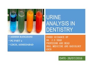
- 1. URINE ANALYSIS IN DENTISTRY • ASHISH RANGHANI • PG PART 2 • GDCH, AHMEDABAD UNDER GUIDANCE OF DR. J.S SHAH PROFESSOR AND HEAD ORAL MEDICINE AND RADIOLOGY GDCH DATE- 28/07/2016
- 2. CONTENTS 1. Processes of Urine Formation 2. Why urinalysis? 3. Collection of urine specimens 4. Types of urine sample 5. Components of urine 6. Urinalysis 7. Physical Examination Volume ,Color, Odor, Turbidity, Reaction (pH), Specific gravity 8. Biochemical Examination Proteins, Sugers , Ketone bodies, Bile salts , Bile Pigments, Blood 9. Microscopic Tests Cells, Crystals ,Casts, Microorganism 10. Urinary changes in Dental Diseases
- 3. Urine is the excretory waste product formed by the kidney It reflects the overall metabolic and kidney functions of the body In normal urine sample many substances such as glucose, proteins, amino acids, are present in trace amounts. Essentials of Medical Physiology Sixth Edition, Sembulingam
- 4. Processes of Urine Formation • When blood passes through glomerular capillaries, the plasma is filtered into the Bowman capsule. This process is called glomerular filtration Glomerular filtration • While passing through the tubule, the filtrate undergoes various changes both in quality and in quantity • Many wanted substances like glucose, amino acids, water and electrolytes are reabsorbed from the tubules Tubular reabsorption • Unwanted substances are secreted into the tubule from peritubular blood vessels Tubular secretion Essentials of Medical Physiology Sixth Edition, Sembulingam
- 5. Why urinalysis? Monitoring of patients with diabetes General evaluation of health Diagnosis of disease or disorders of the kidneys or urinary tract Diagnosis of other systemic disease that affect kidney function
- 6. Collection of urine specimens • Improper collection-- may invalidate the results • Containers for collection of urine should be wide, clean and dry. • Analysed within 2 hours of collection else requires refrigeration. • All specimens must be properly labeled • The patient’s name • The patient’s identification number • The date • The time of collection • The type of specimen • The attending physician’s name • The label should be affixed on the container, not the lid.
- 7. Types of urine sample Sample type Sampling Purpose Random specimen No specific time most common, taken anytime of day Routine screening Morning sample First urine in the morning, most concentrated Pregnancy test, microscopic test Clean catch midstream Discard first few ml, collect the rest Culture 24 hours All the urine passed during the day and night and next day 1st sample is collected. used for quantitative and qualitative analysis of substances Postprandial 2 hours after meal Determine glucose in diabetic monitoring Supra-pubic aspired Needle aspiration Obtaining sterile urine
- 8. Components of urine textbook of routine urinalysis and body fluids
- 9. URINALYSIS 1. Volume 2. Color 3. Odor 4. Turbidity 5. Reaction (pH). 6. Specific gravity. 1. Proteins. 2. Sugers. 3. Ketone bodies. 4. Bile salts. 5. Bile Pigments. 6. Blood. 1. Cells. 2. Crystals. 3. Casts. 4. Microorganism 5. Parasites. 6.Contamination A. Physical Examination B. Biochemical Examination C. Microscopic Tests
- 10. PHYSICAL EXAMINATION • Volume – Normal – 1- 1.5 L /day. Polyuria >3000ml / day increased urination • Diabetes mellitus & insipidus, • Chronic nephritis • After administration of certain drugs like digitalis, salicylates or diuretics Anuria <100 ml per day total suppression of urination • Severe hypotension • Crush injury, • Mercurial poisoning, • After a mismatch transfusion Oliguria <400ml / day Decreased urination • Acute & chronic glomerulonephritis, • Shock, • Congestive cardiac failure, • Dehydration
- 11. APPEARANCE • COLOUR • Normal - amber yellow due to the presence of 1. Urobilin 2. Uroerythrin 3. Urochromes Colorless - Very dilute urine • Diabetes • Polyuria Yellow orange (high colored) • Concentrated urine • Excess urobilin • Bile pigments • Intake of carrots Red/ smoky • RBC • Myoglobin • Aniline dyes • Menstrual contamination
- 12. Milky Pyuria Fat Brown black • Methemoglobin • Alkaptonuria • Melanin Orange • Bile pigments,Drugs like • Rifampicin- orange red • Levodopa -brown to black • Amitryptyline - green or blue-green • Imipenem–cilastatin - brown urine . - Cloudy - Phosphates & Carbonates, Urates & Uric acid, Pus cells, Bacteria, Spermatozoa bacteria, Yeast, Spermatozoa.
- 13. Specific Gravity •It is directly proportional to the concentration of solute & inversely proportional to the volume •Ranges between 1.003 to 1.030
- 14. LOW SPECIFIC GRAVITY HYPOSTHENURIA :indicates dilute urine, which may be caused by 1. Diabetes insipidus ( can be as low as 1.001) 2. Drinking excessive amounts of liquid. 3. Pyelonephritis, glomerulonephritis 4. Use of diuretics. HYPERSTHENURIA : indicates very concentrated urine, which may be caused by 1. Dehydration 2. Diabetes mellitus 3. Adrenal insufficiency. 4. Toximea of pregnancy (protein in the urine). HIGH SPECIFIC GRAVITY
- 15. TURBIDITY Clear and transparent Freshly voided form white precipitate Pus cells gives uniform cloudiness.Bacteria growth Red cells • Gives turbid smoky urine Mucus • it forms bulky deposits
- 16. ODOUR OF URINE After prolonged standingAmmonia smell: • Rancid : Tyrosinaemia. Due to urinary infectionFecal smell: • Mousy order : phenylketonuria Ketone bodies is seen in diabetesFruity smell • Maple syrup odour : MSUD Normal odour Fresh urine has aromatic odor
- 17. pH • Normal pH for urine ranges from 4.5 – 8.0 (average pH 6) • Some foods (such as citrus fruits and dairy products) and medications (such as antacids) can affect urine pH. • In a diet high in protein the urine is more acidic, while a diet high in vegetable material a urine that is more alkaline. • Tested by: • litmus paper • pH paper • dipsticks
- 18. pH CAUSES OF ACIDIC URINE 1. Acidosis 2. Uncontrolled diabetes 3. Diarrhea 4. Starvation and dehydration 5. Respiratory Acidosis CAUSES OF ALKALINE URINE 1. UTI with urease producing org 2. After Meal 3. Salicylate intoxication 4. Urinary retention due to obstruction 5. Chronic renal failure 6. Respiratory alkalosis 7. Renal tubular acidosis
- 19. Chemical examination • Proteins • Sugars • Ketone bodies • Bilirubin • Bile salts • Urobilinogen • Blood 1. Text book of practicle pathology & microbiology V.H. Talib
- 20. Tests for proteins • Principle-proteins are denatured & coagulated on heating to give white cloud precipitate. • Method-take 2/3 of test tube with urine, heat only the upper part keeping lower part as control. • Presence of phosphates, carbonates, proteins gives a white cloud formation. Add acetic acid 1-2 drops, if the cloud persists it indicates it is protein(acetic acid dissolves the carbonates/phosphates) HEAT COAGULATION TEST 1. Text book of practicle pathology & microbiology V.H. Talib
- 21. Other tests SULPHOSALICYLIC ACID TEST • Mix equal volume of clear urine & 3 to 5% acid • Cloudiness indicate presence of proteins HELLER’S NITRIC ACID TEST • White ring at the point of contact of conc. HNO3 and urine indicate presence of albumin Text book of practicle pathology & microbiology v.H. Talib
- 22. Causes of proteinuria • Normally there is a very scanty amount of protein in urine (< 150mg/day) HEAVY PROTEINURIA (>3gm/day) • SLE • Diabetes mellitus • Nephrotic syndrome • Renal vein thrombosis MODERATE PROTEINURIA (1- 3gm/day) • Multiple myeloma • Pyelonephritis • Chronic glomerulonephritis • Nephrosclerosis MILD PROTEINURIA (<1gm/day) • Hypertension • Polycystic kidney • UTI • Fever • Chronic pyelonephritis Pathology practicle book, harsh mohan
- 23. Bence Jones proteins • These are light chain globulins seen in multiple myeloma & lymphoma. • Test- Thermal method(waterbath): Proteins has unusual property of precipitating at 400 -600c & then dissolving when the urine is brought to boiling(1000c) & reappears when the urine is cooled. 1. Text book of practicle pathology & microbiology V.H. Talib
- 24. Test for sugar • Blue-green= negative • Yellow-green=+(<0.5%) • Greenish yellow=++(0.5-1%) • Yellow=+++(1-2%) • Brick red=++++(>2%) 1. Text book of practicle pathology & microbiology V.H. Talib • Test-BENEDICT’S TEST(semiquantitative) • Principle-benedict’s reagent contains cuso4.In the presence of reducing sugars cupric ions are converted to cuprous oxide which is hastened by heating, to give the color. • Method- take 5ml of benedict’s reagent in a test tube, add 8drops of urine. Boil the mixture.
- 25. Benedict’s test • Detects all reducing substances like glucose, fructose, & other reducing sustances. • To confirm it is glucose, dipsticks can be used (glucose oxidase)
- 26. Causes of glycosuria • Glycosuria with hyperglycaemia- 1. Diabetes, 2. Acromegaly, 3. Cushing’s Disease, 4. Hyperthyroidism, 5. Drugs Like Corticosteroids. • Glycosuria without hyperglycaemia- Renal tubular dysfunction Text book of practicle pathology & microbiology v.H. Talib
- 27. KETONE BODIES • 3 types Acetone Acetoacetic acid β-hydroxy butyric acid They are products of fat metabolism
- 28. Rothera’s test • Principle-acetone & acetoacetic acid react with sodium nitroprusside in the presence of alkali to produce purple colour. • Method- take 5ml of urine in a test tube & saturate it with ammonium sulphate. Then add one crystal of sodium nitroprusside. Then gently add 0.5ml of Strong ammonium hydroxide along the sides of the test tube. • Appearance permanganate colored ring at the junction of the two fluids indicates a positive test 1. Text book of practicle pathology & microbiology V.H. Talib
- 29. Causes of ketonuria • Diabetes • Non-diabetic causes- 1. High Fever, 2. Starvation, 3. Severe Vomiting/Diarrhea 4. After General Anaesthesia Text book of practicle pathology & microbiology v.H. Talib
- 30. Blood in urine • Test- BENZIDINE TEST • Method- mix 2ml of benzidine solution with 2ml of hydrogen peroxide in a test tube. Take 2ml of urine & add 2ml of above mixture. A blue color indicates + reaction Text book of practicle pathology & microbiology v.H. Talib
- 31. Causes of hematuria • Acute & Chronic Glomerulonephritis, • Chronic Passive Congestion Of The Kidney • Renal TB, • Leukaemias • Severe UTI, • Urinary Calculi • Benign & Malignant Tumors Of The Kidney & Urinary Tract Text book of practicle pathology & microbiology v.H. Talib
- 32. BILE SALTS Hay’s test The test depends on the surface activity of bilirubin as it lowers the surface tension of urine. Sprinkle a little of precipitated sulfur powder on the surface of 2 ml urine. If bilirubin is present, sulfur powder will sink to the bottom of urine. If bile is absent, sulfur will remain on the surface of urine. Text book of practicle pathology & microbiology v.H. Talib
- 33. Bilirubin • Test- fouchet’s test. • Causes Liver diseases-injury,hepatitis Obstruction to biliary tract
- 34. Urobilinogen • Test- ehrlich test • 5ml fresh urine + 0.5 ml Ehrilch's reagent, allow to stand for 5 min → • pink color on cold → normal trace. • red color on cold → increased amounts. • red color after heating → normal traces. • Causes-hemolytic anemia's Cause- obstruction to bile flow (obstructive jaundice)
- 35. Microscopic examination of urine • A sample of well-mixed urine (usually 10-15 ml) is centrifuged in a test tube at relatively low speed (about 2000-3,000 rpm) for 5-10 minutes which produces a concentration of sediment (cellular matter) at the bottom of the tube. • A drop of sediment is poured onto a glass slide, a coverslip is place over it & observed under microscope Urinalysis: a comprehensive review,
- 36. A variety of normal and abnormal cellular elements may be seen in urine sediment such as 1. Red blood cells 2. White blood cells 3. Mucus 4. Various epithelial cells 5. Various crystals 6. Bacteria 7. Casts
- 37. Hematuria is the presence of abnormal numbers of red cells in urine due to any of several possible causes • Renal stone • Kidney tumors • kidney trauma, • Upper and lower urinary tract infections, • Polycystic kidney WBC in high numbers indicate inflammation or infection somewhere along the urinary or genital tract • UTI • Prostatitis • Chronic pyelonephritis • Renal stone • Renal tumours • Cystitis
- 38. • The most common type of cast- hyaline casts • Seen in fever, exercise, damage to the glomerular capillary. • Red blood cells may stick together and form red blood cell casts. Such casts are indicative of glomerulonephritis, with leakage of RBC's from glomeruli, or severe tubular damage • White blood cell casts • Their presence indicates inflammation of the kidney. TYPES OF CAST Acellular cast • Hyaline casts • Granular casts • Waxy casts • Fatty casts • Pigment casts • Crystal casts Cellular cast • Red cell casts • White cell casts, • Epithelial cell cast
- 39. Crystals & amorphous materials
- 40. URINE ANALYSIS IN DIABETES • Diabetes mellitus (DM) also known as a group of metabolic diseases in which there are high blood sugar levels over a prolonged period. • This high blood sugar produces the symptoms of frequent urination, increased thirst, and increased hunger. • Untreated, diabetes can cause many complications. Acute complications include diabetic ketoacidosis and nonketotic hyper osmolar coma. • Serious long-term complications include heart disease, stroke, kidney failure, foot ulcers and damage to the eyes A study on abnormal constituents of urine in diabetic patients
- 41. • In diabetes mellitus mainly glucose and ketone bodies are elevated • Glucosuria occurs in mainly during diabetis mellitus and renal diabetes. • These ketone bodies are present in the urine this may be due to diabetic ketoacidosis • It occurs when the body cannot use sugar (glucose) as a fuel source because there is little or no insulin. Fat is used for fuel instead
- 42. Diagnosis of Multiple Myeloma Two of the 4 following criteria are generally required for diagnosis of multiple myeloma: 1. Radiographic evidence of osteolytic bone lesions 2. >20% plasma cells in bone marrow aspirates or biopsy specimens. 3. Demonstration of monoclonal or biclonal gammopathy with serum electrophoresis 4. Demonstration of Bence-Jones proteinuria Systemic Lupus Erythematosus • Heavy proteinuria (> 3gm/day) Pathology practicle book, harsh mohan
- 43. Mercury concentrations in urine • Urine levels of mercury less than 20 ng/mL are considered safe. • The mercury body burden of dental personnel is normally higher than in the general population. • This increased body burden is attributed to dental personnel mixing and applying dental amalgam and removing amalgam restorations
- 44. References 1. Text book of practicle pathology & microbiology v.H. Talib 2. Pathology practicle book, harsh mohan 3. Urinalysis in clinical practice, sekhar chakraborty 4. Graff’s textbook of routine urinalysis and body fluids 5. Salma mahaboob, madan mohan rao m, a study on abnormal constituents of urine in diabetic patients, ujmds 2014, page 64-67 6. Urinalysis: a comprehensive review, jeff Simerville, m.D., Georgetown university school of medicine, washington, d.C 7. Essentials of Medical Physiology Sixth Edition, Sembulingam
Hinweis der Redaktion
- 9
- 16