1. Template provided by: “posters4research.com”
Using Drosophila as a Tumor Model to Study Oncogenes
Ashia S. Williams, Amanda Simcox
Department of Molecular Genetics, The Ohio State University, Columbus, OH 43210
Goal Growth of GSR6 Cell +/- RU486
Methods
Results
Future Research
•Flies injected with cells expressing a drug inducible Ras gene
produced tumors in presence of the drug RU486and killed the
hosts.
•Flies injected with cells expressing a drug inducible Ras gene
produced tumors in absence of the drug RU486 failed to express
GFP and hosts survived.
•This indicates we have developed a system to examine
tumorigenesis in an inducible system
The Ohio State University, Columbus OH 43201
Simcox.1@osu.edu phone: (614)292-8857
Figure 2. - Properties of GS-R6-expressing cell cultures. (A, B) Phase
contrast images. (A'-B') GFP images. (A, A') GS-R6 with no RU486 drug. Only
some cells express GFP at low levels by 7 days without drug. (B, B') GS-R6 in the
presence of RU486 at day 7. The cells have strong GFP expression levels and
form foci characteristic of transformed cells. Pictures courtesy of A. Simcox.
Developing Protocol
Defining optimal anesthesia conditions for the flies.
Before transplanting the cells into the flies I have to etherize
them. To determine the best dose for keeping the flies
unconscious for the transplant (that takes about 15 minutes), I
set up an experiment, in which I used 0.5mL of ether and placed
8 flies in a vial for etherization. I determined how long it took the
flies to recover from different lengths of exposure to ether. The
graph shows my results. From this I concluded 3 minutes as
optimal.
•We need larger numbers and direct evidence that the tumors
are not growing in absence of RU486- we assumed this based
on lack of expression of the GFP marker.
The Purpose
The purpose of this study is to test the ability of cells with a drug
inducible Ras oncogene to produce tumors in flies.
The Significance
In cells exposed to the drug RU486, oncogenic Ras gene is
expressed. This is called the GeneSwitch system. My project was
to determine if these cells, when transplanted, form tumors in
flies. With this system we were able to determine the importance
of oncogenic Ras in tumor metastasis by controlling its
expression with RU486. Flies injected with cells in absence of the
drug RU486 failed to produce tumors and survived. However,
flies injected with cells and fed the drug RU486 did produce
tumors, which killed the hosts. This indicates we have developed
a model to examine tumorigenesis in an inducible system.
Tumor cell transplantation:
Host flies. I set up a cross to generate yw/ovoD2 sterile ♀ flies by
mating virgin yellow-white (yw) females with ovoD2 male flies.
-OvoD2 is a female sterile mutation and females have
very small ovaries so that when we transplant the cells into the fly
the tumors have more space to grow.
Cells. I cultured Gene Switch:Ras expressing cells in medium
with the drug RU486, Ras 7, and Ras 3 cell lines.
Transplantation method. First I harvest the tissue culture cells
into a concentrated pellet. To transplant the cells, I first
anesthetize the flies with ether. Next I line the flies in a straight
line with their abdomen facing up and wings flat down on
microscope slide. Using a pipette I transfer the cells under a drop
of oil. (The oil prevents the cells from drying out). I make a small
mark a ‘fill line’ on the needle and fill cells to that line. This allows
me to inject about the same number of cells each time.I
transplant the cells into the abdomen of the flies.
Experiment
-Transplant Ras3 cells into ovoD2/yw and analyze (positive control).
-Transplant Ras7 cells into ovoD2/yw and analyze (positive control).
-Transplant Gene Switch-R6 cells into host flies, feed the flies
RU486, and analyze.
- Transplant Gene Switch-R6 cells into host flies, without the
presence of RU486, and analyze (negative control).
Results
Conclusions
Test: A group of 140 adult flies that were fed 500ul of 100ug/ml
RU486 drug starting at 3rd
larval stage were injected with GS-R6
and observed over 10 days. The fraction represents the number of
host flies that showed fluorescence indicative of tumor growth.
QuickTime™ and a
decompressor
are needed to see this picture.
Obtaining Optimal Expression through Feeding Protocols
Ras3 control+
GS control-
Ras7 control+
A A’
B B’
GS-R6 Day 7
GS-R6 + RU486 Day 7
QuickTime™anda
decompressor
areneededtoseethispicture.
QuickTime™anda
decompressor
areneededtoseethispicture.
Etherization
19.2
-5
0
5
10
15
20
25
30
35
0 1 2 3 4 5 6
Minutes in Ether
MinutestoAwaken
Funding
QuickTime™ and a
decompressor
are needed to see this picture.
QuickTime™ and a
decompressor
are needed to see this picture.
QuickTime™ and a
decompressor
are needed to see this picture.
QuickTime™anda
decompressor
areneededtoseethispicture.
QuickTime™ and a
decompressor
are needed to see this picture.
QuickTime™ and a
decompressor
are needed to see this picture.
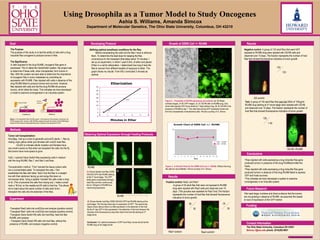
Figure 3. -A Growth Chart for the GSR6 Cell Line +/- RU486. Without the drug
the cells do not proliferate. Picture courtesy of A. Simcox.
Positive control: Ras3, and Ras7
-A group of 35 adult flies that were not exposed to RU486
drug were injected with Ras3 cells and observed over 10
days. (This process was repeated for Ras7 line) The fraction
represents the number of host flies that showed fluorescence
indicative of tumor growth.
25
35
30
30
Negative control: A group of 133 adult flies that were NOT
exposed to RU486 drug were injected with GS-R6 cells and
observed over 10 days. The fraction represents the number of host
flies that showed fluorescence indicative of tumor growth.
GS-R6 +RU486
Uninduced
RU486
(inducer)
GSR6 GFP
Induced
GSR6 GFP
Growth Chart of GSR6 Cell +/- RU486
1
10
0 1 2 3 4 5 6 7 8 9 10 11 12 13
Days
CellNumberX10
5
GS6 + RU486
GS6 - RU486
RU486
RU486
A
B(A) Shows female host flies (255B
GS/UAS-GFP) fed RU486 starting
at the 3rd
larval stage. The GFP
tester gene is expressed strongly.
Adult flies are then continuously
fed on 100ug/ml of RU486 thus
maximizing expression.
(B) Shows female host flies (255B GS/UAS-GFP) fed RU486 starting at the
adult stage. The first day there was no expression of GFP. The second day
(figure shown above) there is a little expression in the abdomen of the host.
By the 6th
day GFP is fully expressed in the abdomen of the host however the
intensity of the fluorescence is less than that of the host fed starting at 3rd
stage larval.
Conclusion: For optimal expression of GFP (and Ras), larvae should be fed
RU486 drug at 3rd stage larvae
Research Experiences for Undergraduates
133
133
135
1 40
Inactive Protein
Figure 1.-The GeneSwitch Ras (GS-R6) system. In the absence of the activator (uninduced), the
GS-R6 remains silent. However, after RU486 is applied (induced) the binding of the RU486 ligand
causes GS-R6 to become transcriptionally active, resulting in the expression of GFP.
Contact Information