1. 1
Genetic screening of CRISPR edited human-derived induced pluripotent stem cells
Apostolos Katsiaunis
Abstract
Stem cells are the origin of every cell, tissue, organ and organ system in the body. There
are different types of stem cells and various levels of differentiation. Stem cells are useful tools
in medicinal and scientific research because their versatility allows for the generation of specific
cell types. Induced pluripotent stem cells (iPSC) are mature, specialized stem cells that are
reprogrammed to respecialize. In this project, we used CRISPR (Clustered Regularly Interspaced
Short Palindromic Repeat) to induce mutations in iPSCs formed from skin fibroblasts. CRISPR
is a robust system that has been shown effective in genome editing previously. We found that
CRISPR can be used to mutate the HRAS gene in iPSCs to create cardiomyocytes with the
multifocal atrial tachycardia that is typical of patients with Costello syndrome. These
cardiomyocytes could be used in the future for drug screens as models in Costello syndrome.
Introduction
Costello syndrome is a disorder that negatively affects a variety of body parts. It is
autosomal dominant with frequent new mutations in the HRAS gene (Wey et al. 2013). It is a
rare disorder, affecting around 1 in 1.25 million, and was first described by Costello in 1971.
Despite not having any formal diagnostic criteria, the disorder causes a distinct craniofacial
appearance, cardiac and musculoskeletal abnormalities, neurologic problems, joint flexibility
abnormalities, developmental psychomotor delay, and intellectual disability. Coarse facial
features are found--large mouth, cheeks and lips, with sparse, curly hair, and a wide nasal bridge.
Fingers are splayed, and the wrists and feet are aligned incorrectly. Cardiac hypertrophy,
2. 2
multifocal atrial tachycardia, and cardiomyopathy are common (Gripp et al. 2012). Infants who
have Costello are born large, but don’t grow as fast as other children. Patients with Costello
usually are of shorter stature and can have reduced levels of human growth hormone. Children
and adults with Costello syndrome are at a higher risk for developing both cancerous and
noncancerous tumors. Papillomas, small growths similar to warts, are usually found near the
mouth or anus. The most frequent tumors are rhabdomyosarcomas, which originate in muscle
tissue (Peixoto et al. 2014).
In order to study Costello syndrome, a model is required. These models are ultimately
acquired using Induced Pluripotent Stem Cells (iPSC). Stem cells are undifferentiated cells that
are capable of becoming different kinds of cells. Totipotent stem cells have the ability of
becoming either placental or embryonic cells. Pluripotent embryonic stem cells have the ability
to specialize into any kind of cell type. There are two types of pluripotent stem cells: human
embryonic stem cells (ESCs), which come from embryos (Thomson et al., 1998; Reubinoff et al.,
2000) and iPSCs. iPS cells are grown using genetic reprogramming of human adult somatic
cells. These adult cells at first have very little potential for differentiation, but can become any
cell type after being reprogrammed to regain their plasticity (Hirschi et al. 2014).
Reprogrammed IPSCs are very similar to ESCs in proliferation rate, morphology, epigenetic
status of pluripotent genes, surface antigen expression, and telomerase activity. IPSCs can
differentiate into cell types of all three germ layer in vivo and in vitro (Takahashi et al. 2007).
iPSCs are generated using ‘reprogramming factors’ into fibroblasts or other differentiated
somatic cell types (Takahashi et al., 2007; Yu et al., 2007; Park et al., 2008a; Nakagawa et al.,
2008). iPSCs are derived from a skin biopsy (Dimos et al., 2008; Park et al., 2008b) or by blood
sample (Seki et al., 2010; Loh et al., 2010;Staerk et al., 2010). This versatility is convenient, as
3. 3
derivation, expansion, and differentiation of somatic cells that are genetically matched to the
patient is otherwise impossible.
Genomic editing tools have been adapted from bacterial immune systems. These tools are
called clustered regularly interspaced short palindromic repeats (CRISPRs). CRISPR-associated
systems (Cas) use a combination of proteins and short guide RNAs to recognize and cleave
complementary DNA sequences. CRISPR is a robust system that has been used to edit the
genomes of multiple eukaryotic organisms before and substantially improves the ease of
genomic editing and regulation (Cho et al. 2013).
Using CRISPR, we planned to edit the HRAS gene in iPSCs in order to cause multifocal
atrial tachycardia. This would cause iPSCs to behave like cells that are afflicted by Costello
syndrome. These cells can then be used as a model for Costello syndrome for drug screens and to
help develop treatments. This will ultimately help patients around the world who are suffering
from Costello syndrome. We hypothesized that using CRISPR, gene mutation and modification
can be generated in HRAS.
Materials and Methods
We received IPSCs from the laboratory that had been engineering prior to the
experiment. Cells were sorted via Fluorescent Assisted Cell Sorting (FACS). After gene editing,
samples were sorted and isolated clones were picked. Then, DNA extraction was done and then
PCR was done on the samples. Sorted cells were then plated and ten days later, clones were
picked into individual wells of a plate for expansion and DNA extraction. A 96-well plate was
used for DNA extraction, where each well contained a clone for screening. After DNA
extraction, a high-fidelity PCR was completed on the samples using specific primers to amplify
the HRAS gene region of interest. After that, a Restriction fragment length polymorphism
4. 4
(RFLP) analysis was performed and the samples were digested with SgrAI, which would help in
differentiating between edited and unedited gene regions, or mutant and wild-type clones.
Finally, gel electrophoresis was performed in order to test whether the samples were mutated or
not. Finally, gel electrophoresis (1% agarose) was performed in order to visually test and confirm
whether the samples were mutated or not.
Results
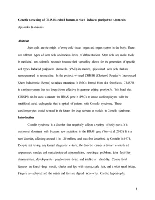
In the RFLP screen, we expected to either see a wild-type band set (around 400bp (base
pairs) and 200bp), a heterozygous mutant band set (around 600bp, 400bp and 200bp), or a
homozygous mutant band (around 600bp). Based on previous results from the lab, we expected
to see 5-10% of clones to contain an edited version of HRAS (heterozygous or homozygous
mutant). In a screen of twelve clones, only ten were readable. Of the ten, two clones possessed a
heterozygous mutation for HRAS G12S (20%) and one clone possessed a homozygous mutation
for HRAS G12S (10%). The remaining seven were homozygous wild-type (70%).
Illustrations
Fig. 1 Gel electrophoresis results. Red signifies a homozygous mutant. Cyan signifies a
heterozygous mutant. Lavender signifies homozygous wild-type
5. 5
Figure 2. Sequencing chromatogram
Discussion
Given the results, our hypothesis that gene editing using CRISPR in iPSCs was
supported. Of our 12 samples, we could only read 10. One reason for the inability to read the 2
clones is that the DNA extraction was not efficient. Samples could have denatured during the
6. 6
ethanol washes. A second reason was that the clones did not grow successfully before DNA was
extracted. A third possible reason for this was that the PCR mix could not have been prepared or
added correctly to the two samples. For example, a primer could be missing or the wrong amount
could have been administered.
However, of those 10, 3 carried the mutation. This means that they were successfully
mutated. 1 was mutated to have two copies of the gene, or be homozygous. The other 2 were
heterozygous. Since the mutations are autosomal dominant, these two are mutants.
After identifying the mutant clones, we will expand the clones and differentiate the iPSC
clones into cardiomyocytes. Then we will check the physiology of the cardiomyocytes. We will
check calcium handling, beats per minute, and electrophysiology. We expect the cardiomyocytes
with the G12S mutation to be abnormal and irregular. We also expect to see similar physiologic
features in the CRISPR edited iPSC-derived cardiomyocytes, as like the actual patients who
suffer from Costello, were normal before developing it. Future tests will include verification of
gene expression profiles for the genes associated with HRAS and calcium handling. These can
be performed using quantitative PCR, to measure gene expression by amplifying gene
transcripts. Protein analyses to be done include western blots, which look at specific proteins and
their activation or inactivation by phosphorylation, and phosphoproteomics, which looks at the
global activation or inactivation of proteins via phosphorylation and regulatory events within the
cell. These tests will show the similarities between edited iPSC clones and patient-derived
Costello syndrome cells. We will then use these results to verify whether HRAS mutations can
contribute to the arrhythmic phenotype visible in Costello patients.
After these mutated stem cells will be passed on to be differentiated into mutants and
non-mutants and grown on plates to become and differentiate into cardiomyocytes, there is great
7. 7
potential with what can be achieved using them. These cardiomyocytes will practically be cells
with Costello syndrome. Therefore, they are usable as models for Costello syndrome without
having to utilize a patient. The iPSCs can be used to perform drug screens and develop
treatments for Costello syndrome, saving scientists’ and patients’ resources and time. These
tools can one day help patients who suffer from Costello.
Human embryonic stem cells are obtained via methods that are ethically controversial.
On the other hand, using iPSCs does have any ethical issues. Engineering fully grown somatic
cells to reprogram into pluripotency bypasses that issue. Donated embryonic cells and tissues are
not needed for experimentation or utilization when iPSCs are used. One limitation, however, was
a small sample size of only 12. In future studies, more samples would be helpful in order to find
the true success rate. As of right now, the rate was 30%, or 3 out of 10, but the rate isn’t entirely
accurate because it is solely out of 10 samples. However, this shows that there is great potential
for future studies to be done with larger sample sizes. In addition, further research is needed. A
goal for one day is to take cells inflicted with Costello syndrome and genetically alter them using
CRISPR to make them healthy. There is great promise with the study of iPSCs.
References
1. Wey M, Lee J, Jeong SS, Kim J, Heo J. 11/2013, Kinetic mechanisms of mutation-
dependent Harvey Ras activation and their relevance for the development of Costello
syndrome. Biochemistry. 2013 Nov 26;52(47):8465-79
2. Sammon MR, Doyle D, Hopkins E, Sol-Church K, Stabley DL, McGready J, Schulze K,
Alade Y, Hoover-Fong J, Gripp KW. 8/7/2012, Normative growth charts for individuals
with Costello syndrome Am J Med Genet A. 2012 Nov;158A(11):2692-9
8. 8
3. Peixoto IL, Carreno AM, Prazeres VM, Chirano CA, Ihara GM, Akel PB. Syndrome in
question. Costello syndrome. An Bras Dermatol. 2014 Nov-Dec;89(6):1005-6.
4. Thomson JA, Itskovitz-Eldor J, Shapiro SS, Waknitz MA, Swiergiel JJ, Marshall VS,
Jones JM. Embryonic stem cell lines derived from human blastocysts Science. 1998 Nov
6;282(5391):1145-7.
5. Reubinoff, B. E., Pera,M. F., Fong, C. Y.,Trounson, A.,and Bongso, A. (2000). Embryonic
stem cell lines from human blastocysts: Somatic differentiation in vitro. Nat. Biotechnol. 18,399–
404.
6. Karen K. Hirschi, Song Li, and Krishnendu Roy, Induced Pluripotent Stem Cells for
Regenerative Medicine Annu Rev Biomed Eng.2014 Jul 11; 16: 277–294.
7. Takahashi K1, Tanabe K, Ohnuki M, Narita M, Ichisaka T, Tomoda K, Yamanaka S.Induction of
pluripotent stem cells from adult human fibroblasts by defined factors. Cell. 2007 Nov
30;131(5):861-72.
8. Yu, J., Vodyanik, M. A.,Smuga-Otto, K., Antosiewicz-Bourget, J.,Frane, J. L., Tian, S., Nie, J.,
Jonsdottir, G. A., Ruotti, V.,Stewart, R.,et al. (2007). Induced pluripotent stem cell lines derived
from human somatic cells. Science 318,1917–1920.
9. Park,I. H., Zhao, R.,West, J. A.,Yabuuchi, A.,Huo, H., Ince,T. A.,Lerou, P. H.,Lensch,M. W.
and Daley, G. Q. (2008a). Reprogramming of human somatic cells to pluripotency with defined
factors. Nature 451, 141–146.
10. Nakagawa,M.,Koyanagi, M., Tanabe,K., Takahashi, K., Ichisaka, T., Aoi, T., Okita, K.,
Mochiduki, Y.,Takizawa, N. and Yamanaka, S. (2008). Generation of induced pluripotent stem
cells without Myc from mouse and human fibroblasts. Nat. Biotechnol.26,101–106.
11. Dimos, J. T.,Rodolfa, K. T., Niakan, K. K.,Weisenthal, L. M., Mitsumoto, H.,Chung, W., Croft,
G. F., Saphier, G., Leibel, R.,Goland, R., et al. (2008). Induced pluripotent stem cells generated
from patients with ALS can be differentiated into motor neurons. Science 321,1218–1221.
9. 9
12. Park,I. H., Arora, N.,Huo, H.,Maherali, N.,Ahfeldt, T., Shimamura, A., Lensch, M. W., Cowan,
C., Hochedlinger, K. and Daley, G. Q. (2008b). Disease-specific induced pluripotent stem cells.
Cell 134, 877–886.
13. Seki, T., Yuasa,S., Oda,M., Egashira, T., Yae,K., Kusumoto, D., Nakata,H.,Tohyama, S.,
Hashimoto, H.,Kodaira, M., et al. (2010). Generation of induced pluripotent stem cells from
human terminally differentiated circulating T cells. Cell, StemCell 7, 11–14.
14. Loh, Y. H., Hartung, O., Li, H., Guo, C., Sahalie, J. M., Manos, P. D.,Urbach,A., Heffner,G. C.,
Grskovic, M., Vigneault, F., et al. (2010). Reprogramming of T cells from human peripheral
blood. Cell, StemCell 7, 15–19.
15. Staerk, J., Dawlaty, M. M., Gao, Q.,Maetzel, D., Hanna,J., Sommer, C. A., Mostoslavsky, G.
and Jaenisch, R. (2010). Reprogramming of human peripheral blood cells to induced pluripotent
stem cells. Cell, StemCell 7, 20–24.
16. Seung Woo Cho, Sojung Kim, Jong Min Kim & Jin-Soo Kim, Targeted genome engineering in
human cells with the Cas9 RNA-guided endonuclease 1/14/2014 Nature Biotechnology 31,230–
232 (2013) doi:10.1038/nbt.2507