Ankle valgus and its management
•
8 gefällt mir•1,883 views

ankle valgus in children- clinical features and its management

Empfohlen
Weitere ähnliche Inhalte
Was ist angesagt?
Was ist angesagt? (20)
Ähnlich wie Ankle valgus and its management
Ähnlich wie Ankle valgus and its management (20)
Mehr von Dr. Anurag Mittal
Mehr von Dr. Anurag Mittal (18)
Kürzlich hochgeladen
Kürzlich hochgeladen (20)
Ankle valgus and its management
- 1. Ankle Valgus Dr Anurag Mittal MS ortho GTBH
- 2. Introduction • The normal fibula is approximately equal in length to the tibia, but its distal tip extends more caudad. Thus, the fibula acts as a lateral buttress, bearing approximately 15% of the body weight during gait. • Ankle valgus is an insidious deformity that results in pronation of the foot and medial malleolar prominence. • The causes are varied and include neuromuscular disorders, skeletal dysplasias, and clubfeet.
- 3. • Left untreated, this deformity may progress, despite the use of orthotics or corrective shoes, resulting in the medial collapse of the ankle and foot. • After skeletal maturity, the only remedy is to perform a supramalleolar osteotomy. • However, in growing children, there is the opportunity to intervene by means of guided growth or hemi-epiphysiodesis of the distal medial tibia.
- 4. Normal ankle alignment • The lateral distal tibial angle (LDTA) is 87º, and the fibular physis is at or distal to the level of the plafond, which is horizontal and, thus, perpendicular to gravity.
- 5. Problem • In the normally aligned extremity, the mechanical axis bisects the knee and ankle, at an angle of 3º with respect to the vertical (gravity). • The physes of the tibia and fibula, along with the ankle plafond, are parallel to the floor and perpendicular to gravity. This permits the physeal and articular cartilage chondrocytes to resist compression—a task that they are well suited for—while sparing them from shear forces.
- 6. Malhotra classification- applicable to children older than 2 years
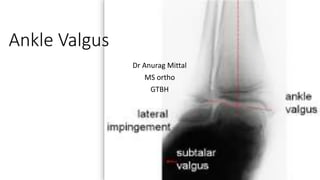
- 7. • The described triad of • fibular physis elevation, • wedging of the lateral tibial epiphysis, and • ankle tilt • may be accompanied by • horizontal expansion of the fibular epiphysis (impingement), • medial clear space widening, and avulsion injuries of the tip of the medial malleolus.
- 8. Ankle valgus, which is rare at birth, may gradually develop because of a variety of conditions, including the following (but not limited to these) : • Cerebral palsy • Spina bifida • Arthrogryposis • Down syndrome • Congenital clubfoot12 • Neurofibromatosis • Hereditary multiple exostoses • Postaxial hypoplasia (fibular hemimelia) • Skeletal dysplasia • Posttraumatic events • Ball-and-socket ankle
- 9. Clinical presentation • In the standing position, the medial malleolus is unduly prominent, the heel and hindfoot are angled laterally, relative to the calf • A common finding is subfibular tenderness due to impingement. • There may be concomitant hindfoot deformity, more commonly planovalgus than cavovarus. • Proximally, there may be concomitant genu valgum with a corresponding increase in the intermalleolar distance . • When the etiology is neuromuscular, the patient may have muscle weakness, imbalance, or contractures
- 10. • One needs to differentiate between ankle valgus (shown here) and hindfoot valgus. It is imperative to obtain a standing AP radiograph of the ankle when evaluating foot problems
- 11. Management • The indications for treatment of ankle valgus are the presence of • related discomfort, • excessive shoe wear, and • documented progression. • Distal tibiofibular synostosis Commonly performed (temporarily) with a transdesmosis screw, distal tibiofibular synostosis is a prophylactic strategy to prevent the complication of iatrogenic ankle valgus during lengthening of the tibia and fibula or during fibular harvest for vascularized bone graft procedures. • Fibular lengthening The technology exists to accomplish isolated fibular lengthening, either acutely with an intercalary graft or gradually with distraction osteogenesis
- 12. • Bracing: While one may temporize and treat mild deformities with lateral heel wedges, or orthoses of varying designs, the underlying growth disturbance will persist and, likely, will progress. As the child grows and gains body mass, these measures will eventually prove inadequate.
- 13. • Osteotomy: One surgical option is to perform a supramalleolar osteotomy. Considering the deformities are often bilateral, the patient will need to be immobilized and non-weight-bearing for 6 weeks. For deformities less than 20 º, a closing wedge osteotomy, leaving the fibula intact, is relatively simple and well tolerated. When the deformity is more than 20 º, it is necessary to cut the fibula and translocate the distal tibia-fibula to restore the mechanical axis. This requires more fixation and carries higher risks. • Unfortunately, depending on the age and etiology, recurrent ankle valgus is common with further growth, and the procedure(s) may need to be repeated.
- 14. • Guided growth: A transmalleolar screw or an 8-plate is necessary. The screw is economical and simple to insert; however, there may be major challenges when it comes time to remove the implant. • The 8-plate offers some advantages: It is simple to apply; the flexible tension band offers a fulcrum that is medial to the physis; the correction is more rapid; and the 8-plate is simpler to remove
- 15. 8 plate
- 17. THANK YOU