3. 1
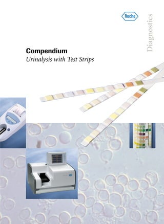
Urine Examination with Test Strips
History of urinalysis with test strips 3
Indications for urine test strips 7
Pre-analytical treatment and test performance 11
Characteristics of Urine Test Strips
from Roche Diagnostics 19
Specific gravity 23
pH 25
Leukocytes 27
Nitrite 29
Protein (Albumin) 33
Glucose 37
Ketones 41
Urobilinogen 43
Bilirubin 47
Blood (erythrocytes/hemoglobin) 49
Microscopical and Bacteriological Examination
The test strip sieve 55
Microscopic assessment of the sediment 57
Urine culture 60
Urine cytology with Testsimplets 63
Automated Urinalysis 64
Detection of Microalbuminuria
with Micral-Test 77
Appendix
The kidneys and the efferent urinary tract 81
Cell atlas – urine sediment/urine cytology 86
Glossary of specialist medical terms 95
Further reading 107
Contents
2
3
4
5
6
4.
5. 3
History of urinalysis with test strips
In many cultures urine was once regarded
as a mystical fluid, and in some cultures it
is still regarded as such to this day. Its uses
have included wound healing, stimulation
of the body’s defences, and examinations
for diagnosing the presence of diseases.
Modern medicine has at its disposal a
variety of quick and hygienic test methods
permitting safe and reliable analysis of
urine test specimens. The starting point
for diagnosing a wide range of patholo-
gical conditions is, however, simple visual
examination of the urine, and a long path
had to be travelled to the development of
the modern test strips now used routinely
for determining the urine status. Let us
now take a quick look at this long devel-
opment process.
It all started over 2000 years ago
The origin of visual urine diagnostics, the
oldest method of examining body fluids,
can be traced back to ancient Egypt, where
polyuria and haematuria are mentioned as
states of disease in old medical papyri.
Hippocrates (ca. 400 BC) observed certain
changes in the odour and color of urine in
the presence of fever, and pointed out the
importance of examining the patient’s
urine. The Indian physician Caraka (ca.
100 AD) described ten pathological kinds
of urine, including urines that contained
sugar and bacteria.
No medical teaching of the past was, how-
ever, so important, and none had such
lasting influence, as that of Claudius
Galenus of Pergamum, also known as
Galen, who in the second century AD
combined the medicine of his day, divided
into a number of groups, into one major
system with his doctrine of humoral
pathology: “It is not solid organs that are
the seat of disease but the four body fluids
or humours: blood, phlegm, black bile,
and choler or yellow bile. Disease is due to
an imbalance of these fluids, and the
nature and site of the disease can be
established from the composition and
appearance of the humours. An illness
Fig. 1: Uroscopy in the 15th century
1
6. History of urinalysis with test strips
therefore also shows itself in the urine.”
This doctrine dominated medical thinking
up to the 16th century. In pathology the
teaching of Galen of Pergamum was in
fact abandoned only in the 19th century.
In the 10th century the Arab physician
Isaac Judaeus, basing himself on Galen’s
humoralism, developed a scheme of hu-
mours with which he raised the urine
findings to the level of an almost infallible
diagnostic criterion for all states of dis-
ease. The extreme consequence of this the-
ory was so-called uromancy or uroscopy
practiced in the Middle Ages (Fig. 1),
which according to modern views was
devoid of any scientific basis. Over 20
shades of color were distinguished in the
urine (from crystal clear via camel hair
white, blackberry red, and pale green to
black), and corresponding conclusions
were drawn about the patient’s illness
(Fig. 2). The development went so far that
all that was wrong with the human body
was believed to be reflected as in a mirror
in the urine specimen. This view served as
a basis for the “urine fortune-telling,”
which was so caustically criticized by
humanistic physicians in the 16th century.
In the 16th century Paracelsus prompted
examination of the urine by the methods
of alchemy, but the thinking of his time,
tinged by ideas of magic and astrology,
prevented his proposals from developing
into forerunners of medical and chemical
analysis of the urine.
4
Fig. 2: A urine glass disc with
20 color nuances (1491 AD)
7. History of urinalysis with test strips
pathological urine constituents. Criticisms
were voiced at the time that doctors active
in general practice had to do too much
chemistry, since the tests were all based on
wet chemistry. The first “test strips” were
developed by the Parisian chemist Jules
Maumené (1818–1898) when, in 1850, he
impregnated a strip of merino wool with
“tin protochloride” (stannous chloride).
On application of a drop of urine and
heating over a candle the strip immediate-
ly turned black if the urine contained sug-
ar. Despite its simplicity the test was not
widely accepted, and it took another 70
years or so before the Viennese chemist
Fritz Feigl (1891–1971) published his
technique of “spot analysis.”
In the intervening years prominent physi-
cians, above all in Britain, concerned
themselves with the development of the
forerunners of modern test strips. Thus,
English physiologist George Oliver
(1841–1915) marketed his “Urinary Test
Papers” in 1883. The principle in this case
was to fix the reagents required for the
preparation of solutions in high concen-
trations on filter paper or cloth, to facili-
tate the work of the practitioner.
Reagent papers were already commercially
obtainable at the beginning of this centu-
ry from the chemical company of Helfen-
berg AG. A test for the presence of blood
by a wet-chemical method using benzi-
dine became known in 1904, and it was
not long before an analogous benzidine
paper test appeared on the market.
From uromancy to the idea of clinical
chemistry of the urine
It was only towards the end of the 18th
century that doctors interested in chemis-
try turned their attention to a scientific
basis of urinalysis and to its use in practi-
cal medicine. Writing in 1797, the physi-
cian Carl Friedrich Gärtner (1772–1850)
expressed a wish for an easy way of testing
urine for disease at the patient’s bedside.
In the same year a work appeared in
Britain in which the chemist William
Cruikshank (1745–1800) described for the
first time the property of coagulation on
heating, exhibited by many urines. This
observation led English physician Richard
Bright to speak of the “albuminous nature
of urine” and to describe this clinical
symptom of nephritis in 1827 in “Reports
of Medical Cases.” This marked the break-
through of qualitative urine chemistry
into medicine.
In the decades that followed a number of
chemical urinalyses were introduced into
clinical general practice, such as examina-
tions of the urine for protein, sugar, and
acetone. However, these examinations
were associated with considerable time
and effort, and the results were not very
specific, e.g. the reduction methods of
Fehling or Nylander for the detection of
sugar in urine.
With the arrival of chemical urine diag-
nostics the year 1840 marked a true boom
for methods aimed at the detection of
5
8. 6
History of urinalysis with test strips
Triumph of the test strips
All these “dry reagents” still did not de-
serve the designation of “dry chemistry”in
the modern sense of the term, but they
must be regarded as rudimentary fore-
runners of the modern test systems. Even
if the basic principle of reagent drying for
a time did not undergo any change, urine
diagnostics made major progress in the
1930s. The informative power and the reli-
ability in particular were distinctly
improved, and test performance itself
became progressively easier.
Urine test strips in the sense used today
were first made on industrial scale and
offered commercially in the 1950s. The
company Boehringer Mannheim, today a
top leader on the world market under the
name of Roche Diagnostics, launched its
first Combur test strips in 1964. Even
though the test strips have changed their
external appearance little since the 1960s,
they now contain a number of revolution-
ary innovations. New impregnation tech-
niques, more stable color indicators, and
the steady improvement in color grada-
tion have all contributed to the fact that
the use of urine test strips has now become
established in clinical and general practice
as a reliable diagnostic instrument.
The parameter menu offered has steadily
grown longer in the intervening decades.
Today Combur-Test product line from
Roche Diagnostics can be used for the
recognition of the early symptoms of
the following three major disease cate-
gories:
Ⅲ diseases of the kidneys and the uro-
genital tract
Ⅲ metabolic diseases (diabetes mellitus)
Ⅲ liver diseases and haemolytic disorders
Diabetic and hypertension-determined
nephropathies have been diagnosed early
with the aid of Micral-Test in the presence
of microalbuminuria.
9. Indications for urine test strips
Ⅲ the result is obtained quickly
Ⅲ the test is easy and inexpensive
Ⅲ high sensitivity (diagnostic sensitivity)
Ⅲ sufficiently high diagnostic specificity
A field study carried out in seven Euro-
pean countries with over 11,000 urine
samples illustrates the value of screening
with urine test strips (Fig. 3). A patholog-
ical urine finding (after checking for
nitrite, protein, glucose, ketones, uro-
bilinogen, and blood) was diagnosed in
16% of “normal healthy persons,” in 40%
of outpatients, and in 57% of hospitalized
patients.
With the aid of routine examinations early
symptoms of the following three groups
are identified:
Ⅲ diseases of the kidneys and the urinary
tract
Ⅲ carbohydrate metabolism disorders
(diabetes mellitus)
Ⅲ liver diseases and haemolytic disorders
Diseases of the kidneys and
urogenital tract
Screening parameters:
Ⅲ leukocytes
Ⅲ nitrite
Ⅲ protein
Ⅲ blood
Ⅲ specific gravity
Ⅲ pH
Urine test strips are a central diagnostic
instrument, their ease of use yielding
quick and reliable information on patho-
logical changes in the urine. Their signi-
ficance lies primarily in first-line diag-
nostics. Routine testing of the urine with
multiparameter strips, allowing a deter-
mination of the complete urine status, is
therefore the first step in the diagnosis of a
very wide range of disease pictures.
Indications for urine test strips:
Ⅲ screening within the framework of
routine examinations
Ⅲ treatment monitoring
Ⅲ self-monitoring by patients
Ⅲ general preventive medicine
Screening within the framework
of routine examinations
Within the framework of routine exami-
nations urine test strips are used for
screening both in hospitals and in general
practice. The aim of screening is early
identification of likely patients by exami-
nation of large groups of the population.
No direct diagnoses are established on the
basis of the screening results, which serve
only as a basis for further microscopic,
bacteriological, or clinicochemical exami-
nations of the urine.
Urine test strips can satisfy all the require-
ments for effective screening:
7
10. Indications for urine test strips
Diseases of the kidneys and the urogenital
tract often remain asymptomatic for a
long time. Renal function disturbances
frequently lie dormant for many years,
leading eventually to often irreversible
severe late damage. Kidney failure as the
terminal stage of various primary and sec-
ondary nephropathics (Fig. 4) can only be
treated by renal substitution therapy such
as dialysis or kidney transplantation.
Effects are also possible on other organ
systems, especially on the cardiovascular
system. The cardinal symptom of a uri-
nary tract infection is the detection of sig-
nificant bacteriuria (nitrite positive) and
leukocyturia (leukocytes positive) by
means of test strips.
The following non-specific symptoms
occur time and time again in patients with
urinary tract infections or pyelonephritis
and require further clarification to avoid
possible late consequences such as urae-
mia, hypertension, and cardiovascular
complications:
Ⅲ tiredness and exhaustion
Ⅲ chronic headaches
Ⅲ persistent lack of appetite
Ⅲ loss of weight
Ⅲ nausea and vomiting
Ⅲ intermittent rises in temperature and
fever of unclear origin (in children
some 50% of urinary tract infections
are manifested by fever)
Ⅲ pale yellow skin color,
puffy appearance
8
Importance of urinalysis as a screening procedure
“normal”
persons 16%
outpatients
40%
hospitalized
patients 57%
Frequency of pathological urine in different groups of people.
Parameters: nitrite, protein, glucose, ketones, urobilinogen, blood
A field study carried out in seven European countries with over 11,000 urine samples
Fig. 3: Frequency of pathological urines
11. Indications for urine test strips
Ⅲ in patients with
congenital urological
disorders approx. 57%
Ⅲ in gout patients approx. 65%
Ⅲ in patients after catheterization,
instrumentation, and operations
on the urinary tract
Regular checking for urinary tract in-
fections and infectious kidney diseases,
especially in women and elevated-risk
patients, enables treatment to be started
early as a result of diagnosis in an early
state of the disease, with good prognosis of
the otherwise serious conditions. After the
end of the therapy further control checks
are also necessary to catch any relapses in
good time.
The following characteristic symptoms are
much more rare:
Ⅲ proteinuria
Ⅲ “weak bladder,” a “bladder cold”
Ⅲ burning and pain during micturition
Ⅲ polyuria, dysuria, pollakiuria
Ⅲ bed-wetting in older children
Ⅲ pains in the lumbar and kidney region.
In certain risk groups the danger of uri-
nary tract infections and pyelonephritis is
particularly high:
Ⅲ in pregnant women 4–8%
Ⅲ in hypertensive subjects approx. 14%
Ⅲ in older people 8–18%
Ⅲ in diabetics up to 20%
Ⅲ in patients with urinary
calculi approx. 50%
9
Type II diabetes
2.9%
Cystic/polycystic
kidney diseases
7.5%
Analgesics nephropathy
Glomerulonephritis
25.6%
Pyelonephritis/
interstitial nephritis
18%
Type I diabetes
7.4%
Hypertension
10.2%
Others
10.9%
Unclear
origin
14.6%
Source:
Demography of Dialysis and
Transplantation in Europe, 1993
Fig. 4: Causes of dialysis and kidney transplants
12. Indications for urine test strips
Carbohydrate metabolism disorders
(inter alia diabetes mellitus)
Screening parameters:
Ⅲ glucose
Ⅲ ketones
Around 30–40% of type I diabetics and
around 20% of type II diabetics suffer in
time from a nephropathy, and early recog-
nition of diabetes is therefore of major
significance for the further state of health
of these patients.
Liver diseases and haemolytic
disorders
Screening parameters:
Ⅲ urobilinogen
Ⅲ bilirubin
In many liver diseases the patients often
show signs of pathology only at a late
stage. Early diagnosis allows appropriate
therapeutic measures to be instituted in
good time, avoiding consequential dam-
age and further infections.
Treatment monitoring
Treatment monitoring with the aid of
urine test strips allows the treating doctor
to check on the results of the prescribed
therapy, and if necessary to introduce any
changes into the therapeutic strategy. An
additional benefit of such monitoring is
improved patient compliance.
Monitoring is particularly useful in two
clinical conditions:
In diabetes mellitus combined checks for
glucose and ketones are advisable for the
purpose of early detection of any dietetic
errors by changes in the metabolic status
and for their correction.
Patients suffering from hypertension run
an increased risk of developing kidney
damage in the course of their condition.
Micral-Test allows early detection of inci-
pient nephropathy.
Self-monitoring by patients
Under their doctor’s instructions patients
can benefit directly from the advantages of
urine test strips. This applies particularly
to diabetics, where the idea of self-moni-
toring of the metabolic status (determi-
nations of glucose and ketones) is self-
evident.
General preventive medicine
Spontaneous preventive monitoring at
home has meanwhile become widespread
in the population. For example, a check on
the first morning urine for an asymp-
tomatic urinary tract infection can be
carried out without any problems on a
day-to-day basis. The same applies to an
examination of the urine 2 hours after a
carbohydrate-rich main meal to check for
the presence of diabetes mellitus. The
whole family is often involved in such
preventive monitoring.
10
13. Pre-analytical treatment and test performance
Depending on the time and nature of the
urine specimen collection, a distinction is
drawn between:
Ⅲ spontaneous urine
Ⅲ first morning urine (after a night’s
rest)
Ⅲ second morning urine (collected
before noon)
Ⅲ timed urine (usually 24-hour urine)
Ⅲ midstream urine
Ⅲ bladder puncture urine
The first morning urine has proved its
worth for most test purposes. As a rule it
ensures sufficiently long residence of urine
in the bladder, and its composition is inde-
pendent of the daily variations due to food
and fluid intake and physical activity. For
Reliable analytical results can only be
obtained from a urine specimen that has
been collected, transported and stored
properly. To this day the diagnostic possi-
bilities of urinalysis are often not utilized
to the full because correct pre-analytical
treatment cannot be ensured.
Sample collection
The urine collection and dispatch equip-
ment should always comprise clean and
sterile disposable containers, made as a
rule from plastic. Important patient data
(surname, first name, date of birth, sender,
collection date and time) should be affixed
to the container in a waterproof manner
before the sample collection.
11
Fig. 5: Collecting a mid-stream urine sample
Wash hands. Take lid off
specimen container
and place
with inside surface fac-
ing upwards.
First void a small
amount of urine
into the toilet,
then fill the specimen
container half full,
void remaining urine
into the toilet.
Replace the lid
on the specimen
container being
careful not to
touch the inside;
give the container
to the nurse or
laboratory.
14. 12
Pre-analytical treatment and test performance
checks on glucosuria it is best to use urine
passed about 2 hours after a carbohydrate-
rich meal.
Contamination is frequent in normal
“spontaneous”urine collected without any
special hygienic precautions, especially in
the case of women, and consists of
leukocytes in the presence of discharge
and of erythrocytes in the presence of
menstruation. For this reason no urine
diagnostics should be attempted in
women during and 2–3 days after men-
struation.
Correct sample collection is made easier
by leaflets with a detailed description of
the procedure for patients and medical
personnel.
Sample storage
Examination of the urine with test strips
should be carried out at the latest 2 hours
after micturition, since longer standing
times can lead to false results owing to the
following influences:
Ⅲ disintegration (lysis) of leukocytes and
erythrocytes
Ⅲ proliferation of bacteria
Ⅲ bacterial degradation of glucose
Ⅲ a rise in pH due to ammonia formed
as a result of bacterial degradation of
urea
Ⅲ oxidation of bilirubin and urobili-
nogen, especially in sunlight
These changes in the specimen can be
slowed down if the urine is kept in a sealed
container in a refrigerator.
15. 13
Pre-analytical treatment and test performance
Parameter Stability in urine Influencing Interference Remarks
measured 4–8 °C 20-25 °C factors factors
Specific Fluid intake, pH > 7 Precipitation changes
gravity diuretics the specific gravity
pH Unstable Unstable Diet (meat ↓, Rise on formation
vegetarian ↑) of ammonia
Leukocytes 1–4 h 1–4 h Vaginal secretion Strong color of Fast lysis at
urine ↑ specific gravity < 1.010
High glucose and and pH > 7.
protein values ↓ Mix urine specimen
Certain well
antibiotics ↑ or ↓
Nitrite 8 h 4 h Bacterial count Strong color of Antibiotics inhibit
urine ↑ nitrite formation
Ascorbic acid ↓
Phenazopyridine ↑
Protein 7 days 1 day Physical activity Ejaculate ↑
(albumin) pregnancy Preservatives ↑
Glucose 8 h 2 h Pregnancy, Bacteria ↓
fever, old age
Ketones 6 h 2 h Starvation, Phenylketones ↑ Test is more sensitive
fasting, Phthaleins ↑ to acetoacetic acid
fever SH compounds ↑ than to acetone
Urobilinogen 2 h Light ↓ Oxidation in air
Strong color of
urine ↑
Phenazopyridine ↑
Bilirubin 2 h Light ↓ Oxidation in air
Ascorbic acid ↓
Phenazopyridine ↑
Blood 1–4 h 1–4 h Menstruation, Oxidizing cleaning Fast lysis at
(erythrocytes) strong physical agents ↑ specific gravity < 1.010
activity and pH > 7.
Mix urine specimen
well
Tab. 1: Storage conditions, influencing factors and interference factors
16. 14
Pre-analytical treatment and test performance
Parameter Stability in urine Influencing Interference Remarks
measured 4–8 °C 20-25 °C factors factors
In sediment:
Bacteria 24 h Urinary pH Cells are lysed in
Casts 1–4 h Unstable dependence on pH and
Epithelial cells hours osmolality.
Erythrocytes 1–4 h 1–4 h Osmolality < 300 mmol/L
Leukocytes 1–4 h 1–4 h reduces storage stability
Urine culture Low pH, Results too low or
antibiotics, false-negative results
infections outside
the bladder
(kidney stones,
prostate), fastidious
microorganisms
Indwelling catheter, Results too high or
collection technique false-positive results
(children, old
persons),
delayed
working-up
Tab. 1 (Continued): Storage conditions, influencing factors and interference factors
17. Pre-analytical treatment and test performance
White turbidity can be due to:
Ⅲ phosphates precipitating in alkaline
urine (test: the precipitate redissolves
on acidification with acetic acid)
Ⅲ pyuria in massive bacterial or fungal
infections (microbial count >107
/mL)
Ⅲ lipiduria in the presence of a nephrotic
syndrome or on contamination with
ointments
Ⅲ massive proteinuria
Odour of the urine
Striking odour changes of clinical signifi-
cance include:
Ⅲ odour of fresh fruit or of acetone in
the presence of ketonuria (sign of
possible presence of metabolic aci-
dosis, most often due to fasting or
uncontrolled diabetes mellitus)
Ⅲ “fetor hepaticus,” a musty odour of
urine and breath in the presence of
hepatic encephalopathies
Ⅲ odour of alcohol in the presence of
intoxication due to ethanol
Ⅲ odour of ammonia in urinary tract
infections due to urea-splitting bac-
teria; odour of hydrogen sulfide in uri-
nary tract infections with proteinuria
due to putrefacient bacteria
Ⅲ a wide range of various odours due to
intoxications and after certain foods
Pneumaturia
Pneumaturia (presence of fine gas bubbles)
is a rare symptom pointing to the presence
of a fistula between the urinary tract and
the intestine.
Macroscopic assessment of urine
specimens
Macroscopic assessment of the urine (its
color and odour) is of little diagnostic
value, but within the framework of visual
examination of the specimens any striking
color changes are usually reported as well.
The normal urine volume of an adult is
some 700–2000 mL/day. An output of
more than 2500 mL/day is classified as po-
lyuria, an output of less than 500 mL/day
as oliguria, and an output of less than
100 mL/day as anuria.
Color of the urine
The color of normal urine is due to the
presence of porphyrins, bilirubin, uro-
bilin, uroerythrin, and some other, still
unidentified, compounds. Striking changes
should be reported in terms of definite
colors: “red,”“brown,”“green,” etc.
Color changes are caused most often by
drugs and their metabolites. A brick-red
sediment is usually due to precipitation of
urates in acidic urine (test: the precipitate
redissolves on gentle warming). Haema-
turia is recognized by the presence of
brown-red turbidity with a red-brown
sediment. Darkening can also occur in the
presence of substances other than those
listed in the table below.
15
19. Pre-analytical treatment and test performance
Points to avoid at all times
Ⅲ Residues of cleaning agents or
disinfectants falsify the results (false-
positive findings for blood, protein
and glucose)
Ⅲ Freezing of the urine specimen will
destroy leukocytes and erythrocytes
and hence make the specimen un-
usable for subsequent microscopic
examinations
Ⅲ Specimens must not be centrifuged
prior to test strip analysis
Ⅲ Specimens must not be exposed to
direct sunlight
Quality assurance
Quality assurance in urinalysis includes in
addition to the analysis itself the opera-
tions of sample collection, preservation,
preparation and transport. It is therefore
necessarily interdisciplinary and requires
involvement of the patient.
Test performance
1. Collect the urine specimen in a clean
sterile container (preferably a dispos-
able container).
2. Dip the test strip in the urine for no
longer than 1 second.
3. On drawing the strip out of the sample
run its edge over the rim of the con-
tainer to remove excess liquid.
4. After 60 seconds (60–120 seconds for
leukocytes) compare the reaction color
in the test area against the color scale on
the label.
Colors occurring only on the edges or
ones that develop only after more than
2 minutes are not relevant for diagnostic
purposes.
Points to note
Ⅲ Urine examinations with test strips
should be carried out within 2 hours
at the latest
Ⅲ The urine specimen should be mixed
thoroughly prior to the test
Ⅲ The specimens must always be kept in
a refrigerator (at +4°C) if the tests
cannot be done within 2 hours of the
urine collection
Ⅲ At the time of testing the samples must
be at room temperature
Ⅲ The test strip tubes must be stoppered
again immediately after the removal of
a test strip
Ⅲ Remember to label the urine container
17
20.
21. Characteristics of urine test strips
from Roche Diagnostics
The absorbent paper layer takes up excess
urine and stops the test area colors from
running.
Semiquantitative results
In the event of a pathological finding a
color change occurs in the respective test
area. The color intensity allows a semi-
quantitative evaluation of the result.
Unequivocal color scale
Special colorfast printing colors on the
vial label allow easy and reliable evaluation
of the results.
Safe and hygienic handling
The reagent paper and the underlying
absorbent paper are covered over with a
thin porous nylon mesh and fixed to a
stable white carrier foil (Fig. 6).
The nylon mesh
Ⅲ protects the reagent pad from con-
tamination
Ⅲ fixes the reagent pad reliably to the
carrier foil
Ⅲ ensures uniform color development
through uniform penetration of the
urine into the test area
Ⅲ prevents falsification of the color
by glue
19
Carrier foil
Reagent paper
Absorbent paper
Nylon mesh
Fig. 6: Structure of test strips from Roche Diagnostics
2
22. Characteristics of urine test strips from Roche Diagnostics
Long storage life
A drying agent in the cap of the plastic
tube protects the sensitive test strips from
atmospheric humidity. The test strips are
stable up to the expiry date specified on
the package when stored and used accord-
ing to the directions.
High sensitivity
An important evaluation criterion for the
quality of urine test strips is the practical
detection limit (Fig. 7), i.e. the concen-
tration of the substance determined at
which the test gives a positive result in
90 out of 100 different samples. The lower
the detection limit, the more sensitively
can pathological changes be identified by
the test strip in question.
The practical detection limit is made such
that even slight pathological changes in
the urine are made visible by a clear color
change in the test area.
20
practical
detection limit
concentration
of the analyte
100
90
50
0
% positive results
Fig. 7: Practical detection limit
23. Characteristics of urine test strips from Roche Diagnostics
In a study published in 1992 Brigden et
al.1
showed that an oral dose of as little as
100 mg vitamin C per day, or even a single
glass of fruit juice, may already produce
ascorbic acid concentrations of some
10 mg/dl in the urine. With conventional
urine test strips these concentrations may
be high enough to provoke interference.
Combur-Test strips from Roche Diagnos-
tics remain stable even in the presence of
high concentrations of vitamin C, and
false-negative reactions to blood and glu-
cose are hardly ever observed.
Risks of ascorbic acid interference
It must be borne in mind that over 20% of
all urine specimens may contain sufficient
ascorbic acid concentrations to involve a
risk of interference in testing for blood
and glucose. The risk of false-negative
results increases particularly sharply in the
flu season, when people turn in large
numbers to vitamin supplements, affect-
ing the diagnosis of the following clinical
pictures:
Ⅲ Blood: glomerulonephritis,
pyelonephritis, lithiasis,
tumours
Ⅲ Glucose: diabetes mellitus,
glucosurias determined by
kidney damage
Protection against interference
due to vitamin C
Vitamin C (ascorbic acid) inhibits the oxi-
dation reactions for blood and glucose in
the test area and can therefore lead to
false-negative results in the presence of
haematuria and glucosuria.
The test strips of the Combur-Test
product line are protected against this
interference by incorporation of iodate.
Any vitamin C present in the urine sample
is thus eliminated by oxidation. If the very
wide use of ascorbic acid in the food in-
dustry is taken into account, and the
numbers of people putting their faith in
vitamin supplements, it obviously makes a
decisive difference in haematuria and
glucosuria diagnostics whether the urine
test strip used is protected from vitamin C
interference.
Significance of vitamin C
Vitamin C is added to many foods and
beverages on account of its outstanding
antioxidant and preservation activity; for
example it is added to flour, bread, cakes
and pastries, to sausages, cereal flakes, fruit
and vegetable juices, to beer, and even to
champagne. Many people in addition take
pure vitamin C prophylactically in the
form of vitamin tablets. All this can lead to
elevated vitamin C levels in the urine and
to interference in urinalysis when using
test strips.
21
24. Characteristics of urine test strips from Roche Diagnostics
Additional test areas for ascorbic acid on
strips not protected from vitamin C inter-
ference will reveal an excessive vitamin C
concentration in the patient’s urine, and
the urine examination must then be
repeated at a later time. On account of
their potential falsification, the results are
not used.
Reference
1 High incidence of significant urinary ascorbic
acid concentrations in a West Coast population –
implications for routine urinalysis. Malcolm L.
Brigden et al., Clin. Chem. 38/3, 426–431 (1992).
22
25. 23
Specific gravity
ity of urine is today only of subordinate
importance. Besides, controlled condi-
tions are a prerequisite, such as fluid dep-
rivation for 12 or 24 hours.
The diuresis factor can be used in the
assessment of other urine parameters by
means of the specific gravity; slightly
elevated analyte values, e.g. the levels of
protein, are more meaningful in samples
having a low specific gravity than in
concentrated urine.
Specific gravity is also significant in ana-
lysis of the urine for narcotics or for pro-
scribed drugs in athletes, as it may point to
manipulation of the specimen.
Values below 1.010 g/mL are of analytical
significance, because in such urine ery-
throcytes and leukocytes undergo rapid
lysis. This may explain negative sediment
results with a positive test strip reaction.
Test principle
The test determines the ion concentra-
tions in urine by reaction with a complex
former and detection of the released pro-
tons.
Non-ionic constituents of the urine such
as glucose or urea are not determined.
Sources of error
In the presence of small amounts of pro-
tein (100–500 mg/dL) there is a tendency
to read off high values. The same occurs in
the case of ketoacidotic urine.
An increase in urine specific gravity due to
glucose concentrations >1000 mg/dL
(>56 mmol/L) is not determined by test
strips.
At pH 7 or higher the test result obtained
has to be increased by 0.005 g/mL.
Influencing factors
The specific gravity of urine depends
primarily on the amount of fluids drunk
by the patient, but factors such as heavy
sweating, the effect of low temperatures,
or increased urine output provoked by
diuretically active agents (e.g. coffee or
certain medicines) also exert an influence,
so that even in healthy persons the values
can vary from 1.000 to 1.040 g/mL.
Clinical significance
The diagnosis of kidney function distur-
bances (e.g. a reduced concentration
capacity) by determining the specific grav-
Specific
gravity
26.
27. pH
Sources of error
If the specimen is allowed to stand for too
long, the urine may become alkaline (pH
>7) as a result of bacterial decomposition
of urea. The pH is then diagnostically
meaningless.
Test principle
The pH test relies on a combination of
three indicators, methyl red, bromthymol
blue and phenolphthalein. In the pH
range of 5–9 this gives a color gradation
going from orange to yellow-green and to
blue.
Reference ranges
Course over the day: pH 4.8–7.4
Morning urine: pH 5–6
25
pH
Test principle
Acidic urine: mixed color orange Alkaline urine: mixed color green
O
BrBr
yellow Bromthymol blue
OH –H
O+
+H
O+
SO3
O-
N(CH3
)2
red Methyl red yellow
COOH COOH
= N N = NN
H
N(CH3
)2
–H
O+
+H
O+
O
BrBr
blue
O
SO3
O-
O-
O
OH
R R
-H2
O
+2 NaOH
Phenolphthalein
O
OH
R
O
R
O
O NaO- O+
O NaO- O+
O+
Fig. 8: Principle of the urine pH test
pH
28. pH
Influencing factors
Ⅲ Nutrition
Ⅲ Animal protein leads to acidic urine, a
vegetarian diet to strong alkalization
Ⅲ Metabolic status
Ⅲ Various diseases
Ⅲ Medicines
Clinical significance
Persistently acidic or alkaline urine points
to the possibility of a disturbance of the
acid-base balance. Persistently alkaline pH
values are evidence of an infection in the
urogenital tract. High pH values are also
of analytical significance because erythro-
cytes and leukocytes are lysed faster in
such urine, which can explain the combi-
nation of negative sediment results with a
positive test strip reaction.
Acidosis (pH <7) and alkalosis (pH >7)
can also be due to the following causes:
Metabolic acidosis
Ⅲ diabetic acidosis
Ⅲ fasting
Ⅲ medicines and toxins
Ⅲ kidney failure
Ⅲ renal tubular acidosis (pH rarely below
6.0)
Respiratory acidosis
Ⅲ retention of CO2 (emphysema)
Metabolic alkalosis
Ⅲ severe potassium deficiency
Ⅲ excessive intake of alkalis
Ⅲ diuretics
Ⅲ vomiting
Respiratory alkalosis
Ⅲ infections
Ⅲ fever
26
29. Leukocytes
Leukocytes
Test principle
OH + HOO
NH
SO2
SO2
R1
R2
Indoxyl
Indoxyl ester Indoxyl
Diazonium salt Dye (violet)
Esterase
N
H
OH
N
H
OH
N
H
R1
R2
N
H
+ N N
O
NH
O
O+ O-
N N
Fig. 9: Principle of the leukocyte test
Specificity
Ⅲ The test detects the esterase activity of
granulocytes and histiocytes (histio-
cytes are also produced in the presence
of inflammatory processes and in
microscopic examinations they are
usually not distinguished from leuko-
cytes)
Ⅲ Not only intact but also already lysed
leukocytes are detected, which are not
found by urine sediment microscopy
Ⅲ The test does not react to urine-patho-
genic bacteria and trichomonads
Ⅲ Epithelia, spermatozoa, and erythro-
cytes do not have any effect in the con-
centrations in which they can occur in
urine
Test principle
The leukocytes excreted in the urine are
almost exclusively granulocytes, whose
esterase activity is detected in the test strip
reaction. The test zone contains an indoxyl
ester, which is cleaved by the granulocyte
esterase. The free indoxyl released reacts
with a diazonium salt to form a violet dye.
Practical detection limit
10–25 leukocytes/µL
Reference ranges
Normal <10 leukocytes/µL
Borderline 10–20 leukocytes/µL
Pathological >20 leukocytes/µL
27
Leuko-
cytes
30. Leukocytes
Ⅲ pH values in the range of 4.5–9,
nitrite in the presence of urinary tract
infections, ascorbic acid, and ketones
do not exert any influence
Sources of error
Ⅲ If the urine is strongly colored, this
intrinsic color could mask the color
formed by the strip reaction
Ⅲ Protein excretion in excess of 500 mg/dL
and glucose excretion of over 2 g/dL
could lead to a weaker color develop-
ment, as could high doses of cepha-
lexin and gentamicin
Ⅲ Preservatives falsify the test result
(false-positive reading in the case of
formaldehyde, false-negative in case
of boric acid). Medication with imipe-
nem, meropenem and clavulanic acid)
could lead to false-positive results.
Clinical significance
Leukocyturia is an important guide symp-
tom of inflammatory diseases of the kid-
neys and efferent urinary tract, e.g.
Ⅲ bacterial infections: cystitis, urethritis,
acute and chronic pyelonephritis
Ⅲ abacterial infections due to yeasts,
fungi and viruses
Ⅲ parasite infestations, e.g. schistosomia-
sis
Ⅲ glomerulopathies
Ⅲ analgesics nephropathies
Ⅲ intoxications
Ⅲ urine-voiding disturbances
Leukocyturia occurs substantially more
often in women than in men. This is
explained on the one hand by the more
frequent occurrence of urinary tract in-
fections in women and on the other by the
risk of contamination of the urine spe-
cimens by leukocytes from a vaginal dis-
charge. One must therefore reckon with a
positive leukocyte test in 30–40% of
spontaneous urine specimens from
women.
The great majority of positive leuko-
cyte findings is due to the presence of
a bacterial urinary tract infection.
If an inflammation is chronic or healed
up, in particular, it is not rare to obtain a
positive leukocyte reaction and yet fail to
find any bacteria in the urine. This condi-
tion is known as “abacterial” leukocyturia.
In chronic pyelonephritis leukocyturia is
often the only symptom in the intervals
between the acute episodes – the addi-
tional symptoms associated with the acute
course, such as fever, kidney pains, pro-
teinuria and erythrocyturia, are absent.
Abacterial leukocyturia can in addition
constitute important evidence for the
presence of tuberculosis or tumours.
Clarification of leukocyturia
The following procedure is recommended
for further differential diagnostics:
Ⅲ clarification of proteinuria, haemat-
uria, nitrituria
Ⅲ determination of the microbial count
Ⅲ microscopic examination of the sedi-
ment for leukocyte casts
28
31. 29
Test principle
Nitrite is detected by the same principle as
that of Griess’ test. Any nitrate present in
the urine is converted by bacterial reduc-
tion into nitrite:
Nitrate bacterial reduction in urine
nitrite
The aromatic amine sulfanilamide reacts
with nitrite in the presence of an acid
buffer to form a diazonium compound,
which is coupled with 3-hydroxy-1,2,3,4-
tetrahydrobenzo-(h)-quinoline to form an
azo dye. The intensity of the red color is
a measure of the nitrite concentration
present, but says nothing about the severi-
ty of the infection.
Nitrite
Practical detection limit
11 µmol/L (0.05 mg/dL).
Reference range
Bacteria-free urine does not contain any
nitrite.
Specificity
The nitrite detection is specific for the
presence of bacteriuria; the reaction is
independent of the pH.
A single negative test does not exclude
a urinary tract infection, because the
microbial count and the nitrate content of
the urine can vary. Absence of color on
repeated testing is also not reliable evi-
dence for the absence of a urinary tract
Nitrite
H2
N–SO2
H2
N–SO2
H2
N–SO2
H2
N – SO2
Diazonium salt Azo dye (red)Coupling component
Sulfanilamide Diazonium saltNitrite
Test principle
NH3
+ NO2
N + 2 H2
O
N=N OH
OH
N +N
O+
N
O-
N
H N
H
H
H
O+
O+
O+
O+
Fig. 10: Principle of the nitrite test
Nitrite
32. Nitrite
infection, since a pathogenic microorgan-
ism that does not form nitrite could be
present. If there is clinical suspicion of an
infection, therefore, it is advisable to go on
in all cases to a determination of the
microbial species and the microbial count.
Sources of error
False-negative results may occur as a result
of:
Ⅲ strong diuresis with frequent voiding
of urine (the incubation time of the
urine in the bladder is too short)
Ⅲ fasting states
Ⅲ parenteral nutrition
Ⅲ vegetable-free diet
Ⅲ specimens that have been left standing
for too long (test done more than
4 hours after the specimen collection)
False-positive results may be due to:
Ⅲ bacterial contamination of urine left to
stand for too long
Ⅲ treatment with medicines containing
phenazopyridine
Clinical significance
Presence of nitrite in the urine is one of
the most important symptoms of a bacte-
rial urinary tract infection. A positive test
strip result in the nitrite field is a reliable
pointer to the presence of an acute infec-
tion.
After respiratory tract infections, urinary
tract infections are the most common bac-
terial diseases. Their spread in the popu-
lation varies with age and sex, increasing
strongly with advancing age (Fig. 11).
Women are particularly affected by this
condition. During pregnancy, regular
checking for urinary tract infections is
indispensable. Men suffer from these
infections increasingly after the age of 60.
Recognition and early treatment of uri-
nary tract infections is of decisive impor-
tance, because a progressive infection may
lead to chronic kidney failure,
pyelonephritic atrophic kidneys, and ur-
aemia.
Normal urine does not contain any nitrite.
The ingestion of even large amounts of
nitrite or nitrite-containing therapy does
not result in nitrite excretion. Any nitrite
excreted through the urinary tract can
therefore be attributed exclusively to bac-
terial reduction of nitrate.
Normal nutrition as a rule ensures a suffi-
ciently high content of nitrate in the urine
for the detection of bacteria. The most fre-
quent pathogen responsible for urinary
tract infections, E. coli, and most of the
other urine-pathogenic organisms (Kleb-
siella, Aerobacter, Citrobacter, Salmonella,
and to some extent also enterococci,
staphylococci, and Pseudomonas) reduce
urinary nitrate to nitrite and can therefore
be detected indirectly with test strips.
30
33. Nitrite
Ⅲ Normal vegetable-containing nutrition
on the preceding day. A normal
vegetable-containing diet generally
ensures a urinary nitrate level sufficient
for performance of the test
Ⅲ Exclusion of antibacterial therapy
Ⅲ In the presence of antibiotic treatment
or chemotherapy the enzyme meta-
bolism and the microbial population
are suppressed, so that not enough
nitrite is formed for the test. All anti-
bacterial therapy must therefore be
discontinued at least 3 days before the
urinalysis
On average about 50% of urinary tract
infections can be identified by the nitrite
test, but under the following conditions
the recognition rate can be improved to
more than 90%:
Ⅲ Repeated checking of the first morning
urine. Being a biological process,
nitrite formation requires a reasonably
long residence time of the urine in the
bladder, at least 4–6 hours
31
Fig. 11: Relative frequency of positive nitrite findings in urine samples
% of Positive Nitrite Findings
Age ≤20 21–30 31–40 41–50 51–60 >60
Sex & ( & ( & ( & ( & ( & (
Group
Normal
subjects 2.4 0.6 3.3 0.9 3.7 0.7 5.9 1.7 1.8 0.4 9.8 2.2
Outpatients 8.8 4.4 3.4 4.1 4.7 6.2 8.2 5.9 9.8 9.9 16.7 8.5
Inpatients 10.8 12.9 11.7 10.4 17.3 13.0 17.1 17.2 16.0 13.5 20.6 18.6
34.
35. Protein (Albumin)
Medicines such as quinine, quinidine,
chloroquine, sulfonamides, and penicillin
have virtually no effect on the color re-
action. The same applies to pH values of
5–9 and to various urine specific gravity.
Practical detection limit
A clear color reaction is obtained at a con-
centration of 6 mg/dL albumin and above,
situated somewhere between the negative
color field and 30 mg/dL on the compari-
son scale.
Evaluation
30 mg/dL was selected as the first positive
comparison color, because pathological
proteinurias are normally above this value.
Color changes that do not unequivocally
reach the value of 30 mg/dL are normally
assessed as negative. In patients with clini-
Test principle
The detection reaction relies on the so-
called protein error of pH indicators.
The protein test area contains a buffer
mixture and an indicator which undergoes
a color change from yellow to green in the
presence of protein, even though the pH is
held constant.
Reference range
Below 10 mg/dL (for total protein).
Specificity
The indicator reacts particularly sensitive-
ly to albumin excreted in the presence of
kidney damage. The sensitivity to other
proteins (e.g. γ-globulins, Bence-Jones
protein, proteoses, peptones, mucopro-
teins) is lower.
33
Protein
Yellow
3',3",5',5"-tetrachlorophenol-
3,4,5,6-tetrabromosulfophthalein
(neutral form)
Green
Anion of this compound
Test principle
-H
O+
Protein
SO3
O-
SO3
O-
Br
Br
Br
Br
Cl
O
Cl Cl
Cl
OH O
O-
Br
Br
Br
Br
Cl
O
Cl Cl
Cl
Fig. 12: Principle of the test for protein
Protein
(Albumin)
36. Protein (Albumin)
cally manifest kidney damage, who often
have only low-grade proteinuria, this find-
ing cannot, however, be used for controll-
ing the course of their illness.
Sources of error
False-positive results are obtained under
the following conditions:
Ⅲ infusion of polyvinylpyrrolidone
(a blood substitute)
Ⅲ presence of residues of disinfectants
containing quaternary ammonium
groups or chlorhexidine in the urine
container
Ⅲ phenazopyridine medication
Clinical significance
Proteinuria is a frequent symptom in renal
diseases, but it is also non-specific. It is not
proof of nephropathy, nor does its absence
exclude nephropathy. Detection of protein
in the urine should therefore always be
followed by differential diagnostics.
Benign proteinuria
In persons with healthy kidneys protein-
urias are observed predominantly up to
the age of 30, and account for up to 90%
of the proteinurias observed in this age
group. The causes of these benign protein-
urias are in particular physical stress
(sport), emotional stress, orthostatism
and lordosis. Proteinurias associated with
hypothermia, heat, pregnancy, or the use
34
Daily course of
urinary protein excretion
Greatest physical activity
Protein excretion in urine, mg/h
3
2
1
0
000 400 800 1200 1600 2000 2400
mg/h
Fig. 13: Daily course of urinary protein excretion
37. Protein (Albumin)
Renal proteinuria
An increase in the permeability of the
glomerular capillaries due to pathological
processes leads to the development of
renal proteinuria.
Renally determined proteinurias are as a
rule persistent and are observed in both
nocturnal and daytime urine. In general
the level is in excess of 25 mg/dL, the most
pronounced proteinurias being observed
in nephroses. In glomerulonephritis the
protein excretion is usually 200–300 mg/dL,
but lower values must be reckoned with in
the event of glomerulonephritis associated
with few symptoms. This proteinuria is
usually accompanied by microhaematuria.
Tubular proteinuria can be due to lesions
of the tubule cells and/or to a disturbance
of the tubular uptake of proteins from
glomerular filtrate. This proteinuria is
encountered e.g. in the presence of
pyelonephritis, cystic kidneys, and gouty
kidneys.
Intermittent protein excretion is often
found in chronic pyelonephritis.
Postrenal proteinuria
Postrenal proteinuria can occur following
inflammation of the bladder or prostate
and on bleeding in the urinary tract.
of vasoconstrictively acting drugs are also
as a rule benign. Benign proteinuria has
been observed in 20% of women during
pregnancy.
Benign proteinurias occur intermittently.
While in the morning urine the protein
excretion is normal, values reaching
500 mg/dL may be observed in the course
of the day. On the basis of this property
benign proteinuria is relatively easily
distinguished from the pathological form
by repeated testing of the first morning
urine (Fig. 13).
Additional examinations of the urine for
nitrite, blood, and leukocytes, and mea-
surement of the blood pressure, give nor-
mal findings if the proteinuria is benign. If
a benign proteinuria is diagnosed, how-
ever, it should be monitored in order to
detect the development of a kidney disease
in good time.
Extrarenal proteinuria
Protein is detected in the urine in many
mostly acute clinical pictures, such as col-
ics, epileptic fits, infarcts, strokes, head
injuries, and postoperative states. These
proteinurias disappear after the extrarenal
cause has been eliminated. Proteinurias
due to fever are usually harmless, but they
do require clinical supervision and course
monitoring.
35
38.
39. Glucose
Practical detection limit
For ascorbic-acid-free urine the practical
detection limit is around 2.2 mmol/L
(40 mg/dL), so that even slightly patho-
logical glucosurias can be detected with
high reliability. The upper limit of physio-
logical glucosuria in the first morning
urine is around 0.8 mmol/L (15 mg/dL).
Specificity
The enzymatically catalyzed reaction
sequence ensures that glucose is the only
urinary constituent that will react and give
a positive test result.
Test principle
The detection of glucose is based on
a specific glucose-oxidase-peroxidase re-
action in which D-glucose is oxidized
enzymatically by atmospheric oxygen to
δ-D-gluconolactone. The hydrogen per-
oxide formed oxidizes the indicator TMB
under peroxidase catalysis, to give a blue-
green dye which on the yellow test paper
causes a color change to green.
Reference range
Fasting morning urine <1.1 mmol/L
(<20 mg/dL)
Daytime urine <1.7 mmol/L
(<30 mg/dL)
37
Glucose
OH
O OOH
OH
+ O2
CH2
OH
3,3',5,5'-tetramethylbenzidine
HO H
OH O + H2
O2
OH
CH2
OH
HO
Test principle
H2
O + dye (blue)
POD
GOD
H2
N NH2
+ H2
O2
>-D-Glucose Oxygen Hydrogen
peroxide
Fig. 14: Principle of the glucose test
Glucose
40. Glucose
Ketones do not interfere, and the pH of
the urine similarly does not exert any
influence on the test result. However, be-
fore its analysis with test strips the urine
must not be acidified.
Sources of error
The best known interference factor for
enzymatic glucose detection in urine, the
presence of ascorbic acid (vitamin C), has
been largely eliminated in this test. At
glucose concentrations of 5.5 mmol/L
(100 mg/dL) and higher even high ascor-
bic acid contents practically never lead to
false-negative test results.
Other metabolic products and drug
metabolites which have a reducing action
and can exert an influence on the reaction,
such as the degradation products of salicy-
lates, normally occur only in small
amounts in the urine and cause inter-
ference only in their sum.
False-positive results can occur as a result
of the presence of residues of peroxide-
containing or other strongly oxidizing
cleaning agents in the urine container.
Clinical significance
The determination of glucose in urine has
a high diagnostic value for early detection
of diabetes mellitus and for course control
38
Renal threshold for glucose
Renal threshold
Glucose concentration
in plasma
600
400
200
0
0 100 200 300 mg/100mL
mg/min
Tubular resorption
Urinary glucose excretion
Glomerular filtration
Amount of glucose transported
Fig. 15: Renal threshold
41. Glucose
higher than in healthy persons. In older
patients in particular the renal threshold is
often so high that no glucose appears in
the urine despite diabetic blood glucose
levels.
In spite of all these restrictions, detection
of urinary glucose is of major significance
for early detection, control, and self-mon-
itoring of diabetes.
The following conditions are seen as risk
factors for diabetes mellitus:
Ⅲ obesity
Ⅲ hyperlipoproteinaemia
Ⅲ hyperuricaemia
Ⅲ gout
Ⅲ hypertension
Ⅲ coronary, cerebral, and peripheral
blood flow disorders
Ⅲ diseases of the liver and the bile tract
Ⅲ chronic urinary and respiratory tract
infections
Ⅲ chronic skin diseases
Further risk factors are:
Ⅲ Age over 40
Ⅲ Familial predisposition to diabetes
Ⅲ Mothers of children with a large
weight at birth (over 4.5 kg)
Renal glucosuria
If the renal threshold is significantly
lowered owing to reduced glucose reab-
sorption in the renal tubules, increased
glucose excretion will be found in the
urine even if the blood glucose is normal,
below the diabetic range. The frequently
or self-monitoring by patients. On the
other hand, absence of glucosuria does not
exclude a disturbance of the glucose
metabolism, and in particular it does not
exclude diabetes mellitus. Another point
to note is that glucosuria may also be due
to clinical conditions other than diabetes.
Glucosuria develops when the tubular
reabsorption of glucose in the kidneys (the
renal threshold) is exceeded (Fig. 15). The
renal threshold is normally at a blood
glucose level of 150–180 mg/dL (8.3–10
mmol/L), but it is often elevated in older
people and in persons who have had dia-
betes mellitus for many years.
Diabetes mellitus
Every second to third diabetic has not
been identified. The reason for this is the
paucity of symptoms at the beginning of
the disease. Older diabetics in particular
tend to view their complaints, such as
quick fatigability, frequent passage of
urine, slow healing of wounds, and failing
visual acuity, as part of the “normal ageing
process” and never suspect that the under-
lying cause might be a metabolic disease.
However, early diagnosis is of decisive
importance, because early treatment can
prevent or at least postpone the delayed
damage.
Most diabetics exhibit glucosuria. The
degree of the glucose excretion depends
on the severity of the metabolic distur-
bance and on the individual renal
threshold, which in diabetics is on average
39
42. Glucose
observed glucosuria in the course of preg-
nancy (in 5–10% of cases) is also due nor-
mally to a lowering of the renal threshold.
After the woman has given birth this kind
of glucosuria is no longer observed.
Alimentary glucosuria
This can occur after an extremely large
ingestion of carbohydrates.
Glucosuria in the presence of kidney
damage
Symptomatic renal glucosuria occurs
when kidney function falls to 30% or less
of normal renal performance. This renal
diabetes mellitus is also observed in acute
renal failure.
Diabur-Test 5000
Keto-Diabur-Test 5000
These two test strips have been designed
especially for monitoring the urinary
glucose level in diabetes mellitus. The
measurement range of the glucose field,
extended compared with the multitest
strips, permits an exact differentiation of
the urine’s glucose content from 0 to 5%.
The glucose test in (Keto-)Diabur-Test
5000 is also based on the glucose-oxidase-
peroxidase reaction and is specific for
glucose. Other sugars, like fructose or
galactose, do not react.
40
43. Ketones
Specificity
Glucose, protein, and ascorbic acid do not
interfere.
Sources of error
Phenylketones and phthalein compounds
produce red colors on the test patch. These
are, however, quite different from the
violet colors produced by ketone bodies.
Captopril, mesna (2-Mercaptoethanesul-
fonic acid sodium salt) and other sub-
stances containing sulfhydryl groups may
produce false-positive results.
Clinical significance
Ketones (acetoacetic acid, β-hydroxy-
butyric acid, and acetone) occur in the
urine when an increased fat degradation
takes place in the organism owing to
Test principle
The detection of ketones is based on the
principle of Legal’s test. Acetoacetic acid
and acetone react with sodium nitroprus-
side and glycine in an alkaline medium to
give a violet color complex. The reaction is
specific for these two ketones. β-hydro-
xybutyric acid does not react.
Reference range
Below 0.5 mmol/L (below 5 mg/dL) for
acetoacetic acid.
Practical detection limit
The test is substantially more sensitive for
acetoacetic acid (detection limit 5 mg/dL
= 0.5 mmol/L) than for acetone (detection
limit about 40 mg/dL = 7 mmol/L).
41
Ketones
Na2
[Fe(CN)5
NO] + CH3
–C–R + NaOH Na3
[Fe(CN)5
N=CH–C–R] + H2
O
Sodium nitroprusside Ketone
O
Color complex (violet)
Test principle
O
OH
Fig. 16: Principle of the ketone test
Ketones
44. Ketones
insufficient supply of energy in the form
of carbohydrates.
Predominance of lipolysis over lipogenesis
leads to increased free fatty acid levels in
serum, and on their breakdown in the liv-
er more acetyl-coenzyme A is formed than
can be utilized by other metabolic pro-
cesses, e.g. the tricarboxylic acid cycle.
This excess is converted into acetoacetic
acid, which in turn is partly transformed
into β-hydroxybutyric acid and to a
smaller degree into acetone.
Ketonuria in diabetes mellitus
Detection of ketones in the urine (aceto-
acetic acid and acetone) is particularly
important in diabetes mellitus for check-
ing metabolic decompensation.
Precomatose and comatose states in dia-
betes are almost invariably accompanied
by ketoacidosis, with the exception of
hyperosmolar comas. Relative or absolute
insulin deficiency causes reduced glucose
utilization in fat and muscle cells and trig-
gers increased lipolysis. The resulting
ketones in combination with other patho-
physiological changes of the dysregulated
metabolism (dehydration, electrolyte
shifts) can lead to diabetic coma.
Diabetic coma is a life-threatening event,
and ketonuria is an early symptom of the
metabolic dysequilibrium.
All diabetics should check their urine for
ketones on a regular basis. In juvenile and
insulin-dependent diabetics in particular,
in whom coma may develop within a few
hours, a check for ketones in urine should
therefore always form part of self-
monitoring, side by side with the checks
on urinary glucose.
Progressive ketoacidotic metabolic dys-
regulation may occur if the diabetic
patient is not sufficiently well managed
with insulin. The condition is character-
ized by ketone excretion in urine, an
odour of acetone on the breath, and
increased urinary levels of glucose.
Ketonuria of non-diabetic origin
Ketones are also found in the urine in the
following situations:
Ⅲ Fasting states
Ⅲ Slimming diets low in carbohydrates,
or protein-rich nutrition, or total star-
vation diets. The acid-base turnover,
however, remains fully compensated if
good kidney function is ensured by
sufficient intake of fluids. Checking for
ketones also serves in such cases as a
control of compliance with the diet
Ⅲ Acetonaemic vomiting in small
children
Ⅲ Fever, especially in the presence of
infectious diseases
Ⅲ Pernicious vomiting in pregnancy
Ⅲ Congenital metabolic diseases
42
45. 43
Urobilinogen
Complete absence of urobilinogen in the
urine, perhaps on complete obstruction of
the common bile duct, cannot be detected.
Specificity
The test is specific for urobilinogen and
does not react with other diazo-positive
substances. It is also not subject to the
interference known in the Ehrlich test.
No red color is formed in the presence of
porphobilinogen, indican, p-aminosali-
cylic acid, sulfonamides, sulfonylureas,
and other substances occurring in the
urine.
Test principle
p-methoxybenzenediazonium fluorobo-
rate, a stable diazonium salt, forms a red
azo dye with urobilinogen in an acid
medium.
Reference range
Below 17 µmol/L (below 1 mg/dL).
Practical detection limit
The practical detection limit is around
7 µmol/L (0.4 mg/dL), at which level the
urobilinogen also present in normal urine
gives a pale pink color. Differentiation
between normal and pathological urine is
possible by means of color comparison.
O+ O-
Urobilinogen
H3
C – O Azo dye (red)
Diazonium salt
BF4 + UrobilinogenN N
Test principle
Acid
medium
Fig. 17: Principle of the urobilinogen test
Urobili-
nogen
46. Urobilinogen
Sources of error
False-negative results occur under the
following conditions:
Ⅲ prolonged standing of the urine
specimen, especially in direct sunlight,
leads to oxidation of urobilinogen
Ⅲ formaldehyde preservation with
concentrations in excess of 70 mmol/L
(200 mg/dL) in the urine
False-positive results:
Ⅲ may be due to drugs that color the
urine red or that are red in an acid
medium (e.g. phenazopyridine)
A momentary yellow coloration of the test
paper points to the presence of large
amounts of bilirubin, but the reading is
not impaired. In rare cases a green or
blue color may develop slowly about
60 seconds after immersion in the urine.
Clinical significance
Urobilinogen is formed by bacterial
reduction from bilirubin secreted into the
intestine with the bile. It is then reab-
sorbed into the bloodstream and is sub-
sequently broken down in the liver and
partly excreted in the urine.
Detection of urobilinogen in the urine is
indicative of two possible causes:
Ⅲ a disturbance of liver function due to a
primary or secondary liver disease
Ⅲ an increased degradation of haemo-
globin due to a primarily haemolytic
disease or secondarily to other disease
pictures
Urobilinogen is excreted in increased
amounts in the urine when in the entero-
hepatic circulation of the bile pigments
the functional capacity of the liver is
impaired or overloaded, or when the liver
is bypassed.
Overloading of the functional
capacity of the liver
If large amounts of bilirubin, and there-
fore also of urobilinogen, are formed as a
result of increased degradation of haemo-
globin, the functional capacity of the liver
may be exceeded even though the liver can
deal with 2–3 times the normal amounts
of urobilinogen. The urobilinogen not
processed further in consequence of such
overloading of the liver’s capacity passes
into the bloodstream and is finally ex-
creted in the urine via the kidneys.
The causes of overloading of the func-
tional capacity of the liver may be listed as
follows:
Ⅲ Increased degradation of haemoglobin
in:
– haemolytic anaemia
– pernicious anaemia
– intravasal haemolysis, e.g. as a result
of intoxications, infectious diseases,
or transfusion accidents
– polycythaemia
– reabsorption of large extravasations
of blood
44
47. Urobilinogen
Ⅲ Incomplete obstructions of the bile
ducts (depending on the degree of
parenchyma cell damage)
In viral hepatitis in particular urobilino-
genuria is very often encountered, while
the cardinal symptom proper, namely
jaundice, is mostly absent.
Bypassing of the liver
In certain pathological conditions, e.g. in
liver cirrhosis, the influx of portal vein
blood and therefore also of urobilinogen
to the liver is reduced. Part of the uro-
bilinogen then bypasses the liver and is ex-
creted in increased amounts in the urine.
The causes of liver bypass include the
following conditions:
Ⅲ liver cirrhosis with portal hypertension
Ⅲ portal vein thrombosis
Ⅲ occlusion of the hepatic vein
Ⅲ Increased formation of urobilinogen in
the intestine due to:
– pronounced constipation
– enterocolitis
– ileus
– increased fermentation processes
Ⅲ Increased urobilinogen formation and
reabsorption in bile tract infections,
e.g. in cholangitis.
Impairment of the functional capacity
of the liver
Liver diseases exert an adverse effect on
its functional capacity. The urobilinogen
coming in through the portal vein can
no longer be completely processed and
appears in increased amounts in the urine.
The functional capacity of the liver may be
reduced in the following situations:
Ⅲ Viral hepatitis (except in very severe
forms or for a short time at the peak
of the illness)
Ⅲ Chronic hepatitis and liver cirrhosis
(depending on the degree of damage
to the parenchyma cells)
Ⅲ Toxic liver damage (e.g. due to alcohol,
fungal poisons, organic solvents,
medicines, and toxins produced in the
course of infections or sepsis)
Ⅲ Congestion of the liver (e.g. after a
cardiac infarct, acute heart failure, or
cardiac insufficiency)
Ⅲ Liver hypoxia (e.g. after severe
anaemias or intoxications with carbon
monoxide)
Ⅲ Liver tumours (depending on their size
and location)
45
48. Urobilinogen
Absence of urobilinogen in the urine
Urobilinogen is absent in the urine in sit-
uations comprising failure of bile produc-
tion in the liver cells, disturbances of bile
secretion into the intestine, and absence of
bilirubin reduction in the intestine, even
though a severe disease may be present.
Possible causes of failure of urobilinogen
formation:
Ⅲ complete obstruction of the common
bile duct in the absence of a bile tract
infection
Ⅲ complete stoppage of bile production
in the liver (very severe viral hepatitis,
severe toxic liver damage)
Ⅲ absence of intestinal flora (physiologi-
cal in newborn babies, rarely observed
after intensive antibiotic therapy)
46
49. Bilirubin
False-negative results:
Ⅲ prolonged standing of the urine,
particularly in direct sunlight, leads to
oxidation of the bilirubin.
False-positive results:
Ⅲ medicines that color the urine red
or that are themselves red in an acid
medium, e.g. phenazopyridine.
Clinical significance
As a result of conjugation (esterification)
with glucuronic acid bilirubin becomes
water-soluble and therefore susceptible to
excretion by the renal route. The bilirubin
present in urine is always conjugated
(direct) bilirubin.
In all pathological processes that increase
the concentration of conjugated bilirubin
in plasma, the excretion of bilirubin in
urine can reach considerable levels. Con-
jugated bilirubin is found in the following
Test principle
The test reaction is coupling of the bili-
rubin with a stable diazonium salt (2,6-
dichlorobenzenediazonium fluoroborate)
in an acid medium of the test paper. A red-
violet azo dye is formed, causing a color
change to violet.
Reference range
Adults below 3.4µmol/L(below 0.2mg/dL).
Practical detection limit
The practical detection limit in urine
free from ascorbic acid lies at 9 µmol/L
(0.5 mg/dL). In favourable cases as little as
3–7 µmol/L (0.2–0.4 mg/dL) may give a
positive reaction.
Sources of error
The sensitivity is degraded by large
amounts of ascorbic acid in the urine
specimen.
47
O+
Bilirubin
Cl
Cl
Azo dye (red-violet)
Diazonium salt
BF4
+ BilirubinN N
Test principle
Acid
medium
O-
Fig. 18: Principle of the bilirubin test
Bilirubin
50. Bilirubin
situations as a result of its increased pas-
sage from the liver into plasma:
Ⅲ increased intracanalicular pressure due
to an extrahepatic or intrahepatic
obstruction
Ⅲ periportal inflammation or fibrosis
Ⅲ swelling or necrosis of the liver cells
Bilirubinuria can be encountered in the
following clinical conditions:
Damage to the liver parenchyma
This is the commonest cause of bili-
rubinuria. The cell membrane permea-
bility increases and conjugated bilirubin
appears in the blood and via the kidneys in
urine:
Ⅲ acute and chronic viral hepatitis
Ⅲ alcoholic hepatitis
Ⅲ fatty liver hepatitis
Ⅲ liver cirrhosis
Ⅲ toxic liver cell damage
Bilirubin excretion disturbances
When conjugated bilirubin can no longer
be secreted into the bile ducts as a result
of an excretion disturbance of the liver,
various amounts of it pass through the cell
membranes or via the lymph into blood,
depending on the severity of the hepatic
disturbance, and escape via the kidneys
into urine:
Ⅲ the Dubin-Johnson syndrome
Ⅲ Rotor’s syndrome
Ⅲ intrahepatic cholestasis and drug icterus
Ⅲ icterus of pregnancy
Ⅲ cholestasis occurring after infectious
diseases
Impedance of bile flow
If bile flow is impeded within or outside the
liver or interrupted altogether, the result is
cholestasis and with it a rise in conjugated
bilirubin in the serum and bilirubin ex-
cretion in urine:
Ⅲ cholangitis
Ⅲ cholangiolitis
Ⅲ cholecystitis
Ⅲ intrahepatic bile duct carcinoma
Ⅲ extrahepatic obstructive jaundice due
to stone occlusions, tumours, and
strictures
Absence of bilirubinuria in
hyperbilirubinaemia
Diseases in which only unconjugated
bilirubin is increased in the serum proceed
without bilirubinuria, because uncon-
jugated bilirubin is not excreted by the
renal route. The cause may be an over-
supply of bilirubin in the liver cells or a
disturbance of uptake or conjugation:
Ⅲ haemolytic jaundice
Ⅲ jaundice of the newborn
Ⅲ Gilbert-Meulengracht disease
Ⅲ Crigler-Najjar syndrome
On account of the differentiated liver
diagnostics in serum, detection of bili-
rubin in urine has now lost much of its
former significance.
48
51. Blood (erythrocytes/hemoglobin)
acid concentrations have virtually no
influence on the test result.
Intact erythrocytes are lysed on the test
paper and the haemoglobin released sets
off the color reaction. Visible green points
are formed. In contrast, haemoglobin
dissolved in the urine (erythrocytes in
lysed form) leads to the development of a
uniform green color.
Reference range
0–5 erythrocytes/µL.
Detection limit
The practical detection limit for intact
erythrocytes is about 5 erythrocytes/µL
and for haemoglobin the amount corre-
Test principle
The test is based on the peroxidative
action of haemoglobin or myoglobin
which catalyzes the oxidation of the color
indicator TMB by an organic hydroper-
oxide (2,5-dimethylhexane-2,5-dihydro-
peroxide) to give a blue-green dye which
on the yellow test paper causes a co-
lour change to green. High sensitivity is
achieved by the addition of an activator
to the reagent mixture.
The risk of interference from ascorbic acid
known for this oxidation reaction has
been eliminated; the test area has an
iodate-impregnated mesh laid over the
actual reagent paper, which oxidizes any
ascorbic acid present. Even high ascorbic
49
Blood
OOH OOH
CH3
CH3
3,3',5,5'-Tetramethylbenzidine2,5-Dimethylhexane-
2,5-dihydroperoxide
NH2
+ H2
NCH3
CH2
CH2
C CH3
C
OH OH
CH3
CH3
CH3
CH2
CH2
C CH3
C
Test principle
+ Dye (blue-green)
CH3
CH3
CH3
CH3
Hemoglobin
Myoglobin
Fig. 19: Principle of the test for blood (erythrocytes/hemoglobin)
Blood
52. Blood (erythrocytes/hemoglobin)
sponding to around 10 erythrocytes/µL.
The practical detection limit of the test
reaches the limit of the normal range.
Specificity
The test is specific for haemoglobin and
myoglobin. Other cellular constituents of
the urine, such as epithelia, leukocytes,
and spermatozoa, have no effect. The
rarely encountered green coloration of
leukocyte-containing urine specimens is
probably attributable to haemoglobin.
Sources of error
Ⅲ In test strips from Roche Diagnostics
the interference due to ascorbic acid
(vitamin C) in the detection of blood
has been widely eliminated so that
ascorbic acid has virtually no influence
on the test results.
Ⅲ False-positive results can be obtained if
the urine contains residues of strongly
oxidizing cleaning agents from the
container.
Evaluation
Erythrocytes
The observation of individually separated
to closely set green dots on the test paper
points to the presence of intact erythro-
cytes.
At higher concentrations the dots may be
so close together that the test area appears
almost uniformly green. Dilution of the
urine 1:10 or 1:100 with 0.9% (physiolog-
ical) saline and repetition of the test with
another strip then makes it possible to
decide whether intact erythrocytes or free
haemoglobin is present.
A finding of 5–10 erythrocytes/µL re-
quires repeated checks on the urine, and
if it is obtained again must be clinically
clarified.
Haemoglobin
A homogeneous green color of the test
area points to the presence of free haemo-
globin or lysed erythrocytes, or to the
presence of myoglobin.
A weaker green color, as a first sign of a
positive reaction, requires a repetition of
the test with a fresh urine specimen. This
second test may reveal, inter alia, intact
erythrocytes which at the time of the first
test had already become haemolyzed. Per-
sistence of the finding requires a clinical
clarification in all cases.
50
53. Blood (erythrocytes/hemoglobin)
Clinical significance
Haematuria, excretion of erythrocytes in
the urine, is encountered in many patho-
logical conditions, and careful clarification
of its cause is therefore absolutely neces-
sary.
This applies both to microhaematuria and
to macrohaematuria, red coloration of the
urine perceptible to the naked eye due to
the presence of more than 0.5 mL blood
per litre of urine, corresponding to
approximately 2500 erythrocytes per µL.
The principal causes of haematuria are
conditions relating to the kidneys and the
urogenital tract (Fig. 20).
In the event of a weakly positive haemo-
globin reaction the cause may also be
simply heavy physical exertion. This can
be easily excluded from the anamnesis.
Partial haemolysis
Partial haemolysis of erythrocytes present
in the urine leads to the appearance on the
test area of individual green dots against a
diffuse green background. An exact
assignment of the comparison color is
then impossible, because the degree of
haemolysis can be very variable as a
function of age, concentration, and pH of
the urine. A repeat test with a fresh urine
specimen is indicated.
51
Important renal and postrenal
causes of haematuria
Glomerulonephritis Renal tumour
Pyelonephritis
Ureteral stone
Cystitis
Prostata adenoma
Kidney stone
Ureteral tumour
Bladder carcinoma
Bladder stone
Fig. 20: Important renal and postrenal causes of haematuria
54. Blood (erythrocytes/hemoglobin)
Stone formation
Approximately 1–3% of the population
suffers from urolithiasis or is at a risk of
urolithiasis. Oxalate-containing stones
(about 60%), phosphate-containing
stones (about 20%), and uric acid calculi
(about 25%) are particularly frequent.
Most of these patients also exhibit hyper-
uricaemia. Men are affected by the con-
dition much more often than women.
Stones in the efferent urinary tract as a
rule provoke acute colic-like pains, though
the initial stage of stone formation may be
completely painless. Detection of haema-
turia is then often the first symptom.
Tumour
The possibility of a tumour should always
be suspected until the cause of the haema-
turia has been clarified.
Microhaematuria is decisive for early
detection of malignant tumours in the
kidneys, the efferent urinary tract, and the
bladder. Determination of this symptom is
therefore very important, since tumours
often remain painless for a long time.
Glomerulonephritis
In the presence of glomerulonephritis
haematuria acts as a detectable leading
symptom in approximately 90% of the
cases. Proteinuria and hypertension are
also often observed in addition. Glomeru-
lonephritis is mostly encountered in chil-
dren and in adults under the age of 30.
Most forms of glomerulonephritis can be
attributed directly or indirectly to an
earlier infection. The important fore-
runner diseases of glomerulonephritis and
other glomerular damage may be listed as
follows:
Ⅲ sore throat, chronic tonsillitis
Ⅲ colds, influenzal infections, pneu-
monia
Ⅲ scarlet fever, diphtheria, measles,
mumps
Ⅲ sinusitis, otitis
Ⅲ skin infections, particularly in children
Ⅲ lupus erythematosus
Ⅲ relapsing dental foci
Ⅲ chronic appendicitis
Ⅲ endocarditis lenta
Ⅲ polyarteritis nodosa
Elimination of these typical forerunner
conditions is necessary to prevent the
subsequent development of glomerular
damage. The possibility of a renal involve-
ment is checked most easily by the deter-
mination of microhaematuria.
Pyelonephritis
According to clinical data the incidence of
pyelonephritis is estimated at 5–8%. The
population groups affected most often are
women and older men. Haematuria is
present in one-third of these patients.
52
55. Blood (erythrocytes/hemoglobin)
occurs as a rule from plasma haemoglo-
bin concentrations of around 60 µmol/L
(100 mg/dL).
Myoglobinuria is generally due to muscu-
lar injury or muscular necrosis, when
the myoglobin level in plasma exceeds
9–12 µmol/L (15–20 mg/dL).
Since haemoglobin and myoglobin are dif-
ficult to distinguish in a urine specimen,
some of the positive test results obtained
after heavy physical exertion must be
attributed to myoglobinuria.
The causes of haemoglobinuria or myo-
globinuria are as follows:
Ⅲ severe haemolytic anaemia
Ⅲ severe intoxications
Ⅲ severe infectious diseases
Ⅲ burns
Ⅲ intensive physical exertion, e.g. in
training athletes
Ⅲ muscle injuries
Ⅲ progressive muscle diseases
Haemorrhagic diatheses
The following causes of haemorrhagic
diatheses come into consideration:
Ⅲ treatment with anticoagulants
Ⅲ haemophilia
Ⅲ coagulopathies
Ⅲ thrombocytopenia
Further diseases
Ⅲ urinary tract infections (cystitis, uro-
genital tuberculosis)
Ⅲ toxic and hypoxic damage and degen-
erative changes in the glomeruli
Ⅲ papillary necrosis
Ⅲ trauma to the kidneys and urinary
tract
Ⅲ renal infarct
Ⅲ renal cysts
Ⅲ gouty kidney
Ⅲ renal congestion in the presence of
right-ventricular failure
Ⅲ hypertension in the presence of vascu-
lar renal involvement
Ⅲ diabetes mellitus
Ⅲ lupus erythematosus
Haemoglobinuria and myoglobinuria
In contrast to haematuria, in which intact
erythrocytes are excreted, in haemo-
globinuria the urine contains free haemo-
globin. This appears in the urine when
erythrocytes have been broken down
within the vascular system. Following
intravasal haemolysis the haemoglobin
passes into the urine as soon as the hapto-
globin-binding capacity of the plasma and
the tubular reabsorption capacity for
haemoglobin have been exceeded. This
53
56.
57. The test strip sieve
differential diagnostics or subjected to a
bacteriological examination.
If the test strip results turn out to be neg-
ative and there is nothing in the patient’s
medical history or clinical picture to raise
suspicion of a pathological process, the
laborious and time-consuming micros-
copy and bacteriology need not be carried
out.
On average every other sample is thus
excluded from subsequent examination of
the sediment, resulting in an appreciable
rationalization of the urinalyses.
The reliability of the “test strip sieve” in
picking out pathological specimens is
Principle of test strip sieve
Several comparison studies have shown
that pathological changes in the urine can
be detected more reliably with the aid of
multitest strips than by examinations of
the sediment. This led to the development
of the “test strip sieve” concept, a stepwise
procedure in which the two examination
methods are efficiently combined.
The urine specimen is first routinely in-
vestigated with test strips for leukocytes,
blood, protein, nitrite, and a pH greater
than 7. If at least one of these parameters
is found to be positive, the urine is desig-
nated as “microscopically relevant.” These
specimens are then immediately checked
for sediment constituents important for
55
The test strip sieve
All results negative, no suspicious signs in the
medical history or the clinical picture:
no further urinalysis necessary.
Microscopic examination of the urine can thus
be avoided in about half of the cases.
pH>7NitrituriaProteinuriaHaematuriaLeukocyturia
One or more of the test strip results are positive:
indication for targeted microscopic
and/or bacteriological examination
of the urine.
Principle
“Filtering out” of microscopically relevant urine specimens
Test-strip findings
Fig. 21: Rational urine diagnostics according to the “test strip sieve” concept
3
58. The test strip sieve
approximately 95%, while in the case of
sediment analyses only about 80–85% of
relevant urine specimens can be recog-
nized as conspicuous.
The “test strip sieve” does not detect vari-
ous types of crystalluria and hyaline casts,
but the diagnostic informative power of
these parameters is low.
Microscopy/test strip comparison
Test strips allow a direct or indirect detec-
tion of the microscopic elements listed in
Fig. 22.
For red and white blood cells the two
methods are in good agreement, as long as
the cells are still intact and microscopically
detectable.
With increasing lysis low or false-negative
results are obtained in the microscopic
examination.
Cell lysis is accelerated by the following
conditions:
Ⅲ low specific gravity or osmolality of
the urine
Ⅲ high pH (pH >7)
Ⅲ long standing time of the urine
(>2 hours)
Ⅲ high room temperature
In contrast, the haemoglobin from ery-
throcytes and the esterase from leukocytes
are still detectable with urine test strips
after several hours. Moreover, the centrifu-
gation of the urine specimen necessary for
subsequent microscopy leads to an appre-
ciable loss of the cells.
The concentration values on the test color
scales and the reflectance photometric
printouts of the results (erythrocytes/µL,
leukocytes/µL) for test strips are based on
comparisons with chamber counting. The
conversion into numbers of cells per high
power field is inexact, because sediment
examination has still not been standard-
ized and its result is influenced by various
factors such as the sample volume or the
duration of centrifuging.
56
Test strips Microscopic elements
Blood Erythrocytes, erythrocyte casts
Leukocytes Leukocytes, leukocyte casts
Protein Granular casts, waxy casts
Nitrite Bacteria
Fig. 22: Microscopic clarification of pathological test strip findings
59. Microscopic assessment of the sediment
Working up
Ⅲ 10 mL volumes of well-mixed urine
are filled into centrifuge tubes with a
tapered bottom and centrifuged for
5 minutes at about 400 g (1500 revo-
lutions per minute with a radius of
15 cm)
Ⅲ The supernatant is decanted in a
single operation, without swirling up
the sediment or entraining it into the
liquid phase
Ⅲ The sediment is then resuspended in
urine running back down the tube
walls
Performance of the examination
Ⅲ A small droplet (about 20 µL) of the
sediment is placed in the middle of a
clean microscope slide.
Ⅲ A cover slip is then placed on it, avoid-
ing the formation of air bubbles.
Ⅲ Using a ×10 objective, the sample is
checked for casts parallel to the edges
of the cover slip.
Ⅲ Using a ×40 objective, at least 10 fields
are inspected.
Ⅲ The results are documented as follows:
Urinalysis with test strips often requires an
additional microscopic examination of the
sediment in order to support the diagno-
sis. In the following indications additional
examination of the sediment is necessary
to supplement the test strip result:
Ⅲ one or more pathological test strip
findings
Ⅲ the patient exhibits symptoms of a
kidney or efferent urinary tract disease
Ⅲ course control of a kidney or efferent
urinary tract disease
Ⅲ determination of a suspicious result.
A cell atlas showing the most important
urinary sediment constituents is included
in the Appendix to this brochure.
Principle
Sediment analysis consists of a micro-
scopic examination of the precipitate of
a centrifuged specimen of native urine, as
a rule using ×10 and ×40 objectives. The
elements investigated are cells, casts, and
individual microorganisms. It should be
noted that sediment analysis is not stan-
dardized and does not yield valid quanti-
tative results.
Test material
Midstream urine collected not more than
2 hours earlier, without the use of preserv-
atives. The concentrated morning urine is
optimal, because erythrocytes and leuko-
cytes are readily haemolyzed in hypotonic
urine.
57
Cells
(+) 0–1 perfield (corresponds
to about 0,3 µL)
+ 1–5
++ 6–15
+++ 16–50
Abundant >50
60. Microscopic assessment of the sediment
Result
If urinalysis with test strips was carried
out at the same time, the results are inter-
preted in combination and documented as
a joint finding.
Erythrocytes
Round discs without nuclei (diameter 7–8
µm), without granules, with a doubly-
contoured margin; in hypertonic urine
they are shrunken into a thorn apple form;
after emergence of their haemoglobin they
fade to pale shadows. Deformed (dys-
morphic) erythrocytes are of glomerular
origin and are indicative of the presence of
a kidney disease.
Sources of error: Possible confusion with
fat droplets or yeast
cells (smaller, oval,
often germinating).
Reference interval: 0–5 per field of view,
>30% of dysmorphic
erythrocytes points to
their glomerular origin.
Leukocytes
Almost exclusively granulocytes (diameter
10–12 µm).
Sources of error: In the case of women
spontaneous urine
gives up to 40% of
false-positive results
due to vaginal contam-
ination.
Reference interval: 0–5 per field of view.
Epithelial cells
Pavement or squamous epithelium cells
always originate from the urethra or the
external genitals and are regarded as con-
tamination.
Transitional epithelium cells are smaller
than pavement epithelium cells, often have
tail-like processes, and come from the
efferent urinary tract.
Renal epithelium cells are the only ones of
diagnostic significance. They come from
the tubules and resemble leukocytes; they
are distinguished by their large round
nuclei.
Casts
Protein-containing cylindrical casts from
the renal tubules, with a diameter of
15–50 µm.
Hyaline casts are transparent, colorless,
and structureless formations of Tamm-
Horsfall protein, a mucoprotein secreted
by the distal tubules. They often appear in
the urine following physical exertion, pro-
longed standing, or fever, and they have no
diagnostic significance.
Granular casts are observed most often in
the presence of chronic glomerulonephri-
tis. Droplets of plasma proteins or frag-
ments of lysed cells are included in the
matrix.
58
61. Microscopic assessment of the sediment
Artefacts
Recognition of artefacts is essential if
incorrect interpretations are to be avoided.
Fat droplets are as a rule due to contami-
nation with ointments, residues of sup-
positories, or catheter lubricants.
Crystals are usually treated as artefacts
because they are only formed pH-depend-
ently in cooled urine on standing. Diag-
nostic significance is attributed only to
the extremely rare crystals of cystine
(colorless hexagonal plates), leucine (yel-
low-brown spheres with radial banding),
and tyrosine (clusters of fine, colorless,
shiny needles).
Fungi (most often yeasts) are usually the
result of contamination; fungal infections
are rare.
Starch grains are round to oval, variable in
size, and exhibit concentric layering. They
originate from cosmetic powders.
Fibres are frequently observed as contami-
nants.
Pollen grain may be confused with worm
eggs.
Erythrocyte casts are made up of eryth-
rocytes embedded in a homogeneous
matrix. They point to a renal origin of the
haematuria.
Leukocyte casts similarly point to a renal
origin of the leukocyturia, which can be
differentiated from a leukocyturia due to
cystitis or a vaginal discharge.
Epithelial casts consist of desquamating
tubular epithelium and are indicative of
ischaemically or toxically determined tu-
bule cell necroses. With time they degen-
erate to granular and finally wax-like casts.
Renal insufficiency casts are 2–6 times as
wide as the other cylindrical casts. They are
formed in dilated tubules or in collecting
tubules when the flow of urine has been
strongly slowed down.
Microorganisms
Bacteria can only be detected and the
result documented as a “yes” or “no.”
Simultaneous observation of leukocyturia
is indicative of an infection, otherwise the
possibility of contamination should be
considered.
Trichomonads (diameter 10–30 µm) are
best observed live in very fresh urine by
their erratic motion.
Worm eggs or echinococcal constituents are
rarely observed in the urine in Central
Europe, in contrast to the tropical coun-
tries.
59
62. Urine culture
Urine is normally a virtually sterile body
fluid, but it may serve as a very good
nutrient medium for many bacteria.
Diagnostics
The evidence for a urinary tract infection
can be based on a test strip reaction (a
positive nitrite test or leukocyturia) or on
the outcome of microscopic examination
of the sediment (leukocytes, bacteria).
Pre-packed immersion nutrient media are
suitable for primary culture and for
microbial count determinations of gram-
positive and gram-negative bacteria, and
also as a medium for transport between
the doctor and the test laboratory. CLED
agar can be used for growing all organ-
isms, and it is particularly good for uri-
nary tract pathogens. MacConkey agar
largely suppresses the growth of gram-
positives with the exception of entero-
cocci. Proliferation of Proteus is largely
suppressed on both these agars. Other
nutrient media are also available, e.g. for
selective growth of Pseudomonas. Gono-
cocci, mycobacteria, and other relevant
organisms do not grow.
Sample material
Midstream first morning urine.
Analysis
The nutrient medium carrier is dipped in
fresh urine in a sterile container to a depth
such that the agar layer is completely
moistened. If the sample volume is small,
the agar layer is carefully moistened all
over.
Ⅲ The excess urine is allowed to flow off
the medium carrier; the last few drops
on the lower edge of the carrier held
vertically are removed by suction using
a swab.
Ⅲ The nutrient medium carrier is next
placed back in its tube and incubated
for 16–24 hours at 35–37°C.
60
63. Urine culture
61
1000/mL 10,000/mL 100,000/mL 1,000,000/mL
Contamination Doubtful range Infection
(Midstream urine)
Catheter or bladder
puncture urine: a count of
less than 10,000 bacteria
per mL can already be
indicative of an infection
in this case.
Fig. 23: Microbial counts on MacConkey agar
Repetition of the test
is recommended
because these counts
occur in chronic uri-
nary tract infections,
but they can also
appear in midstream
urine as contami-
nants.
The urine sediment must be
examined. In women high
microbial counts can some-
times be due to external
contamination, e.g. due to
vaginal discharge or vagini-
tis, and the urine sediment
can then show increased
numbers of pavement epi-
thelia without an increase
in leukocytes. A diagnosis of
infection is therefore reliable
only when the test urine has
been obtained by catheter-
ization or by bladder punc-
ture. Subsequent identifica-
tion of the organism and a
determination of its antibi-
otic sensitivity are necessary.
64. Urine culture
Interpretation
Ⅲ If each side of the nutrient medium
carrier shows <10,000 organisms/mL,
the specimen has most probably been
contaminated and the result is not
significant.
Ⅲ The bacteriuria is significant when
both sides of the carrier give a count of
over 100,000 organisms/mL.
Ⅲ This reference value applies when only
one organism is present; if there are
two or more species the bacteriuria is
again usually due to contamination.
Further procedure
Ⅲ The nutrient medium carriers must
be dispatched to the test laboratory
within 24 hours.
Ⅲ As a rule the frequently encountered
local pathogens such as enterobacte-
riaceae, non-fermenting gram-negative
bacilli, staphylococci, streptococci, and
fast growing Candida strains are rou-
tinely cultured. Less frequent micro-
organisms, such as Mycoplasma, My-
cobacteria, and anaerobes are cultured
only when this is specially requested.
Resistance tests are done in the usual
way.
Remark
Ⅲ The nutrient medium carriers must be
kept in unopened tubes at 15–25°C
until the expiry date. They must not
be frozen. Carriers showing signs of
mould or bacterial growth must not be
used. Water of condensation does not
interfere as long as the nutrient layer is
not appreciably shrunken.
62
65. Urine cytology with Testsimplets
Evaluation
It is best to start screening the prepara-
tions at ×100 magnification and then to
assess specific suspect cells or cell clusters
at ×200 to ×400 magnification or at
×1000 magnification using oil immersion.
The vital staining means that stained cells
appear at their natural size, so – by con-
trast to other techniques – details such as
the nuclear structures and nuclear mem-
branes in particular are extremely clear.
Clinical significance
Testsimplets permit extraordinarily good
differentiation and classification of nu-
clear structures, nuclear membranes,
chromatin networks, and nucleoli as well
as structures in the cytoplasm.
Urinary cytodiagnosis is helpful in:
Ⅲ all forms of micro- and macrohaemat-
uria
Ⅲ treatment-resistant cystitis
Ⅲ cystalgia
Ⅲ dysuria of unclear origin
Ⅲ follow-up after urothelial tumour
operations
Ⅲ early identification and follow-up of
bladder cancer
Assessment of the degree of malignancy is
carried out in exactly the same way as with
conventional staining techniques.
Testsimplets*)
are ready-to-use stain-
coated microscope slides which produce
excellent color differentiation of cells in
body fluids for microscopy and in cytolog-
ical screening of urine. Testsimplets per-
mit rapid differential staining of suspected
cancerous cells in urinary sediment and
replace the time-consuming Papanicolaou
and Pappenheim staining procedures.
The simple, clean procedure and rapid
staining with standard stains make Test-
simplets easy to use in the doctor’s office
or hospital.
Procedure
The method of working up the urine is
similar to that used when preparing nor-
mal urinary sediment:
1. Centrifuge the urine at 3000 rpm for
approx. 10 minutes
2. Carefully decant all the supernatant
3. Resuspend the sediment in 3 drops of
physiological saline
4. Put 1 drop of this suspension in the
middle of one of the enclosed cover
slips and lay it on the color field
5. The staining process is complete after
5–10 minutes
6. The preparation can then be examined
The preparation can be evaluated up to
5 hours after staining if kept at room tem-
perature.
63
*) Distributed by: Diagonal GmbH & Co. KG
Havixbecker Straße 62, D-48161 Münster
www.diagonal.de, info@diagonal-online.com
66. Automated Urinalysis
Instrumental urinalysis
Because of their ease of use, high sensiti-
vity and specificity, Combur-Test urine
test strips permit rapid and reliable con-
clusions about pathological changes in the
urine. However, it is not really possible to
standardize visual evaluation of urine test
strips, and a number of environmental
factors such as
Ⅲ differences in light conditions at the
workplace
Ⅲ strongly colored urine samples
Ⅲ individual differences in color differ-
entiation ability of users
Ⅲ declining concentration when examin-
ing long series of samples, and
Ⅲ differences in the accuracy of compli-
ance with the specified test strip reac-
tion time
can have a negative effect on the quality of
the result.
Instrumental evaluation of urine test
strips virtually eliminates the above fac-
tors and guarantees a rapid, standardized
measurement and immediate reliable doc-
umentation of the result.
Urinalysis systems can be divided into
3 categories:
Instruments intended for single
measurements
One test strip is inserted at a time, manu-
ally. The test strip is measured automati-
cally, and the result is delivered after about
1 minute. The test strip then has to be
removed manually.
Semi-automatic urinalysis systems
Test strips can be inserted manually at
short intervals. Transport, measurement,
and disposal of the used test strips into an
64
Urisys 1100 Miditron Junior II
Parameter
Specific gravity 610 nm 620 nm
pH 610 nm / 565 nm 620 nm / 557 nm
Leukocytes 565 nm 557 nm
Nitrite 565 nm 557 nm
Protein 565 nm 557 nm
Glucose 565 nm 557 nm
Ketone 565 nm 557 nm
Urobilinogen 565 nm 557 nm
Bilirubin 565 nm 557 nm
Erythrocytes 610 nm 620 nm / 557 nm
Compensation 565 nm 557 nm
Table 3: Wavelengths
67. Automated Urinalysis
improved accuracy near the limit of detec-
tion.
In calculating the result, a correction for
interference by the intrinsic color of the
urine is made by measuring a blank field
on the test strip (compensation field). In
addition, the result of the Specific Gravity
test is automatically corrected if the pH is
high.
Although urinalysis systems using reflec-
tance photometry evaluate the test field
color changes with high precision, it is not
possible to completely eliminate all differ-
ences in the composition of the sample
material which could have an effect on
color development, so urinalysis systems –
unlike instruments for the measurement
of blood glucose or Reflotron – only yield
semi-quantitative results.
in-built container are automatic. The
results are automatically saved in the
memory and printed out.
Fully automatic urinalysis systems
Manual dipping and insertion of the test
strips is not necessary. The urine samples
are applied from sample tubes using a
rotor or rack. Sample identification, the
test procedure and disposal of the used
test strips into an in-built container are
fully automatic. The results are automati-
cally saved in the memory and printed out.
Urinalysis systems evaluate test strips by
reflectance photometry using selective
light-emitting diodes with a wavelength
and measurement time tailored exactly to
the chemical reaction and color develop-
ment of the test field concerned. Compar-
ed with visual assessment, this produces
65
Urisys 1800 Urisys 2400
620 nm 650 nm
620 nm / 555 nm 620 nm / 555 nm
555 nm 555 nm
555 nm 555 nm
620 nm 555 nm
555 nm 555 nm
555 nm 555 nm
555 nm 555 nm
555 nm 555 nm
620 nm / 555 nm 620 nm / 555 nm
555 nm 555 nm
4
68. Automated Urinalysis
As well as correct determination of the test
strip result with the objective of achieving
a high level of standardization, urinalysis
systems must fulfil the data-processing
requirements of the laboratory environ-
ment. The urinalysis systems supplied by
Roche Diagnostics can be connected to
bar-code scanners for automatic sample
identification and to external printers to
print the findings, and the results of the
measurements can be transferred to
the laboratory computer system or PC.
Fully automatic urinalysis systems such as
Urisys 2400 and semi-automatic systems
such as Miditron M and Miditron Junior II
have already proved themselves over a
period of years in the hospital laboratory.
As a logical development of Miditron M a
new system, Urisys 1800, will be launched
in 2004.
The compact Urisys 1100 system enables
even the medical practice, the small hospi-
tal laboratory or hospital ward to make
use of the advantages of instrumental
urinalysis.
66
Measuring Head (Sensor)
Detector
orange
Test strip
Scheme
green
Fig. 24: Measuring Head (schematic)