SlideShare ist ein Scribd-Unternehmen logo
1 von 6
Downloaden Sie, um offline zu lesen
Ahmad Amro Baradee
ContentsForamina & Openings
1- Nasal emissary vein (to superior sagittal sinus)
2- Prolongation of dura mater
Foramen Cecum
Olfactory nerve bundles
Foramina Of Cribriform
Plate
On each side of crista galli formed by the cribriform plate ,
it helps to attach the olfactory lobe to the cribriform plate
Olfactory Incisure
Prolongation of dura materEthmoidal Hiatus
Anterior ethmoid nerve & arteryAnterior Ethmoid Foramen
1- Posterior ethmoid nerve & artery ( sphenoethmoidal
nerve) (Luschka nerve) innervates the ethmoidal cells and
the sphenoid sinus
2- Orbital branches of the pterygopalatine ganglion to
innervate the sphenoid sinus and the posterior ethmoidal
cells , and they innervate the periorbita and muscles of eye
by their sympathetic fibers
Posterior Ethmoid Foramen
1- Optic nerve
2- Ophthalmic artery(and the central retinal artery branch of the ophthalmic
artery)
Optic Canal
1- Lacrimal nerve
2- Frontal nerve
3- Trochlear nerve
4- Superior ophthalmic vein (to the cavernous sinus)
The Superior Narrow Lateral
Part Of The Superior Orbital
Fissure
5- Abducent nerve
6- Nasocilliary nerve
7- Oculomotor nerve
8- Sympathetic branches of the cavernous plexus to the
ciliary ganglion
The Inferior Wide Medial
Part Of The Superior Orbital
Fissure
9- Orbital branch of the middle meningeal artery
10- Recurrent meningeal branch of lacrimal artery
11- One of the two branches of inferior ophthalmic vein
(the other is the infraorbital vein) the inferior ophthalmic
vein ends in the cavernous sinus with the superior
ophthalmic vein
Superior Orbital Fissure
Ahmad Amro Baradee
Surrounds the optic canal and the inferior part of the
superior orbital fissure , formed by the tendons of the eye's
muscles , pass through it:
1- Optic nerve
2- Ophthalmic artery
3- Abducent nerve
4- Nasocilliary nerve
5- Oculomotor nerve
Zinn's Ring
(Common Tendinous Ring)
1- Infraorbital nerve
2- Zygomatic nerve
3- Orbital branches of the pterygopalatine ganglion to
innervate the sphenoid sinus and the posterior ethmoidal
cells , and they innervate the periorbita and muscles of eye
by their sympathetic fibers
4- Infraorbital artery
5- Infraorbital vein to connect with the pterygoid plexus
(infraorbital vein is a branch of inferior ophthalmic vein)
6- Terminal branches of the deep anterior temporal artery
which is branch of maxillary artery
Inferior Orbital Fissure
(Lies in the sphenoid bone , medial to the foramen ovale by
an internal aspect of the base of the skull , lateral to the
scaphoid fossa by an external aspect ( exists in 17% of the
skulls )
A small emissary vein passes through this foramen (it
connects the extracranial pterygoid plexus with the
intracranial cavernous sinus )
Foramen Vesalii (Canaliculus
Sphenoidalis)
Maxillary nerveForamen Rotundom
1- Mandibular nerve
2- Accessory meningeal artery (branch of maxillary to
nourish gasser's ganglion)
3- Lesser petrosal nerve (occasionally)
4- An emissary vein connects the extracranial pterygoid
plexus with the intracranial cavernous sinus
Foramen Ovale
1- Middle meningeal artery & vein
2- Meningeal branch of mandibular nerve
Foramen Spinosum
Lesser petrosal nerve (occasionally)Sphenotemporal Fissure
Ahmad Amro Baradee
The foramen is occluded by cartilage , the internal carotid
artery travels superiorly to the cartilage. However , some
nerves,arteries and veins do pass through the cartilage:
1- Greater petrosal nerve (the deep & superficial then they combine in
the entrance of the pterygoid canal forming the nerve of pteryoid canal)
2- The artery of pterygoid canal
3- Meningeal branch of the ascending pharyngeal artery
4- Emissary veins connect the extracranial pterygoid plexus
with the intracranial cavernous sinus
Foramen Lacerum
Nerve and blood vessels of pterygoid canal (Vidian nerve)
Pterygoid Canal(Vidian
Canal)
1- Internal carotid artery
2- Carotid sympathetic plexus
3- Venous plexus
Carotid Canal
1- Greater petrosal nerve
2- Petrosal branch of middle meningeal artery
Greater Petrosal Groove
Lesser petrosal nerveLesser Petrosal Groove
1- Medulla oblongata surrounded by meninges
2- Two vertebral arteries
3- Meningeal branches of vertebral arteries
4- Anterior spinal artery
5- Two posterior spinal arteries
6- Spinal root of accessory nerve
7- Ascending sympathetic branches
8- Emissary veins of basilar plexus
9- Apical ligament and tectorial membrane
Foramen Magnum
1- Anterior part: Glossopharyngeal nerve + inferior petrosal
sinus
2- Middle part: Vagus and accessory nerve + branch of posterior
meningeal artery (which is branch of ascending pharyngeal
artery ) + meningeal branch of occipital artery
3- Posterior part: Sigmoid sinus + superior bulb of jugular vein
Jugular Foramen
Located on the lateral wall of jugular foramen (on the
tympanomastoid fissure ) passes through it: The auricular branch of
vagus nerve
Mastoid Canaliculus
1- In the superior part: Facial nerve and it's branch , the
intermediate nerve >> pass together through the facial
canal (Fallopii canal)
2- In the inferior part: Vestibulocochlear nerve ( the
cochlear nerve runs antero-inferiorly & the vestibular nerve
runs postero-inferiorly )
Internal Auditory Meatus
Ahmad Amro Baradee
3- Labyrinthine artery
1- Endolymphatic duct
2- Vestibular vessels
3- Prolongation of dura mater
External Aperture Of The
Vestibular Aqueduct
Prolongation of dura mater with a small veinSubarcuate Fossa
Divided by an osteoid septum into two canals:
1- The superior canal: For the tensor muscle of the
tympanic membrane
2- The inferior canal: Is the auditory tube (Eustachian tube)
(which presents the osteoid part of auditory tube , the other
is chondroitic )
Musculotubal Canal
1- Exit: Chorda tympani nerve
2- Enter: Anterior tympanic artery (branch of maxillary) +
posterior tympanic artery (branch of stylomastoid)
Glasser Fissure
(Petrotympanic Fissure)
1- Posterior meningeal artery
2- Occipital emissary vein
Condylar Canal
1- Hypoglossal nerve
2- Meningeal branch of the ascending pharyngeal artery
3- Emissary vein of basilar plexus
Hypoglossal Canal (Anterior
Condyloid Canal )
Gives origion to the posterior belly of the digastric muscleMastoid Notch
For the occipital artery
Notch Medial To Mastoid
Notch
Gives origin to the tensor veli palatini muscleScaphoid Fossa
1- Parietal emissary vein
2- Branch of occipital artery
Parietal Foramen
1- Mastoid emissary vein (to sigmoid sinus)
2- Mastoidal branch of occipital artery
Mastoid Foramen
1- Exit: Facial nerve
2- Enter: Stylomastid artery (branch of either occipital
artery 66% or posterior auricular artery 33%)
Stylomastoid Foramen
The inferior ganglion of glossopharyngeal nerve (Andersch
ganglion) is situated in this fossula
Petrosal Fossula
Opens in the bottom of tympanic fossula , through it enters
the tympanic nerve (Jacobson's nerve) branch of
glossopharyngeal nerve
Tympanic Canaliculus
(Jacobson Canaliculus)
1- Nasopalatine nerve
2- Nasal nervous branches of pterygopalatine ganglion
3- Sphenopalatine artery
Sphenopalatine Foramen
Ahmad Amro Baradee
"Connects the nasal cavity with the pterygopalatine fossa"
1- Pharyngeal artery branch of maxillary artery
2- Pharyngeal nerve (bock's nerve) branch of
pterygopalatine ganglion and it carries postsynapatic
parasympathetic fibres to mucus glands of nasopharynx
Palatovaginal Canal
(Pterygopalatine Canal)
(Pharyngeal Canal)
Pharyngeal branch of sphenopalatine arteryVomerovaginal Canal
Greater and lesser palatine nerves and blood vessels
Greater Palatine Canal
(Pterygopalatine Canal)
1- Greater palatine nerve
2- Greater (descending) palatine artery (branch of
maxillary)
Greater Palatine Foramen
1- Lesser palatine nerve
2- Lesser palatine artery (branch of greater palatine artery)
Lesser Palatine Foramen
Greater palatine artery – greater palatine nerve –and
palatine veins .. In this exact order
Two Or Three Grooves In
The Posterior Part Of Hard
Palate
1- Nasopalatine nerve
2- Terminal branch of greater palatine artery
Incisive Foramen
Posterior superior alveolar nerves and arteries
Alveolar Foramina Of
Maxilla
Supraorbital nerve and blood vessels
Supraorbital Foramen
(Notch)
Supratrochlear nerve and blood vessels
Frontal Notch
(Supratrochlear Notch)
Infratrochlear nerve passes by itTrochlear Fossula
Located within the supero-lateral wall of the orbit (of the
frontal bone)
Lacrimal Gland's Fossa
Located within the infero-medial wall of the orbit (of the
lacrimal and maxillary bones)
Lacrimal Sac's Fossa
Carries tears from the lacrimal sac into the inferior meatus
of the nasal cavity
Nasolacrimal Duct (Tear
Duct)
Infraorbital nerve and blood vesselsInfraorbital Foramen
Zygomaticofacial nerveZygomaticofacial Foramen
Zygomaticotemporal nerve
Zygomaticotemporal
Foramen
Located infero-medial the inferior border of the orbit ,
through it passes a small artery branch of angular artery to
nourish the canine (exists in 1% of the skulls)
Parinaud Canal
Ahmad Amro Baradee
I hope this was helpful 
By: Ahmad Amro Baradee
Mental nerve and blood vesselsMental Foramen
Nervous branches of the cervical plexus C1-C4 , innervate
the medial root of the first inferior molar
Foramina Opposite The
Mental Foramen (On The
Lingual Side )
Inferior alveolar nerve and blood vesselsMandibular Canal (Foramen)
Mylohyoid nerve and blood vesselsMylohyoid Groove
This foramen is formed by the free space between the
sphenomandibular ligament and the tympanomandibular
ligament* , through it passes the mylohyoid neurovascular
bundle (*the tympanomandibular ligament: Is a fibrous ligament
strengthens the posterior part of the sphenomandibular ligament)
Mylohyoid Foramen
Located in the intermediate line of the lingual surface of the
mandible , near the mental spines . Small blood vessels exit
through it
Lingual Foramen
Attachment for lateral pterygoid musclePterygoid Fovea
The maxillary artery passes through it to reach the
pterygomaxillary region
Juvara Groove (
Retrocondylar Groove )

Weitere ähnliche Inhalte

Was ist angesagt?

Triangles of the neck ppt year 1
Triangles of the neck ppt year 1Triangles of the neck ppt year 1
Triangles of the neck ppt year 1
farhan_aq91
 
Dural folds and cavernous sinus
Dural  folds and cavernous sinusDural  folds and cavernous sinus
Dural folds and cavernous sinus
Aravind Murugesan
 
venous supply of head & neck
venous supply of head & neckvenous supply of head & neck
venous supply of head & neck
Ashish Soni
 

Was ist angesagt? (20)

Dural venous sinuses
Dural venous sinusesDural venous sinuses
Dural venous sinuses
 
Parotid gland
 Parotid gland  Parotid gland
Parotid gland
 
15 dural venous sinuses
15 dural venous sinuses15 dural venous sinuses
15 dural venous sinuses
 
Muscles of tongue
Muscles of tongueMuscles of tongue
Muscles of tongue
 
Development of Thyroid Gland (Special Embryology)
Development of Thyroid Gland (Special Embryology)Development of Thyroid Gland (Special Embryology)
Development of Thyroid Gland (Special Embryology)
 
Triangles of the neck ppt year 1
Triangles of the neck ppt year 1Triangles of the neck ppt year 1
Triangles of the neck ppt year 1
 
Carotid Arteries (Anatomy of the Neck)
Carotid Arteries (Anatomy of the Neck)Carotid Arteries (Anatomy of the Neck)
Carotid Arteries (Anatomy of the Neck)
 
Cavernous sinus
Cavernous sinusCavernous sinus
Cavernous sinus
 
Veins of the neck
Veins of the neckVeins of the neck
Veins of the neck
 
Pterygopalatine fossa
Pterygopalatine  fossaPterygopalatine  fossa
Pterygopalatine fossa
 
Dural folds and cavernous sinus
Dural  folds and cavernous sinusDural  folds and cavernous sinus
Dural folds and cavernous sinus
 
Larynx Anatomy
Larynx AnatomyLarynx Anatomy
Larynx Anatomy
 
Dural venous sinuses & cavernous sinus - Dr.N.Mugunthan.
Dural venous sinuses & cavernous sinus - Dr.N.Mugunthan.Dural venous sinuses & cavernous sinus - Dr.N.Mugunthan.
Dural venous sinuses & cavernous sinus - Dr.N.Mugunthan.
 
venous supply of head & neck
venous supply of head & neckvenous supply of head & neck
venous supply of head & neck
 
Arteries of head and neck; subclavian vessels
Arteries of head and neck; subclavian vesselsArteries of head and neck; subclavian vessels
Arteries of head and neck; subclavian vessels
 
Posterior triangle of the neck
Posterior triangle of the neckPosterior triangle of the neck
Posterior triangle of the neck
 
VENOUS DRAINAGE OF HEAD, FACE, NECK AND BRAIN
VENOUS DRAINAGE OF HEAD, FACE, NECK AND BRAINVENOUS DRAINAGE OF HEAD, FACE, NECK AND BRAIN
VENOUS DRAINAGE OF HEAD, FACE, NECK AND BRAIN
 
Temporal & infra temporal region
Temporal & infra temporal regionTemporal & infra temporal region
Temporal & infra temporal region
 
Scalp
Scalp Scalp
Scalp
 
External carotid artery, branches and ligation
External carotid artery, branches and ligationExternal carotid artery, branches and ligation
External carotid artery, branches and ligation
 

Ähnlich wie Foramina of the Skull and the Structures that Pass Through

The cranial bones
The cranial bonesThe cranial bones
The cranial bones
Doc Lorie B
 
anatomy of Tmj
anatomy of Tmjanatomy of Tmj
anatomy of Tmj
ddert
 

Ähnlich wie Foramina of the Skull and the Structures that Pass Through (20)

Trigeminal Nerve
Trigeminal NerveTrigeminal Nerve
Trigeminal Nerve
 
Parasympathetic ganglia of Head and Neck
 Parasympathetic ganglia of Head and Neck Parasympathetic ganglia of Head and Neck
Parasympathetic ganglia of Head and Neck
 
Orbit Anatomy
Orbit AnatomyOrbit Anatomy
Orbit Anatomy
 
Anatomy for local anesthesia
Anatomy for local anesthesiaAnatomy for local anesthesia
Anatomy for local anesthesia
 
Anatomy of pterygopalatine fossa, infra temporal space
Anatomy of pterygopalatine fossa, infra temporal spaceAnatomy of pterygopalatine fossa, infra temporal space
Anatomy of pterygopalatine fossa, infra temporal space
 
Abhijith head and neck bs
Abhijith head and neck bsAbhijith head and neck bs
Abhijith head and neck bs
 
Trigeminal nerve
Trigeminal nerveTrigeminal nerve
Trigeminal nerve
 
The cranial bones
The cranial bonesThe cranial bones
The cranial bones
 
Parotid Gland
Parotid GlandParotid Gland
Parotid Gland
 
Blood supply of head and neck
Blood supply of head and neckBlood supply of head and neck
Blood supply of head and neck
 
Anatomy of skull base
Anatomy of skull baseAnatomy of skull base
Anatomy of skull base
 
anatomy of trigeminal nerve.pptx by dr. payal
anatomy of trigeminal nerve.pptx by dr. payalanatomy of trigeminal nerve.pptx by dr. payal
anatomy of trigeminal nerve.pptx by dr. payal
 
Innervation of teeth
Innervation of teethInnervation of teeth
Innervation of teeth
 
ix x.pptx
ix x.pptxix x.pptx
ix x.pptx
 
Temporal & infratemporal regions II
Temporal & infratemporal regions IITemporal & infratemporal regions II
Temporal & infratemporal regions II
 
Facialnerve
FacialnerveFacialnerve
Facialnerve
 
Parotid gland anatomy and applied aspects .pptx
Parotid gland anatomy and applied aspects .pptxParotid gland anatomy and applied aspects .pptx
Parotid gland anatomy and applied aspects .pptx
 
anatomy of Tmj
anatomy of Tmjanatomy of Tmj
anatomy of Tmj
 
Anatomy facial nerve
Anatomy facial nerveAnatomy facial nerve
Anatomy facial nerve
 
cranial nerves by rabia zulfiqar
 cranial nerves by rabia zulfiqar cranial nerves by rabia zulfiqar
cranial nerves by rabia zulfiqar
 

Mehr von Ahmad Amro Baradee

Mehr von Ahmad Amro Baradee (9)

The Maxilla - Anatomy تشريح عظم الفك العلوي
The Maxilla - Anatomy تشريح عظم الفك العلويThe Maxilla - Anatomy تشريح عظم الفك العلوي
The Maxilla - Anatomy تشريح عظم الفك العلوي
 
Failure of Dental Implants أسباب فشل الغرسات السنية
Failure of Dental Implants أسباب فشل الغرسات السنيةFailure of Dental Implants أسباب فشل الغرسات السنية
Failure of Dental Implants أسباب فشل الغرسات السنية
 
Todd Snyder's Provisionalization Technique طريقة لصنع التعويض المؤقت
Todd Snyder's Provisionalization Technique طريقة لصنع التعويض المؤقت Todd Snyder's Provisionalization Technique طريقة لصنع التعويض المؤقت
Todd Snyder's Provisionalization Technique طريقة لصنع التعويض المؤقت
 
Mandibular growth & development نمو وتطور الفك السفلي
Mandibular growth & development نمو وتطور الفك السفليMandibular growth & development نمو وتطور الفك السفلي
Mandibular growth & development نمو وتطور الفك السفلي
 
مراجعة لتشريح الرأس والعنق
مراجعة لتشريح الرأس والعنقمراجعة لتشريح الرأس والعنق
مراجعة لتشريح الرأس والعنق
 
ثقوب الجمجمة والعناصر التشريحية التي تمر عبرها
ثقوب الجمجمة والعناصر التشريحية التي تمر عبرهاثقوب الجمجمة والعناصر التشريحية التي تمر عبرها
ثقوب الجمجمة والعناصر التشريحية التي تمر عبرها
 
مرادفات خاصة بتشريح الرأس والعنق
مرادفات خاصة بتشريح الرأس والعنقمرادفات خاصة بتشريح الرأس والعنق
مرادفات خاصة بتشريح الرأس والعنق
 
Review of Head and Neck Anatomy
Review of Head and Neck AnatomyReview of Head and Neck Anatomy
Review of Head and Neck Anatomy
 
Epithelial Remnants Derivatives
Epithelial Remnants DerivativesEpithelial Remnants Derivatives
Epithelial Remnants Derivatives
 

Kürzlich hochgeladen

The basics of sentences session 3pptx.pptx
The basics of sentences session 3pptx.pptxThe basics of sentences session 3pptx.pptx
The basics of sentences session 3pptx.pptx
heathfieldcps1
 

Kürzlich hochgeladen (20)

How to Give a Domain for a Field in Odoo 17
How to Give a Domain for a Field in Odoo 17How to Give a Domain for a Field in Odoo 17
How to Give a Domain for a Field in Odoo 17
 
HMCS Max Bernays Pre-Deployment Brief (May 2024).pptx
HMCS Max Bernays Pre-Deployment Brief (May 2024).pptxHMCS Max Bernays Pre-Deployment Brief (May 2024).pptx
HMCS Max Bernays Pre-Deployment Brief (May 2024).pptx
 
SOC 101 Demonstration of Learning Presentation
SOC 101 Demonstration of Learning PresentationSOC 101 Demonstration of Learning Presentation
SOC 101 Demonstration of Learning Presentation
 
Google Gemini An AI Revolution in Education.pptx
Google Gemini An AI Revolution in Education.pptxGoogle Gemini An AI Revolution in Education.pptx
Google Gemini An AI Revolution in Education.pptx
 
Plant propagation: Sexual and Asexual propapagation.pptx
Plant propagation: Sexual and Asexual propapagation.pptxPlant propagation: Sexual and Asexual propapagation.pptx
Plant propagation: Sexual and Asexual propapagation.pptx
 
Wellbeing inclusion and digital dystopias.pptx
Wellbeing inclusion and digital dystopias.pptxWellbeing inclusion and digital dystopias.pptx
Wellbeing inclusion and digital dystopias.pptx
 
The basics of sentences session 3pptx.pptx
The basics of sentences session 3pptx.pptxThe basics of sentences session 3pptx.pptx
The basics of sentences session 3pptx.pptx
 
Holdier Curriculum Vitae (April 2024).pdf
Holdier Curriculum Vitae (April 2024).pdfHoldier Curriculum Vitae (April 2024).pdf
Holdier Curriculum Vitae (April 2024).pdf
 
How to setup Pycharm environment for Odoo 17.pptx
How to setup Pycharm environment for Odoo 17.pptxHow to setup Pycharm environment for Odoo 17.pptx
How to setup Pycharm environment for Odoo 17.pptx
 
Graduate Outcomes Presentation Slides - English
Graduate Outcomes Presentation Slides - EnglishGraduate Outcomes Presentation Slides - English
Graduate Outcomes Presentation Slides - English
 
Mehran University Newsletter Vol-X, Issue-I, 2024
Mehran University Newsletter Vol-X, Issue-I, 2024Mehran University Newsletter Vol-X, Issue-I, 2024
Mehran University Newsletter Vol-X, Issue-I, 2024
 
Single or Multiple melodic lines structure
Single or Multiple melodic lines structureSingle or Multiple melodic lines structure
Single or Multiple melodic lines structure
 
Python Notes for mca i year students osmania university.docx
Python Notes for mca i year students osmania university.docxPython Notes for mca i year students osmania university.docx
Python Notes for mca i year students osmania university.docx
 
How to Add New Custom Addons Path in Odoo 17
How to Add New Custom Addons Path in Odoo 17How to Add New Custom Addons Path in Odoo 17
How to Add New Custom Addons Path in Odoo 17
 
Interdisciplinary_Insights_Data_Collection_Methods.pptx
Interdisciplinary_Insights_Data_Collection_Methods.pptxInterdisciplinary_Insights_Data_Collection_Methods.pptx
Interdisciplinary_Insights_Data_Collection_Methods.pptx
 
Accessible Digital Futures project (20/03/2024)
Accessible Digital Futures project (20/03/2024)Accessible Digital Futures project (20/03/2024)
Accessible Digital Futures project (20/03/2024)
 
On_Translating_a_Tamil_Poem_by_A_K_Ramanujan.pptx
On_Translating_a_Tamil_Poem_by_A_K_Ramanujan.pptxOn_Translating_a_Tamil_Poem_by_A_K_Ramanujan.pptx
On_Translating_a_Tamil_Poem_by_A_K_Ramanujan.pptx
 
Unit 3 Emotional Intelligence and Spiritual Intelligence.pdf
Unit 3 Emotional Intelligence and Spiritual Intelligence.pdfUnit 3 Emotional Intelligence and Spiritual Intelligence.pdf
Unit 3 Emotional Intelligence and Spiritual Intelligence.pdf
 
Application orientated numerical on hev.ppt
Application orientated numerical on hev.pptApplication orientated numerical on hev.ppt
Application orientated numerical on hev.ppt
 
TỔNG ÔN TẬP THI VÀO LỚP 10 MÔN TIẾNG ANH NĂM HỌC 2023 - 2024 CÓ ĐÁP ÁN (NGỮ Â...
TỔNG ÔN TẬP THI VÀO LỚP 10 MÔN TIẾNG ANH NĂM HỌC 2023 - 2024 CÓ ĐÁP ÁN (NGỮ Â...TỔNG ÔN TẬP THI VÀO LỚP 10 MÔN TIẾNG ANH NĂM HỌC 2023 - 2024 CÓ ĐÁP ÁN (NGỮ Â...
TỔNG ÔN TẬP THI VÀO LỚP 10 MÔN TIẾNG ANH NĂM HỌC 2023 - 2024 CÓ ĐÁP ÁN (NGỮ Â...
 

Foramina of the Skull and the Structures that Pass Through

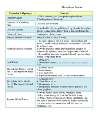
  • 1. Ahmad Amro Baradee ContentsForamina & Openings 1- Nasal emissary vein (to superior sagittal sinus) 2- Prolongation of dura mater Foramen Cecum Olfactory nerve bundles Foramina Of Cribriform Plate On each side of crista galli formed by the cribriform plate , it helps to attach the olfactory lobe to the cribriform plate Olfactory Incisure Prolongation of dura materEthmoidal Hiatus Anterior ethmoid nerve & arteryAnterior Ethmoid Foramen 1- Posterior ethmoid nerve & artery ( sphenoethmoidal nerve) (Luschka nerve) innervates the ethmoidal cells and the sphenoid sinus 2- Orbital branches of the pterygopalatine ganglion to innervate the sphenoid sinus and the posterior ethmoidal cells , and they innervate the periorbita and muscles of eye by their sympathetic fibers Posterior Ethmoid Foramen 1- Optic nerve 2- Ophthalmic artery(and the central retinal artery branch of the ophthalmic artery) Optic Canal 1- Lacrimal nerve 2- Frontal nerve 3- Trochlear nerve 4- Superior ophthalmic vein (to the cavernous sinus) The Superior Narrow Lateral Part Of The Superior Orbital Fissure 5- Abducent nerve 6- Nasocilliary nerve 7- Oculomotor nerve 8- Sympathetic branches of the cavernous plexus to the ciliary ganglion The Inferior Wide Medial Part Of The Superior Orbital Fissure 9- Orbital branch of the middle meningeal artery 10- Recurrent meningeal branch of lacrimal artery 11- One of the two branches of inferior ophthalmic vein (the other is the infraorbital vein) the inferior ophthalmic vein ends in the cavernous sinus with the superior ophthalmic vein Superior Orbital Fissure
  • 2. Ahmad Amro Baradee Surrounds the optic canal and the inferior part of the superior orbital fissure , formed by the tendons of the eye's muscles , pass through it: 1- Optic nerve 2- Ophthalmic artery 3- Abducent nerve 4- Nasocilliary nerve 5- Oculomotor nerve Zinn's Ring (Common Tendinous Ring) 1- Infraorbital nerve 2- Zygomatic nerve 3- Orbital branches of the pterygopalatine ganglion to innervate the sphenoid sinus and the posterior ethmoidal cells , and they innervate the periorbita and muscles of eye by their sympathetic fibers 4- Infraorbital artery 5- Infraorbital vein to connect with the pterygoid plexus (infraorbital vein is a branch of inferior ophthalmic vein) 6- Terminal branches of the deep anterior temporal artery which is branch of maxillary artery Inferior Orbital Fissure (Lies in the sphenoid bone , medial to the foramen ovale by an internal aspect of the base of the skull , lateral to the scaphoid fossa by an external aspect ( exists in 17% of the skulls ) A small emissary vein passes through this foramen (it connects the extracranial pterygoid plexus with the intracranial cavernous sinus ) Foramen Vesalii (Canaliculus Sphenoidalis) Maxillary nerveForamen Rotundom 1- Mandibular nerve 2- Accessory meningeal artery (branch of maxillary to nourish gasser's ganglion) 3- Lesser petrosal nerve (occasionally) 4- An emissary vein connects the extracranial pterygoid plexus with the intracranial cavernous sinus Foramen Ovale 1- Middle meningeal artery & vein 2- Meningeal branch of mandibular nerve Foramen Spinosum Lesser petrosal nerve (occasionally)Sphenotemporal Fissure
  • 3. Ahmad Amro Baradee The foramen is occluded by cartilage , the internal carotid artery travels superiorly to the cartilage. However , some nerves,arteries and veins do pass through the cartilage: 1- Greater petrosal nerve (the deep & superficial then they combine in the entrance of the pterygoid canal forming the nerve of pteryoid canal) 2- The artery of pterygoid canal 3- Meningeal branch of the ascending pharyngeal artery 4- Emissary veins connect the extracranial pterygoid plexus with the intracranial cavernous sinus Foramen Lacerum Nerve and blood vessels of pterygoid canal (Vidian nerve) Pterygoid Canal(Vidian Canal) 1- Internal carotid artery 2- Carotid sympathetic plexus 3- Venous plexus Carotid Canal 1- Greater petrosal nerve 2- Petrosal branch of middle meningeal artery Greater Petrosal Groove Lesser petrosal nerveLesser Petrosal Groove 1- Medulla oblongata surrounded by meninges 2- Two vertebral arteries 3- Meningeal branches of vertebral arteries 4- Anterior spinal artery 5- Two posterior spinal arteries 6- Spinal root of accessory nerve 7- Ascending sympathetic branches 8- Emissary veins of basilar plexus 9- Apical ligament and tectorial membrane Foramen Magnum 1- Anterior part: Glossopharyngeal nerve + inferior petrosal sinus 2- Middle part: Vagus and accessory nerve + branch of posterior meningeal artery (which is branch of ascending pharyngeal artery ) + meningeal branch of occipital artery 3- Posterior part: Sigmoid sinus + superior bulb of jugular vein Jugular Foramen Located on the lateral wall of jugular foramen (on the tympanomastoid fissure ) passes through it: The auricular branch of vagus nerve Mastoid Canaliculus 1- In the superior part: Facial nerve and it's branch , the intermediate nerve >> pass together through the facial canal (Fallopii canal) 2- In the inferior part: Vestibulocochlear nerve ( the cochlear nerve runs antero-inferiorly & the vestibular nerve runs postero-inferiorly ) Internal Auditory Meatus
  • 4. Ahmad Amro Baradee 3- Labyrinthine artery 1- Endolymphatic duct 2- Vestibular vessels 3- Prolongation of dura mater External Aperture Of The Vestibular Aqueduct Prolongation of dura mater with a small veinSubarcuate Fossa Divided by an osteoid septum into two canals: 1- The superior canal: For the tensor muscle of the tympanic membrane 2- The inferior canal: Is the auditory tube (Eustachian tube) (which presents the osteoid part of auditory tube , the other is chondroitic ) Musculotubal Canal 1- Exit: Chorda tympani nerve 2- Enter: Anterior tympanic artery (branch of maxillary) + posterior tympanic artery (branch of stylomastoid) Glasser Fissure (Petrotympanic Fissure) 1- Posterior meningeal artery 2- Occipital emissary vein Condylar Canal 1- Hypoglossal nerve 2- Meningeal branch of the ascending pharyngeal artery 3- Emissary vein of basilar plexus Hypoglossal Canal (Anterior Condyloid Canal ) Gives origion to the posterior belly of the digastric muscleMastoid Notch For the occipital artery Notch Medial To Mastoid Notch Gives origin to the tensor veli palatini muscleScaphoid Fossa 1- Parietal emissary vein 2- Branch of occipital artery Parietal Foramen 1- Mastoid emissary vein (to sigmoid sinus) 2- Mastoidal branch of occipital artery Mastoid Foramen 1- Exit: Facial nerve 2- Enter: Stylomastid artery (branch of either occipital artery 66% or posterior auricular artery 33%) Stylomastoid Foramen The inferior ganglion of glossopharyngeal nerve (Andersch ganglion) is situated in this fossula Petrosal Fossula Opens in the bottom of tympanic fossula , through it enters the tympanic nerve (Jacobson's nerve) branch of glossopharyngeal nerve Tympanic Canaliculus (Jacobson Canaliculus) 1- Nasopalatine nerve 2- Nasal nervous branches of pterygopalatine ganglion 3- Sphenopalatine artery Sphenopalatine Foramen
  • 5. Ahmad Amro Baradee "Connects the nasal cavity with the pterygopalatine fossa" 1- Pharyngeal artery branch of maxillary artery 2- Pharyngeal nerve (bock's nerve) branch of pterygopalatine ganglion and it carries postsynapatic parasympathetic fibres to mucus glands of nasopharynx Palatovaginal Canal (Pterygopalatine Canal) (Pharyngeal Canal) Pharyngeal branch of sphenopalatine arteryVomerovaginal Canal Greater and lesser palatine nerves and blood vessels Greater Palatine Canal (Pterygopalatine Canal) 1- Greater palatine nerve 2- Greater (descending) palatine artery (branch of maxillary) Greater Palatine Foramen 1- Lesser palatine nerve 2- Lesser palatine artery (branch of greater palatine artery) Lesser Palatine Foramen Greater palatine artery – greater palatine nerve –and palatine veins .. In this exact order Two Or Three Grooves In The Posterior Part Of Hard Palate 1- Nasopalatine nerve 2- Terminal branch of greater palatine artery Incisive Foramen Posterior superior alveolar nerves and arteries Alveolar Foramina Of Maxilla Supraorbital nerve and blood vessels Supraorbital Foramen (Notch) Supratrochlear nerve and blood vessels Frontal Notch (Supratrochlear Notch) Infratrochlear nerve passes by itTrochlear Fossula Located within the supero-lateral wall of the orbit (of the frontal bone) Lacrimal Gland's Fossa Located within the infero-medial wall of the orbit (of the lacrimal and maxillary bones) Lacrimal Sac's Fossa Carries tears from the lacrimal sac into the inferior meatus of the nasal cavity Nasolacrimal Duct (Tear Duct) Infraorbital nerve and blood vesselsInfraorbital Foramen Zygomaticofacial nerveZygomaticofacial Foramen Zygomaticotemporal nerve Zygomaticotemporal Foramen Located infero-medial the inferior border of the orbit , through it passes a small artery branch of angular artery to nourish the canine (exists in 1% of the skulls) Parinaud Canal
  • 6. Ahmad Amro Baradee I hope this was helpful  By: Ahmad Amro Baradee Mental nerve and blood vesselsMental Foramen Nervous branches of the cervical plexus C1-C4 , innervate the medial root of the first inferior molar Foramina Opposite The Mental Foramen (On The Lingual Side ) Inferior alveolar nerve and blood vesselsMandibular Canal (Foramen) Mylohyoid nerve and blood vesselsMylohyoid Groove This foramen is formed by the free space between the sphenomandibular ligament and the tympanomandibular ligament* , through it passes the mylohyoid neurovascular bundle (*the tympanomandibular ligament: Is a fibrous ligament strengthens the posterior part of the sphenomandibular ligament) Mylohyoid Foramen Located in the intermediate line of the lingual surface of the mandible , near the mental spines . Small blood vessels exit through it Lingual Foramen Attachment for lateral pterygoid musclePterygoid Fovea The maxillary artery passes through it to reach the pterygomaxillary region Juvara Groove ( Retrocondylar Groove )