Empfohlen

Empfohlen
Weitere ähnliche Inhalte
Ähnlich wie Posterpresentasjon_Treimo KER og JTS forslag
Ähnlich wie Posterpresentasjon_Treimo KER og JTS forslag (20)
Posterpresentasjon_Treimo KER og JTS forslag
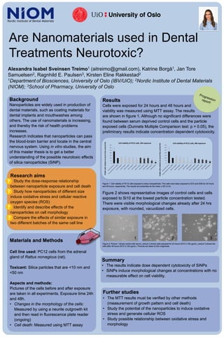
- 1. Are Nanomaterials used in Dental Treatments Neurotoxic? Alexandra Isabel Sveinsen Treimo1 (aitreimo@gmail.com), Katrine Borgå1, Jan Tore Samuelsen2, Ragnhild E. Paulsen3, Kirsten Eline Rakkestad2 1Department of Biosciences, University of Oslo (IBV/UiO); 2Nordic Institute of Dental Materials (NIOM); 3School of Pharmacy, University of Oslo Background Nanoparticles are widely used in production of dental materials, such as coating materials for dental implants and mouthwashes among others. The use of nanomaterials is increasing, and thereby the risk of health problems increases. Research indicates that nanoparticles can pass the blood-brain barrier and locate in the central nervous system. Using in vitro studies, the aim of this master thesis is to get a better understanding of the possible neurotoxic effects of silica nanoparticles (SiNP). Research aims Study the dose-response relationship between nanoparticle exposure and cell death Study how nanoparticles of different size induce oxidative stress and cellular reactive oxygen species (ROS) Identify and describe effects of the nanoparticles on cell morphology Compare the effects of similar exposure in two different batches of the same cell line Materials and Methods Cell line used: PC12 cells from the adrenal gland of Rattus norvegicus (rat). Toxicant: Silica particles that are <10 nm and <50 nm Aspects and methods: Pictures of the cells before and after exposure are taken in all experiments. Exposure time 24h and 48h. • Changes in the morphology of the cells: Measured by using a neurite outgrowth kit and then read in fluorescence plate reader (ongoing) • Cell death: Measured using MTT assay Results Cells were exposed for 24 hours and 48 hours and viability was measured using MTT assay. The results are shown in figure 1. Although no significant differences were found between serum deprived control cells and the particle exposed cells (Dunnets Multiple Comparison test: p > 0.05), the preliminary results indicate concentration dependent cytotoxicity. Figure 1: Cell viability of PC12 cells exposed to silica nanoparticles. The cells have been exposed to Si10 and Si50 for 24 hours and 48 hours, respectively. The results are presented as the mean ± SD (n=3). Figure 2 shows representative images of control cells and cells exposed to Si10 at the lowest particle concentration tested. There were visible morphological changes already after 24 hrs exposure, with rounded, vacuolized cells. Figure 2: Picture 1 shows control with serum, picture 2 shows cells exposed for 24 hours (Si10 3,125 μg/mL), picture 3 shows the cells after 48 hours (Si10 3,125 μg/mL). Pictures are taken at 20x magnitude. Summary • The results indicate dose dependent cytotoxicity of SiNPs • SiNPs induce morphological changes at concentrations with no measurable effect on cell viability. 0 50 100 150 200 250 300 350 Viablecellsrelativetoserumfreecontrol(%) Cell viability of PC12 cells, 48h exposure 0 50 100 150 200 250 300 350 Viablecellsrelativetoserumfreecontrol(%) Cell viability of PC12 cells, 24h exposure 1 2 3 Further studies • The MTT results must be verified by other methods (measurement of growth pattern and cell death) • Study the potential of the nanoparticles to induce oxidative stress and generate cellular ROS • Study possible relationship between oxidative stress and morphology