Paediatric operative dentistry
•Als PPTX, PDF herunterladen•
152 gefällt mir•24,456 views

this presentation includes fundamental of cavity preparation and modification of cavity design for different restorative materials

Empfohlen
Weitere ähnliche Inhalte
Was ist angesagt?
Was ist angesagt? (20)
Andere mochten auch
Andere mochten auch (18)
Ähnlich wie Paediatric operative dentistry
Ähnlich wie Paediatric operative dentistry (20)
Mehr von Dr. Akash Ardeshana
Kürzlich hochgeladen
Kürzlich hochgeladen (20)
Paediatric operative dentistry
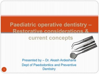
- 1. Presented by – Dr. Akash Ardeshana Dept of Paedodontics and Preventive Dentistry Paediatric operative dentistry – Restorative considerations & current concepts 1
- 2. Content Introduction History Objectives Epidemiology indication Importance of primary teeth Primary v/s permanent teeth Classifications Armamentarium Principles of cavity preparation Bibliography 2
- 3. Introduction Operative dentistry is the art and science of the diagnosis, treatment, and prognosis of defects of teeth that do not require full coverage restorations for correction. Such treatment should result in the restoration of proper tooth form, function, and esthetics while maintaining the physiologic integrity of the teeth in harmonious relationship with the adjacent hard and soft tissues, all of which should enhance the general health and welfare of the patient. - Sturdevant Operative dentistry is a subject that deals with the diagnosis, prevention and treatment of problems and conditions of natural teeth, both vital and non vital, so as to preserve the natural dentition and restore it to the best state of teeth, function and esthetics. - Gilmore 3
- 4. History Early dentists- barbers Later, cavity preparation and tooth restoration became widely popular. The first successful tooth restorations were developed in the United States. G.V.Black (1924) –” Father of operative dentistry” Charles E. Woodbury, E.K. Wedelstaedt, Waldon . Ferrier, and George Hollenback made significant contributions to the early development of operative dentistry. M.E.J.Curzon, J.F.Roberts, D.B.Kennedy.Kennedy’s Paediatric Operative Dentistry.4th edition. Reed publishing house;1996.4
- 5. Objectives (AAPD) To repair or limit the damage from caries, To protect and preserve the tooth structure, To reestablish adequate function, restore esthetics (where applicable), To provide ease in maintaining good oral hygiene Pulp vitality should be maintained whenever possible AAPD Guideline On Restorative Dentistry. American Academy Of Pediatric Dentistry . V 36 / NO 6 14 / 155
- 6. Epidemiology Primary dentition • 0-2yrs – 8% - sites lingual, interproximal areas of maxillary incisors, occlusal surfaces of 1st primary molars • 3 yrs- occlusal surfaces of 2nd primary molars > 1st primary molars; mand> maxillary Mixed dentition • Occlusal surfaces of permanent molars • Lingual pits in maxillary permanent incisors – lateral incisors Early permanent dentition • 1st permanent molar > 2nd permanent molar > premolars > maxillary anterior teeth > canine and mandibular incisors M.E.J.Curzon, J.F.Roberts, D.B.Kennedy.Kennedy’s Paediatric Operative Dentistry.4th edition. Reed publishing house;1996. 6
- 7. indication Dental caries Tooth wear Trauma Developmental defects Nikhil Marwah. Textbook of Pediatric Dentistry.3rd edition. Jaypee Brothers Medical Publishers Private Limited;20147
- 8. Importance of primary teeth Mastication Impairment of speech Esthetics Maintenance of arch length Prevents development of oral habits Prevent associated psychological effects Nikhil Marwah. Textbook of Pediatric Dentistry.3rd edition. Jaypee Brothers Medical Publishers Private Limited;2014 8
- 10. Primary v/s permanent General considerations 20 in number More white in color Morphological Considerations (crown ) Smaller and Bulbous crowns Crowns are wider mesiodistally General considerations 32 in number Darker, yellowish in color Morphological considerations (crown ) Crowns are wider Crowns are wider occlusogingivally M. S. Duggal,M. E. J. Curzon,S. A. Fayle, K. J. Toynba. Restorative Techniques in Paediatric Dentistry: An Illustrated Guide to the Restoration of Extensive Carious Primary Teeth.2nd edition. CRC Press:2002. 10
- 11. Primary v/s permanent Narrow bucco-lingual occlusal table. Mamelons are absent Contact areas in primary teeth Broader, flatter, situated farther gingivally Occlusal table is not narrow Mamelons are present Contact points in permanent teeth are placed more occlusally M. S. Duggal,M. E. J. Curzon,S. A. Fayle, K. J. Toynba. Restorative Techniques in Paediatric Dentistry: An Illustrated Guide to the Restoration of Extensive Carious Primary Teeth.2nd edition. CRC Press:2002.11
- 12. Primary v/s permanent Enamel is thin, (1mm) Dentin thickness between pulp and enamel is less hence: Caries progress is faster. Chances of pulpal exposure are more Enamel rod orientation More thicker than the primary teeth Dentine thickness is uniform Enamel rod orientation 12
- 13. Primary v/s permanent Roots are long and slender Roots have short trunk They are more divergent and flaring Undergo physiologic resorption Roots are short and robust Large undivided portion of root is seen They are less divergent and do not flare Only pathologic changes are seen 13
- 14. Classifications G.V.Black’s classification Class 1 - All pit and fissure lesions on occlusal surfaces of premolars and molars, lesions on the occlusal 2/3rds of facial and lingual surfaces of molars, and lesions on the lingual surfaces of maxillary incisors 14
- 15. Class 2 – Lesions on the proximal surface of the posterior teeth Theodore M. Roberson, Harald 0. Heymann Edward J. Swift, Jr.Sturdevant’s Art and Science of Operative Dentistry. 4th edition. St. Louis: Mosby; 2002. 15
- 16. Class 3 – Lesions on the proximal surface of the anterior teeth without involving the incisal edge Theodore M. Roberson, Harald 0. Heymann Edward J. Swift, Jr.Sturdevant’s Art and Science of Operative Dentistry. 4th edition. St. Louis: Mosby; 2002. 16
- 17. Class 4 – Lesions on the proximal surface of the anterior teeth involving the incisal edge Theodore M. Roberson, Harald 0. Heymann Edward J. Swift, Jr.Sturdevant’s Art and Science of Operative Dentistry. 4th edition. St. Louis: Mosby; 2002. 17
- 18. Class 5 – Lesions on the gingival third of the facial or lingual surfaces of all the teeth Theodore M. Roberson, Harald 0. Heymann Edward J. Swift, Jr.Sturdevant’s Art and Science of Operative Dentistry. 4th edition. St. Louis: Mosby; 2002. 18
- 19. Class 6 – Lesions on the incisal edge of the anterior teeth or the occlusal cusp tips of posterior teeth (Simon’s modification) Theodore M. Roberson, Harald 0. Heymann Edward J. Swift, Jr.Sturdevant’s Art and Science of Operative Dentistry. 4th edition. St. Louis: Mosby; 2002. 19
- 20. Finn’s modification 1. Class I: cavities involving the pits and fissures of the molar teeth and the buccal and lingual pits of all teeth. 2. Class II: cavities involving proximal surface of molar teeth with access established from the occlusal surface. 3. Class III: cavities involving proximal surfaces of anterior teeth which may or may not involve a labial or a lingual extention. M. S. Duggal,M. E. J. Curzon,S. A. Fayle, K. J. Toynba. Restorative Techniques in Paediatric Dentistry: An Illustrated Guide to the Restoration of Extensive Carious Primary Teeth.2nd edition. CRC Press:2002. 20
- 21. 4. Class IV: Cavities of the proximal surface of an anterior tooth which involve the restoration of an incisal angle. 5. Class V : Cavities present on the cervical third of all teeth including proximal surface where the marginal ridge is not included in the cavity preparation M. S. Duggal,M. E. J. Curzon,S. A. Fayle, K. J. Toynba. Restorative Techniques in Paediatric Dentistry: An Illustrated Guide to the Restoration of Extensive Carious Primary Teeth.2nd edition. CRC Press:2002.21
- 22. Sturdevant’s classification: CAVITY FEATURE Simple cavity A cavity involving only one tooth surface Compound cavity A cavity involving two surfaces of a tooth Complex cavity A cavity involves more than two surfaces of a tooth. Theodore M. Roberson, Harald 0. Heymann Edward J. Swift, Jr.Sturdevant’s Art and Science of Operative Dentistry. 4th edition. St. Louis: Mosby; 2002. 22
- 23. Mount and Hume’s classification: 1. Site 1- pit and fissure on occlusal surfaces 2. Site 2- proximal areas below contact point 3. Site 3- cervical 1/3rd of the crown M. S. Duggal,M. E. J. Curzon,S. A. Fayle, K. J. Toynba. Restorative Techniques in Paediatric Dentistry: An Illustrated Guide to the Restoration of Extensive Carious Primary Teeth.2nd edition. CRC Press:2002. 23
- 24. Mount and Hume’s classification: 1. Size1- minimal involvement of dentin 2. Size 2- moderate involvement, remaining tooth structure strong enough to support restoration 3. Size 3- large cavity with weakened tooth structure 4. Size 4- extensive caries with loss of bulk of tooth structure M. S. Duggal,M. E. J. Curzon,S. A. Fayle, K. J. Toynba. Restorative Techniques in Paediatric Dentistry: An Illustrated Guide to the Restoration of Extensive Carious Primary Teeth.2nd edition. CRC Press:2002. 24
- 25. Armamentarium G.V.Black’s classification: Cutting instruments Hand Chisels Hatchets Excavators Hoes Rotary Burs Stones Discs 25
- 26. Armamentarium Condensing instruments Hand pluggers Mechanical pluggers Theodore M. Roberson, Harald 0. Heymann Edward J. Swift, Jr.Sturdevant’s Art and Science of Operative Dentistry. 4th edition. St. Louis: Mosby; 2002. Nikhil Marwah. Textbook of Pediatric Dentistry.3rd edition. Jaypee Brothers Medical Publishers Private Limited;2014 26
- 27. Armamentarium Plastic instruments • Spatulas • Carriers • Carvers • Plastic filling instruments • Burnishers Finishing and polishing instruments Hand Rotary Orangewood sticks Finishing burs Polishing points Mounted brushes Finishing strips Rubber cups and discs Theodore M. Roberson, Harald 0. Heymann Edward J. Swift, Jr.Sturdevant’s Art and Science of Operative Dentistry. 4th edition. St. Louis: Mosby; 2002. 27
- 28. Armamentarium Isolation instruments • Rubber dam kit • Saliva ejector • Cotton rolls • High volume ejector Miscellaneous instruments • Mouth mirrors • Probes • Pliers • Cotton tweezers Theodore M. Roberson, Harald 0. Heymann Edward J. Swift, Jr.Sturdevant’s Art and Science of Operative Dentistry. 4th edition. St. Louis: Mosby; 2002. 28
- 29. Armamentarium Marzouk’s classification: Exploring instruments • Mouth mirrors • Explorers • Probes • Cotton tweezers Instruments for tooth structure removal • Hand – chisels, excavators • Rotary – headpieces, burs, abrasives Restoring instruments • Cement spatulas • Plastic filling instrument • Amalgam carriers • Condensers • Burnishers • Carvers Finishing and polishing instruments • Finishing strips • Finishing burs • Brushes • Rubber cups Nikhil Marwah. Textbook of Pediatric Dentistry.3rd edition. Jaypee Brothers Medical Publishers Private Limited;2014 29
- 30. Matrices It is thereby used as a temporary wall, which is created opposite to the axial walls, surrounding areas of the tooth structure that were lost during cavity preparation. Matrix is a device used during the restorative procedures to hold the plastic restorative material within the tooth while it is setting. 30
- 31. Matrices Classification: 1. Place of application Posterior – T-Band, Toffelmire Anterior – Celluloid matrix 2. Constituents Metallic – Ivory no.1, Ivory no.8 Non metallic – Mylar strips 3. Presence / absence of retainer With retainer – Ivory no.1, Ivory no.8 Without retainer – S-band 31
- 32. Matrices 4. Form Anatomical – Celluloid crown form Non anatomical – Ivory no.1 5. Use Universal – Ivory no.8, Toffelmire Unilateral – Ivory no.1 Edwina A. M. Kidd, Bernard G. N., Smith Timothy F, Watson H. M, Pickard. Pickard’s Manual of Operative Dentistry.8th edition. Oxford: University Press; 200332
- 33. Matrices Recent modifications of matrix Sectional matrix Easy to place, gives a large preparation area and reduces working time Mesial and distal proximal area restorations can be accomplished by one matrix Smartview matrix Comes with smartband sectional matrices and titanium instruments Non stick surfaces, anatomical contours Mostly used for composite restorations Edwina A. M. Kidd, Bernard G. N., Smith Timothy F, Watson H. M, Pickard. Pickard’s Manual of Operative Dentistry.8th edition. Oxford: University Press; 200333
- 34. Types of matrices used for tooth restoration Matrices for class I cavity (compound cavity) Double banded tofflemire Matrices for class II Single band tofflemire Ivory matrix no.1 Ivory matrix no.8 Black’s matrices Soldered band or seamless copper band matrix Anatomical matrix Auto matrix S-shaped matrix T-shaped matrix 34
- 35. Types of matrices used for tooth restoration Matrices for cavity preparation for amalgam on distal surface of cuspid S- shaped matrix Tofflemire Matrices for class III tooth colored restorations Celluloid strips Matrices for class IV tooth colored restorations Celluloid strips Aluminium foil Transparent crown form matrices Anatomic matrix Modified S-shaped band of copper, tin, aluminium 35
- 36. Types of matrices used for tooth restoration Matrices for class V amalgam restorations Window matrix S-shaped matrix Matrices for classV tooth colored restorations Anatomic matrix Aluminium or copper collars Celluloid strips 36
- 38. Wedges It is defined as a piece of wood, metal etc. one end of which is an acute angled edge formed by two converging planes used to tighten or exert force in various ways. 1883- wedges of boxwood, orangewood, balsam wood were made. 1st metal wedge- Ottolengui steel wedge. Current wedges – plastic, metal, wood, celluloid Recent wedges – Luci-wedge, Hawe-Neos dental Edwina A. M. Kidd, Bernard G. N., Smith Timothy F, Watson H. M, Pickard. Pickard’s Manual of Operative Dentistry.8th edition. Oxford: University Press; 200338
- 39. Wedges Types Acc. to anatomy Anatomical – in shape of embrassures Non- anatomical – round Acc. to material used Wooden – hard wood or soft wood Plastic – in various shapes Acc. to color Colored Light reflecting – used with composite Edwina A. M. Kidd, Bernard G. N., Smith Timothy F, Watson H. M, Pickard. Pickard’s Manual of Operative Dentistry.8th edition. Oxford: University Press; 2003 39
- 40. Isolation Rubber dam: 1864 – Sanford Christie Barnum 1882 – SS White introduced a rubber dam punch, Dr.Doleus Palmer introduced set metal clamps. • Moisture control, dry field, aseptic • Accessibility • Improved material properties • Prevents aspiration Advantage s • Patient acceptance • Trauma to tissues • Latex allergy • Frame causes indentations Disadvantag es Euphesisms Raincoat Hanger Clip 40
- 41. Armamentarium: Rubber dam sheet Frame Rubber dam punch Clamps Rubber dam retaining forceps Edwina A. M. Kidd, Bernard G. N., Smith Timothy F, Watson H. M, Pickard. Pickard’s Manual of Operative Dentistry.8th edition. Oxford: University Press; 2003 41
- 42. 42
- 43. Procedures for rubber dam placement: 43
- 44. 44
- 45. Recent advances in rubber dam Articulated frame. Safe T frame 45
- 46. Recent advances: Quick dam or insta dam Optra dam Split dam technique Optra dam Insta dam Edwina A. M. Kidd, Bernard G. N., Smith Timothy F, Watson H. M, Pickard. Pickard’s Manual of Operative Dentistry.8th edition. Oxford: University Press; 2003 46
- 47. Handi dam Dry dam. Insti dam 47
- 49. Super clamp Long guard extension clamp Tiger clamp Cushees 49
- 50. Recent alternatives to Rubber Dam Kool dam (Pulpdent Corporation) 50
- 51. Provides hands-free evacuation, retraction, and safety shielding, as well as illumination 51
- 52. Fast dam 52
- 53. Title Efficiency and patient satisfaction with the Isolite system versus rubber dam for sealant placement in pediatric patients. Authors Alhareky MS1, Mermelstein D2, Finkelman M3, Alhumaid J4, Loo C2. Pediatr Dent. 2014 Sep-Oct;36(5):400-4. Level of evidence llA Aim The purpose of this clinical study was to compare the chair time and degree of patient satisfaction after use of the Isolite system (IS) versus rubber dam (RD) during the application of pit and fissure sealants. Materials and methods The patients included in this study ranged from seven to 16 years old. In each subject, pit and fissure sealants were applied to one permanent molar in each quadrant. IS dental isolation was used on one side; RD isolation was used on the other side. Chair time was assessed using a stopwatch, and patient acceptance was evaluated using a questionnaire. Result Forty-two subjects (23 females and 19 males) were enrolled in the study. The average chair time was 19.36 minutes for the application of pit and fissure sealants on the RD side; average chair time was 10 minutes for the IS side (P<.001). Sixty-nine percent of the subjects were more comfortable using IS, while 31 percent found RD to be more comfortable (P=.02). Interpretation Isolite is a viable alternative to the conventional rubber dam. The use of Isolite is associated with reduced chair time and greater patient satisfaction. 53
- 54. Other isolation methods: Edwina A. M. Kidd, Bernard G. N., Smith Timothy F, Watson H. M, Pickard. Pickard’s Manual of Operative Dentistry.8th edition. Oxford: University Press; 54
- 55. Principles of cavity preparation Tooth preparation is defined as the mechanical alteration of a defective, injured, or diseased tooth to best receive a restorative material that will reestablish a healthy state for the tooth, including esthetic corrections where indicated, along with normal form and function. - Sturdevant 55
- 56. Principles of cavity preparation Initial tooth preparation – the mechanical alterations of the tooth are extended to sound tooth structure (sound dentin or enamel supported by noncarious dentin) in all directions (facially, lingually, gingivally, incisally or occlusally, mesially, and distally) while adhering to a specific, limited pulpal or axial depth. Outline form and initial depth Primary resistance form Primary retention form Convenience form Theodore M. Roberson, Harald 0. Heymann Edward J. Swift, Jr.Sturdevant’s Art and Science of Operative Dentistry. 4th edition. St. Louis: Mosby; 2002.56
- 57. Final tooth preparation Removal of any remaining infected dentin and old restoration Pulp protection if needed Secondary resistance and retention form Procedure for finishing the external walls Final procedures Theodore M. Roberson, Harald 0. Heymann Edward J. Swift, Jr.Sturdevant’s Art and Science of Operative Dentistry. 4th edition. St. Louis: Mosby; 2002. 57
- 58. Outline form and initial depth: • placing the preparation margins in the positions they will occupy in the final preparation • preparing an initial depth of 0.2 to 0.8 mm pulpally of the DEJ position Principles involved 1. all friable and/or weakened enamel should be removed 2. all faults should be included 3. all margins should be placed in a position to afford good finishing of the margins of the restoration. Theodore M. Roberson, Harald 0. Heymann Edward J. Swift, Jr.Sturdevant’s Art and Science of Operative Dentistry. 4th edition. St. Louis: Mosby; 2002.58
- 59. Features of outline form: preserving cuspal strength preserving marginal ridge strength minimizing faciolingual extensions using enameloplasty connecting two close (less than 0.5 mm apart) faults or tooth preparations restricting the depth of the preparation into dentin to a maximum of 0.2 mm for pit-and-fissure caries and 0.2 to 0.8 mm for the axial wall of smooth surface caries Theodore M. Roberson, Harald 0. Heymann Edward J. Swift, Jr.Sturdevant’s Art and Science of Operative Dentistry. 4th edition. St. Louis: Mosby; 2002. 59
- 60. Primary resistance form: It is defined as that shape and placement of the preparation walls that best enable both the restoration and the tooth to withstand, without fracture, masticatory forces delivered principally in the long axis of the tooth. Principle: to use the box shape with a relatively flat floor to restrict the extension of the external walls to have a slight rounding (coving) of internal line angles in extensive tooth preparations, to cap weak cusps and envelope or include enough of a weakened tooth to provide enough thickness of restorative material to bond the material to tooth structure when appropriateTheodore M. Roberson, Harald 0. Heymann Edward J. Swift, Jr.Sturdevant’s Art and Science of Operative Dentistry. 4th edition. St. Louis: Mosby; 2002. 60
- 61. A box preparation will prevent restoration from rocking, whereas a rounded cavity preparation will lead to rocking of the restoration within the cavity. Features: Relatively flat floors Box shape Inclusion of weakened tooth structure Preservation of cusps and marginal ridges Rounded internal line angles Adequate thickness of restorative material Reduction of cusps for capping when indicated 61
- 62. Primary retention form: shape or form of the conventional preparation that resists displacement or removal of the restoration from tipping or lifting forces. In class I and class II cavity – the external walls are made converging Theodore M. Roberson, Harald 0. Heymann Edward J. Swift, Jr.Sturdevant’s Art and Science of Operative Dentistry. 4th edition. St. Louis: Mosby; 2002. 62
- 63. Convenience form: shape or form of the preparation that provides for adequate observation, accessibility, and ease of operation in preparing and restoring the tooth. obtaining this form, may necessitate extension of distal, mesial, facial, or lingual walls to gain adequate access to the deeper portion of the preparation occlusal divergence of vertical (longitudinal) walls of tooth preparations for Class II cast restorations extending proximal preparations beyond proximal contacts Theodore M. Roberson, Harald 0. Heymann Edward J. Swift, Jr.Sturdevant’s Art and Science of Operative Dentistry. 4th edition. St. Louis: Mosby; 2002. 63
- 64. Removal of any remaining enamel pit or fissure, infected dentin, and/or old restorative material, if indicated: Affect the esthetics Affect the retention Radiographic evidence indicates presence of caries below the old restoration. The tooth pulp was symptomatic preoperatively The periphery of the remaining old restorative material was not intact Theodore M. Roberson, Harald 0. Heymann Edward J. Swift, Jr.Sturdevant’s Art and Science of Operative Dentistry. 4th edition. St. Louis: Mosby; 2002. 64
- 65. In large preparations with extensive soft caries, the removal of infected dentin may be accomplished early in the initial tooth preparation. Large areas of soft caries usually are best removed with spoon excavators by flaking up the caries around the periphery of the infected mass and peeling it off in layers. For harder caries removal, round steel burs at very low speed, and round carbide burs rotating at high speeds. Removal of remaining old restorative material, when indicated: Also is accomplished with use of a round carbide bur, at slow speed Theodore M. Roberson, Harald 0. Heymann Edward J. Swift, Jr.Sturdevant’s Art and Science of Operative Dentistry. 4th edition. St. Louis: Mosby; 2002. 65
- 66. Pulp protection, if indicated: The reason for using traditional liners or bases is to either protect the pulp or to aid pulpal recovery or both. When the thickness of the remaining dentin is minimal, heat generated by injudicious cutting can result in a pulpal burn lesion, an abscess formation, or pulpal necrosis. Some ingredients of various materials Thermal changes conducted through restorative materials Forces transmitted through materials to the dentin Galvanic shock The ingress of noxious products and bacteria through microleakage. Theodore M. Roberson, Harald 0. Heymann Edward J. Swift, Jr.Sturdevant’s Art and Science of Operative Dentistry. 4th edition. St. Louis: Mosby; 2002. 66
- 67. Secondary resistance and retention forms: 1. Mechanical preparation features 2. Treatments of the preparation walls with etching, priming, and adhesive materials Many compound and complex preparations require these additional features. When a tooth preparation includes both occlusal and proximal surfaces, each of those areas should have independent retention and resistance features. Theodore M. Roberson, Harald 0. Heymann Edward J. Swift, Jr.Sturdevant’s Art and Science of Operative Dentistry. 4th edition. St. Louis: Mosby; 2002. 67
- 68. 1. Mechanical preparation features Retention locks, grooves, and coves Vertically oriented retention locks and retention grooves are used to provide additional retention for proximal portions of some tooth preparations. The locks are for amalgams and the grooves are for cast metal restorations. Horizontally oriented retention grooves are prepared in most Classes III and V preparations for amalgam and in some root surface tooth preparations for composite. Retention coves are appropriately placed undercuts for the incisal retention of Class III amalgams, occlusal portion of some amalgam restorations Theodore M. Roberson, Harald 0. Heymann Edward J. Swift, Jr.Sturdevant’s Art and Science of Operative Dentistry. 4th edition. St. Louis: Mosby; 2002. 68
- 70. 2. Placement of Etchant, Primer, or Adhesive on Prepared Walls In addition to mechanical alterations to the tooth preparation, certain alterations to the preparation walls by actions of various materials also afford increased retention, as well as resistance to fracture. Theodore M. Roberson, Harald 0. Heymann Edward J. Swift, Jr.Sturdevant’s Art and Science of Operative Dentistry. 4th edition. St. Louis: Mosby; 2002. Nikhil Marwah. Textbook of Pediatric Dentistry.3rd edition. Jaypee Brothers Medical Publishers Private Limited;201470
- 71. Procedures for finishing the external walls: Finishing the external walls of the preparation entails consideration of both degree of smoothness and cavosurface design, since each restorative material has its maximum effectiveness when the appropriate conditions are developed for that specific material. When a preparation has extended onto the root surface (no enamel present), the root-surface cavosurface angle should be either 90 degrees (for amalgam, composite, or porcelain restorations) or beveled (for intracoronal cast metal restorations). 71
- 72. The 90-degree root-surface margin provides a butt joint relationship between the restorative material and the cementum /dentin preparation wall, a configuration that provides appropriate strength to both. 72
- 73. Objectives a. Create the best marginal seal possible between the restorative material and the tooth structure b. Afford a smooth marginal junction c. Provide maximum strength of both the tooth and the restorative material at and near the margin Factors a. The direction of the enamel rods b. Support of the enamel rods both at the DEJ and laterally (preparation side) c. The type of restorative material to be placed in the preparation d. The location of the margin e. The degree of smoothness or roughness desired Theodore M. Roberson, Harald 0. Heymann Edward J. Swift, Jr.Sturdevant’s Art and Science of Operative Dentistry. 4th edition. St. Louis: Mosby; 2002. Nikhil Marwah. Textbook of Pediatric Dentistry.3rd edition. Jaypee Brothers Medical Publishers Private Limited;201473
- 74. Final procedures: cleaning, inspecting, and sealing: Removing all chips and loose debris that have accumulated, drying the preparation (do not desiccate), and making a final complete inspection of the preparation for any remaining infected dentin, unsound enamel margins, or any condition that renders the preparation unacceptable to receive the restorative material. Theodore M. Roberson, Harald 0. Heymann Edward J. Swift, Jr.Sturdevant’s Art and Science of Operative Dentistry. 4th edition. St. Louis: Mosby; 2002. Nikhil Marwah. Textbook of Pediatric Dentistry.3rd edition. Jaypee Brothers Medical Publishers Private Limited;201474
- 75. Tooth preparation walls: Internal Wall- An internal wall is a prepared (cut) surface that does not extend to the external tooth surface. Axial wall-An axial wall is an internal wall parallel with the long axis of the tooth. Pulpal wall-A pulpal wall is an internal wall that is both perpendicular to the long axis of the tooth and occlusa of the pulp Externa l walls Internal walls Theodore M. Roberson, Harald 0. Heymann Edward J. Swift, Jr.Sturdevant’s Art and Science of Operative Dentistry. 4th edition. St. Louis: Mosby; 2002. M.E.J.Curzon, J.F.Roberts, D.B.Kennedy.Kennedy’s Paediatric Operative Dentistry.4th edition. Reed publishing house;1996. 75
- 76. External Wall-An external wall is a prepared (cut) surface that extends to the external tooth surface. Floor (or Seat)-A floor (or seat) is a prepared (cut) wall that is reasonably flat and perpendicular to those occlusal forces that are directed occlusogingivally (generally parallel to the long axis of the tooth). Enamel Wall-The enamel wall is that portion of a prepared external wall consisting of enamel. Dentinal Wall-The dentinal wall is that portion of a prepared External walls Internal walls Theodore M. Roberson, Harald 0. Heymann Edward J. Swift, Jr.Sturdevant’s Art and Science of Operative Dentistry. 4th edition. St. Louis: Mosby; 2002. M.E.J.Curzon, J.F.Roberts, D.B.Kennedy.Kennedy’s Paediatric Operative Dentistry.4th edition. Reed publishing house;1996. 76
- 77. Tooth preparation angles: Line Angle- A line angle is the junction of two planal surfaces of different orientation along a line. An internal line angle is a line angle whose apex points into the tooth. An external line angle is a line angle whose apex points away from the tooth. Point Angle - A point angle is the junction of three planal surfaces of different orientation Theodore M. Roberson, Harald 0. Heymann Edward J. Swift, Jr.Sturdevant’s Art and Science of Operative Dentistry. 4th edition. St. Louis: Mosby; 2002. M.E.J.Curzon, J.F.Roberts, D.B.Kennedy.Kennedy’s Paediatric Operative Dentistry.4th edition. Reed publishing house;1996. 77
- 78. Theodore M. Roberson, Harald 0. Heymann Edward J. Swift, Jr.Sturdevant’s Art and Science of Operative Dentistry. 4th edition. St. Louis: Mosby; 2002. M. S. Duggal,M. E. J. Curzon,S. A. Fayle, K. J. Toynba. Restorative Techniques in Paediatric Dentistry: An Illustrated Guide to the Restoration of Extensive Carious Primary Teeth.2nd edition. CRC Press:2002. Edwina A. M. Kidd, Bernard G. N., Smith Timothy F, Watson H. M, Pickard. Pickard’s Manual of Operative Dentistry.8th edition. Oxford: University Press; 2003 M.E.J.Curzon, J.F.Roberts, D.B.Kennedy.Kennedy’s Paediatric Operative Dentistry.4th edition. Reed publishing house;1996. Nikhil Marwah. Textbook of Pediatric Dentistry.3rd edition. Jaypee Brothers Medical Publishers Private Limited;2014 78
- 79. 79
- 80. Presented by – Dr. Akash Ardeshana Dept of Paedodontics and Preventive Dentistry Paediatric operative dentistry – Restorative considerations & current concepts 80
- 81. content Amalgam preparation for primary molars Cavity preparation for adhesive material Modification of cavity design Step for composite restoration Step for GIC restoration Summary Bibliography 81
- 82. amalgam preparation Dental amalgam has been the most commonly used restorative material in posterior teeth for over 150 years and is still widely used throughout the world today. Dental amalgam has declined in use over the past decade, perhaps due to the controversy surrounding perceived health effects of mercury vapor, environmental concerns from its mercury content, and increased demand for esthetic alternatives. 82 AAPD Guideline on Restorative Dentist. REFERENCE MANUAL V 36 / NO 6 14 /15
- 83. With regard to safety of dental amalgam, a comprehensive literature review of dental studies published between 2004 and 2008 found insufficient evidence of associations between mercury release from dental amalgam and the various medical complaints. 83 AAPD Guideline on Restorative Dentist. REFERENCE MANUAL V 36 / NO 6 14 /15
- 84. However, on July 28, 2009, the Food and Drug Administration (FDA) issued a “final rule” that reclassified dental amalgam to a Class II device (having some risk) and designated guidance that included warning labels regarding: (1) possible harm of mercury vapors; (2) disclosure of mercury content; (3) contraindications for persons with known mercury sensitivity. 84 AAPD Guideline on Restorative Dentist. REFERENCE MANUAL V 36 / NO 6 14 /15
- 85. With regard to clinical efficacy of dental amalgam, results comparing longevity of amalgam to other restorative materials are inconsistent. 85 AAPD Guideline on Restorative Dentist. REFERENCE MANUAL V 36 / NO 6 14 /15
- 86. Title The longevity of amalgam versus compomer/composite restorations in posterior primary and permanent teeth: findings From the New England Children's Amalgam Trial. Authors Soncini JA1, Maserejian NN, Trachtenberg F, Tavares M, Hayes C. Level of evidence IIb Aim The authors compared replacement rates of these types of restorations in posterior teeth during the five-year follow-up of the New England Children's Amalgam Trial. Materials and methods The authors randomized children aged 6 to 10 years who had two or more posterior occlusal carious lesions into groups that received amalgam (n=267) or compomer (primary teeth)/composite (permanent teeth) (n=267) restorations and followed them up semiannually. They compared the longevity of restorations placed on all posterior surfaces using random effects survival analys. Result The average+/-standard deviation follow-up was 2.8+/-1.4 years for primary tooth restorations and 3.4+/-1.9 years for permanent tooth restorations. In primary teeth, the replacement rate was 5.8 percent of compomers versus 4.0 percent of amalgams (P=.10), with 3.0 percent versus 0.5 percent (P=.002), respectively, due to recurrent caries. In permanent teeth, the replacement rate was 14.9 percent of composites versus 10.8 percent of amalgams (P=.45), and the repair rate was 2.8 percent of composites versus 0.4 percent of amalgams (P=.02). conclusion Although the overall difference in longevity was not statistically significant, compomer was replaced significantly more frequently owing to recurrent caries, and composite restorations required seven times as many repairs as did amalgam restorations. 86
- 87. The survival rate of the amalgam restorations was 94.4 percent; that of composite restorations was 85.5 percent. Annual failure rates ranged from 0.16 to 2.83 percent for amalgam restorations and from 0.94 to 9.43 percent for composite restorations. Secondary caries was the main reason for failure in both materials. Risk of secondary caries was 3.5 times greater in the composite group. 87 AAPD Guideline on Restorative Dentist. REFERENCE MANUAL V 36 / NO 6 14 /15
- 88. Rounded-end, high-speed carbide burs No. 329, No. 330, No. 245, and No. 256, which may be used for cutting cavity preparations.88
- 89. Modifications of cavity preparation in primary teeth: 1. Class I Narrow occlusal table – buccolingual dimension of cavity is reduced 0.5mm pulpal to DEJ Maximum intercuspal width should be minimum Walls should be parallel or slightly convergent Outline form should limit to the central pit, its buccal and lingual grooves and triangular fossa Pulpal floor should be slightly concave Total depth of the cavity – 1.5 All pits and fissures should be included Paul S. Casamassimo, Henry W. Fields Jr., Dennis J. McTigue, Arthur Nowak. Pediatric Dentistry: Infancy through Adolescence.5th edition. Saunders: Elsevier ; 2013 89
- 90. Steps External outline form Start preparation with a no.330 bur, perpendicular to occlusal surface- mesial to distal Include all deep and defective grooves blend the outline to form a smooth flowing arcs and curves Contour the outline parallel to mesial and distal marginal ridges Width of cavity – 1/3rd intercuspal width Internal outline form 0.50-1.0 mm into the dentin – 330 bur Round line angles – no.330 bur Converging walls Sharp cavosurface angle – 169L bur 90
- 91. Common errors in Class I amalgam restorations 1. Preparing cavity too deep 2. Undercutting marginal ridges 3. Carving the anatomy of amalgam too deep 4. Udercarving that leads to fracture 5. Not including all susceptible fissures 91
- 93. 2. Class II Occlusal box 1st molars – extend the occlusal box half way mesiodistally like dovetail Mandibular 2nd molar – all pits and fissures should be included Maxillary 2nd molars – nearest occlusal pit should be included Sharp cavosurface angle Rounded / beveled/ grooved axiopulpal line angle Proximal box – greater width More buccolingual extension of gingival seat / floor Occlusal convergence Axial wall should follow contour of external wall and 0.5 mm in dentine No retention grooves M. S. Duggal,M. E. J. Curzon,S. A. Fayle, K. J. Toynba. Restorative Techniques in Paediatric Dentistry: An Illustrated Guide to the Restoration of Extensive Carious Primary Teeth.2nd edition. CRC Press:2002. Paul S. Casamassimo, Henry W. Fields Jr., Dennis J. McTigue, Arthur Nowak. Pediatric Dentistry: Infancy through Adolescence.5th edition. Saunders: Elsevier ; 2013 93
- 94. Kennedy (1997) contraindicated the idea of dovetail lock, as in primary teeth occlusal fissures are prepared which produces curved shape that provides retention. Rodda recommended 1mm depth of cavity because the distance between the mesial surface of mandibular 1st molar and pulp horn is only 1.6 mm. M. S. Duggal,M. E. J. Curzon,S. A. Fayle, K. J. Toynba. Restorative Techniques in Paediatric Dentistry: An Illustrated Guide to the Restoration of Extensive Carious Primary Teeth.2nd edition. CRC Press:2002. 94
- 95. Common errors in Class II amalgam restorations 1. Failure to extend occlusal outline into all susceptible pits and fissures 2. Failure to follow the outline of the cusps 3. Isthmus cut too wide 4. Flare of the proximal wall too great 5. Angle formed by the axial, buccal and lingual walls too great 6. Gingival contact not broken 7. Axial wall not confirming to the proximal contour of the tooth 95 Paul S. Casamassimo, Henry W. Fields Jr., Dennis J. McTigue, Arthur Nowak. Pediatric Dentistry: Infancy through Adolescence.5th edition. Saunders: Elsevier ; 2013
- 96. Messer and Levering reported that SSCs placed in 4 your old and younger children showed a success rate approximately twice that of class II amalgam restoration up to 10 years. Roberts and Sherriff reported that after 5 year, one third of class II amalgams placed in primary teeth had failed or required replacement, whereas only 8% of SSCs required retreatment. 96 Paul S. Casamassimo, Henry W. Fields Jr., Dennis J. McTigue, Arthur Nowak. Pediatric Dentistry: Infancy through Adolescence.5th edition. Saunders: Elsevier ; 2013
- 97. Title Factors influencing dentists’ choice of amalgam and tooth- colored restorative materials for Class II preparations in younger patients Authors Simen Vidnes-kopperud, Anne Bjørg Tveit, Torunn Gaarden, Leiv Sandvik & Ivar Espelid. Acta Odontologica Scandinavica, 2009; 67: 74-79 Level of evidence III Aim To identify factors associated with dentists’ decisions on choice of restorative material in children and adolescents. Materials and methods In the period 20012004, 27 dentists in the Public Dental Health Service in Norway placed 4030 Class II restorations in 1912 patients. The reason for placement, previous caries experience (DMFT), oral hygiene, and characteristics of the cavity were recorded. Results The most frequently used material was resin composite (81.5%), followed by compomer (12.7%), amalgam (4.6%), and glass ionomer cement (1.2%). Tooth-colored restorations were more frequently placed than amalgam in younger patients (p0.017). Female patients received fewer amalgam restorations than male patients (p0.006). The findings indicate that in a period when the use of amalgam 97
- 98. Problems with amalgam restorations: Accounts for anatomic or morphologic structural characteristics. Fracture of isthmus in Class II amalgam restorations Marginal failure in the proximal box, due to excessive flare of the cavosurface angle Failure to remove all caries or to extend the cavity into caries susceptible areas Paul S. Casamassimo, Henry W. Fields Jr., Dennis J. McTigue, Arthur Nowak. Pediatric Dentistry: Infancy through Adolescence.5th edition. Saunders: Elsevier ; 2013 98
- 99. Adhesive materials in primary teeth In early mid 1960’s, composite or resin based composites were suggested as replacement for class I and class II amalgam restorations. In due course of time, with the improvement in resin based composites due to smaller filler particle, increase in material strength and improvement of dentin bonding agents, led to improved clinical results. Norman et al reported that both amalgam and resin based composites produced satisfactory results over a long duration, the only difference seen were due to poorer marginal integrity for amalgam and greater wear rate for resins.Paul S. Casamassimo, Henry W. Fields Jr., Dennis J. McTigue, Arthur Nowak. Pediatric Dentistry: Infancy through Adolescence.5th edition. Saunders: Elsevier ; 2013 99
- 100. Class I and Class II preparation: Absolute moisture control is required Unlike amalgam preparations, there is no current consensus about the precise design of Class II preparation for primary teeth to receive an adhesive material. Leinfelder recommended that Class II preparation be primarily restricted to the region of caries, with no occlusal extent A short bevel to the cavosurface margin should be given to increase surface area for bonding Wedges or matrices should be used before restoration is done for better contact M. S. Duggal,M. E. J. Curzon,S. A. Fayle, K. J. Toynba. Restorative Techniques in Paediatric Dentistry: An Illustrated Guide to the Restoration of Extensive Carious Primary Teeth.2nd edition. CRC Press:2002. Paul S. Casamassimo, Henry W. Fields Jr., Dennis J. McTigue, Arthur Nowak. Pediatric Dentistry: Infancy through Adolescence.5th edition. Saunders: Elsevier ; 2013100
- 101. 101
- 102. Title Split mouth randomized controlled clinical trial of beveled cavity preparations in primary molars: an 18-Month follow up. Authors Oliveira CA1, Dias PF, Dos Santos MP, Maia LC J Dent. 2008 Sep;36(9):754-8. doi: 10.1016/j.jdent.2008.05.006. Epub 2008 Jun 25. Level of evidence IB Aim This split-mouth, double-blind, randomized controlled study evaluated the clinical performance of composite restorations in Class I beveled margin cavity preparations in primary molars. Materials and methods A total of 94 Class I cavity preparations were performed in the carious primary molars of 32 children aged 4-10 years. Two cavity designs were used: conventional conservative preparation (G1) and modified preparation with cavosurface bevel (G2). All teeth were restored using TPH Spectrumtrade mark. The restorations were evaluated by two calibrated investigators at baseline, 6, 12 and 18 months using slight modified USPH criteria. The visible plaque index and fiber optic transilumination (FOTI) were also used. result Of the 94 restorations performed and evaluated at baseline, 76 restorations were available after 18 months. Seventy-three restorations were considered as clinical success, 10 were censored for drop out reasons and 5 were lost by natural exfoliation. Four restorations failed by secondary caries and two failed by marginal adaptation discrepancies. For all evaluation there was difference between the baseline, 6, 12 and 18 months (p<0.05) but there were no statistically significant difference in G1 and G2 for any criteria evaluated (p>0.05) at any period of the study. The visible plaque index was correlated to secondary caries.102
- 103. Small Lesions. Very small incipient proximal lesions may be chemically restored with topical fluoride therapy provided by the dentist, along with the judicious use of fluoride products designed for topical application at home. If this treatment regimen is accompanied by improved diet and improved oral hygiene, some incipient proximal lesions may remineralize or remain in an arrested state indefinitely. 103 Dean JA, Averry DR, McDonald RE. McDonald and Avery’s Dentistry for child and adeloscent. Ninth edition, Elsevier, India 2011.
- 104. In otherwise sound teeth free of susceptible pits and fissures, accessing small class II carious lesions via small openings in the marginal ridges or in the facial surfaces of the teeth is becoming a popular technique. 104
- 105. 105
- 107. Tunnel This type of cavity design could be used when the contact area may remain sound and the marginal ridge may be quite strong, provided the lesion is more than 2.5 mm below the crest of the marginal ridge. Access to the lesion through the occlusal surface should be limited to the extent required to achieve visibility and, where possible, should be undertaken from an area that is not under direct occlusal load. For most patients, there is a fossa immediately medial to the marginal ridge that is the most suitable position for initial entry. 107 Mount GJ. Minimal intervention dentistry: Rationale of cavity design. Oper Dent 2003;28:92-9.
- 108. Resin composite is not indicated for restoration of these lesions because it will not be possible to access the proximal lesion to a sufficient degree to be able to reliably remove all demineralized enamel. Also, it will not be possible to provide a beveled margin to ensure proper adaptation of the resin to the enamel. On the other hand, glass ionomer will flow readily into a small cavity and has the ability to remineralized the enamel margins and any dentin on the axial wall that may be demineralized. 108 Mount GJ. Minimal intervention dentistry: Rationale of cavity design. Oper Dent 2003;28:92-9.
- 109. Indications and contraindications • Use of tunnel preparation can be considered when small, proximal carious lesions necessitate restoration • Large carious lesion are diagnosed, where access is particularly difficult. •Overlying marginal ridge is subjected to heavy occlusion or demonstrates a crack 109
- 110. Fig. 1. Initial approach – Enter the lesion from the occlusal fossa aiming towards the lesion. Fig. 2. Gain access – Turn the bur vertical and lean it buccally and lingually to ‘funnel’ the cavity for visibility. Fig. 3. Completed cavity – axial wall left untouched. Fig. 4. GIC Restoration done110
- 111. Advantages Preserves the marginal ridge - conservative approach Less potential for a restorative overhang Perimeter of the restoration is reduced, decreasing the potential for micro leakage. Potential for disturbance of the adjacent tooth is reduced 111
- 112. Disadvantages • Highly technique sensitive, demanding careful control of the preparation by the operator • Angulations of preparation often passes close pulp • Visibility is decreased and caries removal is more uncertain - • Fragile marginal ridge 112
- 113. Slot cavity preparation It is the creation of a small slot on the proximal aspect of posterior teeth. (Mount and Ngo 2000) Indicated if there is a small lesion involving the area of or below the marginal ridge only in deciduous teeth. The outline form will be dictated entirely by the extent of the breakdown of the enamel, removing only that which is friable and easily eliminated without applying undue pressure. Retention will be through adhesion, so it is only necessary to clean the walls around the full circumference of the lesion, leaving the axial wall because it will be affected by dentin only. Hugh Devlin. Operative Dentistry-A Practical Guide to Recent Innovations.1st edition. Springer. Germany:2006. Nikhil Marwah. Textbook of Pediatric Dentistry.3rd edition. Jaypee Brothers Medical Publishers Private Limited;2014 113
- 114. • Whenever required the contact area should be preserved while cavity preparation for maintaining a good contact area. • For such a lesion, resin composite may be a useful material because on many occasions there will be an enamel margin around the full circumference. • However, glass ionomer is still a sound option because the occlusal load will not be great and the ion exchange will remain valuable both for adhesion and remineralization. 114
- 115. Fig. 1. Small carious lesion on the proximal surface of the first bicuspid Fig. 2. Slot cavity preparation Fig. 3. Finished slot cavity Fig . 4 Resin modified GIC Restoration done115
- 116. Proximal Approach A further, very conservative approach to restoring a proximal lesion. It can be achieved on limited occasions only when the proximal surface of a tooth becomes accessible at the time of cavity preparation in an adjacent tooth. The lesion may have been revealed through radiographs or it may be noted only during cavity preparation. When such an approach is possible, it leads to considerable conservation of natural tooth structure. 116 Mount GJ. Minimal intervention dentistry: Rationale of cavity design. Oper Dent 2003;28:92-9.
- 117. Restoration of primary incisor and canine Indication : Caries Trauma Developmental defects 117
- 118. Adhesive material usually resin based composite or resin ionomer products, are placed into class II and class V restorations in primary teeth. class IV restoration may also be done. 118
- 119. Class III restotrations: Preparations should be kept very small, due to large pulp chamber A slot preparation, with a short cavosurface bevel is recommended In children with bruxism when such restorations are done, an additional mechanical retention is required Paul S. Casamassimo, Henry W. Fields Jr., Dennis J. McTigue, Arthur Nowak. Pediatric Dentistry: Infancy through Adolescence.5th edition. Saunders: Elsevier ; 2013 119
- 120. Retentive locks on facial and lingual surfaces and by beveling the cavosurface margin to increase the surface area Restoring the distal surface of canines, a proximal box directed at a different angle towards the gingiva is essential A dovetail maybe also placed on the facial surface for resin restorations and lingual dovetail for amalgam restoration 120
- 121. 121
- 122. 122
- 123. Mandibular primary incisors with small proximal carious lesions may not require conventional restorations at all. Enameloplasty of the affected proximal surface (usually described as "disking") to open the proximal contact and to remove most, if not all, of the cavitation, followed by topical treatments with fluoride varnish, will often suffice until the teeth exfoliate naturally. Extraction is usually indicated when mandibular primary incisors have extensive caries.123 Paul S. Casamassimo, Henry W. Fields Jr., Dennis J. McTigue, Arthur Nowak. Pediatric Dentistry: Infancy through Adolescence.5th edition. Saunders: Elsevier ; 2013
- 124. Class V restorations: Pulpal wall should be convex, parallel to the outer enamelsurface Lateral walls are slightly flared near proximal surfaces to prevent undermining of the enamel The final preparation should include all the carious tooth structure A short bevel is placed around the entire cavosurface margin. Cavity depth approximately 1 mm from the outer enamel surface. Mechanical retention can be given. M. S. Duggal,M. E. J. Curzon,S. A. Fayle, K. J. Toynba. Restorative Techniques in Paediatric Dentistry: An Illustrated Guide to the Restoration of Extensive Carious Primary Teeth.2nd edition. CRC Press:2002. Paul S. Casamassimo, Henry W. Fields Jr., Dennis J. McTigue, Arthur Nowak. Pediatric Dentistry: Infancy through Adolescence.5th edition. Saunders: Elsevier ; 2013 124
- 125. Restoration of proximal incisal caries in primary anterior teeth 125
- 126. McEvoy has described a similar preparation and restoration for primary incisors, except that the retentive locking component is placed on the labial surface only in the gingival one third of the tooth.'' The lock extends minimally across two thirds of the labial surface and may extend even farther to include decalcified enamel in the cervical area. 126
- 127. Tooth preparation for composite restoration Basically, the tooth preparation for a composite restoration includes: 1. Removing the fault, defect, old material, or friable tooth structure 2. Creating prepared enamel margins of 90 degrees or greater (greater than 90 degrees usually preferable) 3. Creating 90-degree (or butt joint) cavosurface margins on root surfaces 4. Roughening the prepared tooth structure (enamel and dentin) with a diamond stone 127 Theodore M. Roberson, Harald 0. Heymann Edward J. Swift, Jr.Sturdevant’s Art and Science of Operative Dentistry. 4th edition. St. Louis: Mosby; 2002.
- 128. Differences from amalgam restoration include: 1. Less outline extension (adjacent suspicious or at-risk areas [grooves or pits] may be "sealed" rather than restored) 2. An axial and/or pulpal wall of varying depth (not uniform) 3. Incorporation of an enamel bevel at some areas (the width of which is dictated by the need for secondary retention) 4. Tooth preparation walls being rough (to increase the surface area for bonding) 5. Use of a diamond stone (to increase the128 Theodore M. Roberson, Harald 0. Heymann Edward J. Swift, Jr.Sturdevant’s Art and Science of Operative Dentistry. 4th edition. St. Louis: Mosby; 2002.
- 129. Steps Administration of anasthesi, rubber dam and wooden wedge in inter proximal area A small round or pear-shaped diamond bur in a high- speed handpiece to gain access to the caries. The occlusal outline should not extend into all the fissures but needs to incorporate a small isthmus and a dovetail for retention. Create access, and remove caries with a no. 330 bur Deeper caries should be removed using a slow-speed round bur. 129
- 130. 130 Place a sectional matrix or plastic matrix. Etch preparation for 15 to 20 second Placed dentine bonding agent With plastic instrument of pressure syringe, place the composite in the preparation Finishing and polishing Remove the rubber dam
- 131. 131
- 132. 132
- 133. Steps for GIC restoration Removal of caries and prepare the cavity surface as smooth as possible. Clean tooth surface using a slurry of plain pumice and water. Condition cavity with cavity conditioner ––20% polyacrylic acid for 10 seconds. Wash vigorously with water spray for 30 seconds. Dry for 5 seconds lightly but not dehydrate the surface. Application of the GIC materials with the help of celluloid strip or matrix. Light cure for RMGIC. 133
- 134. 134
- 135. Class II cavity for GIC restoration 135
- 136. Summary Paediatric dentistry is a dynamic combination of ever improving materials and tried and true techniques. Though many aspects of primary teeth restoration have not changed for decades. Still there has been a tremendous development when in comes to newer materials or techniques in restoration of primary teeth. 136
- 137. Bibliography AAPD Guideline on Restorative Dentist. REFERENCE MANUAL V 36 / NO 6 14 /15 Paul S. Casamassimo, Henry W. Fields Jr., Dennis J. McTigue, Arthur Nowak. Pediatric Dentistry: Infancy through Adolescence.5th edition. Saunders: Elsevier ; 2013 Dean JA, Averry DR, McDonald RE. McDonald and Avery’s Dentistry for child and adeloscent. Ninth edition, Elsevier, India 2011. M. S. Duggal,M. E. J. Curzon,S. A. Fayle, K. J. Toynba. Restorative Techniques in Paediatric Dentistry: An Illustrated Guide to the Restoration of Extensive Carious Primary Teeth.2nd edition. CRC Press:2002. 137
- 138. 138 Soncini JA, Maserejian NN, Trachtenberg F, Tavares M, Hayes C. The longevity of amalgam versus compomer/composite restorations in posterior primary and permanent teeth: findings From the New England Children's Amalgam Trial. The Journal of the American Dental Association. 2007 Jun 30;138(6):763-72. Oliveira CA, Dias PF, dos Santos MP, Maia LC. Split mouth randomized controlled clinical trial of beveled cavity preparations in primary molars: an 18-Month follow up. Journal of dentistry. 2008 Sep 30;36(9):754-8 Mount GJ. Minimal intervention dentistry: Rationale of cavity design. Oper Dent 2003;28:92-9. Hübel S1, Mejàre I.Conventional versus resin- modified glass-ionomer cement for Class II restorations in primary molars. A 3-year clinical study.
- 139. 139 Hugh Devlin. Operative Dentistry-A Practical Guide to Recent Innovations.1st edition. Springer. Germany:2006. Nikhil Marwah. Textbook of Pediatric Dentistry.3rd edition. Jaypee Brothers Medical Publishers Private Limited;2014
- 140. 140
- 141. Differences in amalgam cavity preparation in primary and permanent tooth 141 Primary tooth Permanent tooth Intercuspal width should not more than one third One forth or 1.5 mm Cavity depth : 1.5 mm Minimulm: 1.5 to 2 mm Proximal box •No gingival bevel •Gingival floor incline occlussaly •Retentive groove is not indicated Proximal box: •Gingival bevel should be given •Gingival floor is perpendicular to axial wall •Retentive groove for secondary retention form Width of proximal box : 1mm Proximal box should be 0.2- 0.8 mm in dentine
- 142. Differences in amalgam and composite cavity preparation in primary and permanent tooth 142 Amalgam composite Outline form Include the fault and adjacent suspicious areas Include fault but do not extend to the adjacent suspicious areas. Pulpal depth Minimum 1.5 1-2 pulpar floor usually not uniform Axial depth 0.2 to 0.5 mm inside Dej Only extent of the defect, not uniform Cavosurface margine 900 900 Occlusal bevel No No Primary retention form Occlusal dovetail and convergence Etching and bonding Secondary resistance Box shape cavity, groove, slots, locks Grooves only large or root surface preparations. Box for large cavity
- 143. 143 Different type of beve a. Ultra short / pratial bevel b. Short bevel c. Long bevel d. Full bevel e. Hollow bevel f. Inverted bevel
- 144. 144 Bevel is any abrupt inclination between the two surface of prepared tooth or between the cavity wall and the cavosurface margins in the prepare cavity. Bevel are usually recommended on the labial surface of ant teeth In posterior restoration, bevels are not indicated since the thin layer of composite might chipped off under stresses leading to marginal gaps and sensitivity.
- 145. 145 Heintze SD1, Rousson V. 2012 Meta analysis For class I and class II :The overall success rate of composite resin restorations was about 90% after 10 years, which was not different from that of amalgam. Soncini JA J Am Dent Assoc 2007 Bernardo M. et al. (J Am Dent Assoc 2007) RCT the main reason for restoration failure in composite and amalgam was recurrent caries. Soncini JA, et al. (J Am Dent Assoc 2007) Hickel R, et al. (Am J Dent 2005) RCT Fuks AB, et al. (Pediatr Dent 2000) RCT composite resins are indicated for class II restorations in primary molars that are expected to exfoliate within two years. Heintze SD, Rousson V. (J Adhes Dent 2012) Meta analysis that etching and bonding of enamel and dentin significantly decreases marginal staining and detectable margins in composite
- 146. AAPD Recommendation 146 In primary molars, composite resins are successful when used in Class I restorations. For Class II lesions in primary teeth, the success of composite resin restorations for two years. In permanent molars, composite resins can be used success-fully for Class I and II restorations. Evidence from a meta-analysis shows enamel and dentin bonding agents decrease marginal staining and detectable margins for the different types of composites.
- 147. 147 Qvist V et al. Acta Odontol Scand 2004;62(1):37-45. RCT The overall median time from treatment to failure of glass ionomer restored teeth was 1.2 years. Toh SL, Messer LB. Pediatr Dent 2007;29(1):8-15. Systematic review &Meta- analysis conventional glass ionomers are not recom-mended for Class II restorations in primary molars. Composite restorations were more successful than glass ionomer cements where moisture control was not a problem. Chadwick BL, Evans DJ. (Eur Arch Paediatr Dent 2007;8(1):14-21. ) systematic review A systematic review supports the use of RMGIC in small to moderate sized Class II cavities and GIC cannot be recommended for class II cavities in primary molars. Alves dos Santos MP, Luiz RR, Maia LC. (J Dent 2010;38(6):451-9. ) RCT According to one randomized clinical trial, cavosurface beveling leads to high marginal failure in RMGIC restorations and is not recommended. GIC
- 148. AAPD Recommendation 148 There is evidence in favor of glass ionomer cements for Class I restorations in primary teeth. resin-modified glass ionomer cements for Class I restorations are efficacious, and expert opinion supports Class II restorations in primary teeth. There is insufficient evidence to support the use of conventional or resin-modified glass ionomer cements as long-term restorative material in permanent teeth.
- 149. 149 Interim therapeutic restoration/atraumatic restorative technique (ITR/ART) using high viscosity glass ionomer cements has value as single surface temporary restoration for both primary and permanent teeth. Additionally, ITR may be used for caries control in children with multiple open carious lesions, prior to definitive restoration of the teeth.
- 150. 150 Soncini JA et al (J Am Dent Assoc 2007;138(6):763-72. ) RCT Class I compomer restorations in primary teeth was not statistically different compared to amalgam, but compomers were found to need replacement more frequently due to recurrent caries. Duggal MS et al. (Brit DentJ 2002;193(6):339-42. ) RCT In Class II compomer restorations in primary teeth, the risk of developing secondary caries and failure did not increase over a two-year period in primary molars. Welbury RR et al. ( Br Dent J 2000;189(2):93-7.) RCT The compomer also performed significantly better in terms of anatomical form, marginal integrity, cavo surface discoloration and maintenance of interproximal contact compare to GIC at 42 month period. Compomer
- 151. 151
- 152. 152
- 153. AAPD Recommendation 153 Compomers can be an alternative to other restorative materials in the primary dentition in Class I and Class II restorations. There is not enough data comparing compomers to other restorative materials in permanent teeth of children.
- 154. Referances 154 Composite: 1. Heintze SD, Rousson V. Clinical effectiveness of direct Class II restorations – A meta-analysis. J Adhes Dent 2012;14:407-31. 2. Soncini JA, Meserejian NN, Trachtenberg F, Tavares M, Hayes C. The longevity of amalgam versus compomer/composite restorations in posterior primary and permanent teeth: Findings from the New England Children’s Amal-gam Trial. J Am Dent Assoc 2007;138(6):763-72. 3. Hickel R, Kaaden C, Paschos E, Buerkle V, García- Godoy F, Manhart J. Longevity of occlusally- stressed restorations in posterior primary teeth. Am J Dent 2005;8(3):198-211. 4. Fuks AB, Araujo FB, Osorio LB, Hadani PE, Pinto AS. Clinical and radiographic assessment of Class II esthetic restorations in primary molars. Pediatr Dent
- 155. 155 GIC 1. Qvist V, Laurberg L, Poulsen A, Teglers PT. Eight- year study on conventional glass ionomer and amalgam restorations in primary teeth. Acta Odontol Scand 2004;62(1):37-45. 2. Toh SL, Messer LB. Evidence-based assessment of tooth-colored restorations in proximal lesions of primary molars. Pediatr Dent 2007;29(1):8-15. 3. Chadwick BL, Evans DJ. Restoration of Class II cavities in primary molar teeth with conventional and resin modified glass ionomer cements: A systematic review of the litera-ture. Eur Arch Paediatr Dent 2007;8(1):14-21.
- 156. 156 Compomer: 1. Soncini JA, Meserejian NN, Trachtenberg F, Tavares M, Hayes C. The longevity of amalgam versus compomer/composite restorations in posterior primary and permanent teeth: Findings from the New England Children’s Amal-gam Trial. J Am Dent Assoc 2007;138(6):763-72. 2. Duggal MS, Toumba KJ, Sharma NK. Clinical performance of a compomer and amalgam for the interproximal restoration of primary molars: A 24 month evaluation. Brit DentJ 2002;193(6):339-42.