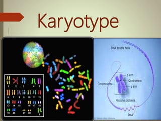
Human Karyotype ( Chromosomes)
•Als PPTX, PDF herunterladen•
69 gefällt mir•35,025 views

Karyoype Cladogram Numer of Chromosomes Types Of Chromosomes Pairs of Chromosome

Empfohlen
Weitere ähnliche Inhalte
Was ist angesagt?
Was ist angesagt? (20)
Ähnlich wie Human Karyotype ( Chromosomes)
Ähnlich wie Human Karyotype ( Chromosomes) (20)
Mehr von Ahmad Raza
Mehr von Ahmad Raza (18)
Kürzlich hochgeladen
Kürzlich hochgeladen (20)
Human Karyotype ( Chromosomes)
- 1. Karyotype
- 2. “Karyotype” Definition: A karyotype is the number and appearance of chromosome in the nucleus of a eukaryotic cell The term is also used for the complete set of chromosomes in a species or in an individual organism and for a test that detects this complement or measures the number
- 3. Karyology & Idiogram Definitions : The study of whole sets of chromosomes is sometimes known as karyology. The chromosomes are depicted in a standard format known as a karyogram or idiogram
- 4. Explanation In pairs, ordered by size and position of centromere for chromosomes of the same size. Derived from Greek word “karyon”, which means "nucleus”, karyotype is represented as Idiogram. • When the haploid set of chromosomes of an organism are ordered in a series of decreasing size, it is said to be an idiogram. • In other sense diagrammatic representation of a karyotype is an Idiogram.
- 5. Chromosome DNA packaging into thread-like structure Is called “chromosomes”. Each chromosome is made up of DNA tightly coiled many times around proteins that support its structure. Only visible during cell division. It has specific number in each species e.g. Humans have 46 chromosomes while dog has 78 & fruit fly has 8 chromosomes.
- 6. Centromere Constricted region of linear chromosome is known as “Centromere”. Divides the chromosome in two regions referred as “arms”. Help to keep aligned on mitotic apparatus during cell division Provide a site for attachment of sister chromatids.
- 8. Division of chromosome according to centromere location Metacentric A chromosome that has a centrally placed centromere.. having centromere in the center such that both sections are of equal length. e.g. Human chromosome 1 – 3 & 16 – 20 Sub metacentric A chromosome whose centromere is placed closer to one end than the other having centromere slightly offset from the center leading to asymmetry. e.g. human chromosomes from 4 – 12. Acrocentric A chromosome whose centromere is placed very close to, but not at, one end having centromere severely offset from center leading one very long and one very short section. e.g. human chromosomes 13,15,21,22. Telocentric :- having centromere at very end of chromosome. Humans don’t have this type of chromosomes but found in other species like mice.
- 11. Human ideogram Metacentric chromosomes in human idogram are shown in red box thy are 9 in number.. Sub metacentric chromosome in human idogram are shown in blue box they are 10 in number. Acrocentric chromosome inhuman are shown in black colure and they are 5 in number. There are no telocentric chromosomes in humans
- 13. Preparation of chromosome for karyotyping Karyotyping is a process of finding a chromosomal characteristics of a cell Karyotyping refers to the analysis of chromosome. The chromosome preparation for karyotyping is generally occur to know about chromosome abnormalities.
- 14. Fig. 6-6, p. 124 Add a few drops of blood. Add phytohemagglutinin to stimulate mitosis. Draw 10 to 20 ml of blood. Incubate at 37°C for 2 to 3 days. Transfer to tube containing fixative. Transfer cells to tube. Add Colcemid to culture for 1 to 2 hours to stop mitosis in metaphase. Centrifuge to concentrate cells. Add low-salt solution to eliminate red blood cells and swell lymphocytes. Drop cells onto microscope slide. Examine with microscope. Digitized chromosome images processed to make karyotype. Stain slide with Giemsa.
- 15. Analysis of karyotyping Karyotype can be analyzed by basic following techniques. Different stains and dyes produce banding patterns specific to each chromosome. And there is chromosome painting technique as well for the analysis of karyotyping.
- 17. BANDING OF CHROMOSOMES G - Banding Q - Banding C - Banding R - Banding
- 19. Q-banding 1. Dehydrate the slides by dipping in alcohol with decreasing concentration 90%, 70% and 50% one min each. 2. Rinse in distilled water. . 3. Wash the slide in phosphate buffer at pH 6.8. 4. Stain the slide in quinacrine mustard (5 mg in 100 mI) or in quinacrine dihydrochloride 5% for 20 min. 5. Rinse in phosphate buffer and mount in the same buffer. 6. Examine under fluorescent microscope.
- 21. METHODOLOGY G- Banding technique Ageing of good slides for 10 days Normal saline Treated with trypsin 0.25% solution 10-15 sec Immersed in 70% ethanol for few minutes Stained with 10% Giemsa for 6-10min Microphotograph good spreads Construction of G-banded karyotype
- 23. N banding technique Take a chromosome from a blood sample. Give dry air to the chromosomes. Treated this dried air chromosome with 5 % solution of Tricholoroacetic acid 95* C for 30 min Treated this solution with 0.1 N of HCl at 60*C for 30 min. After this the banding pattern in structural non histone protein linked to NOR region.
- 25. C-banding 1. Treat the slides in 0.2 N HCI for one hr at room temperature. 2. Rinse in de-ionized water. 3. Immerse in 1% barium hydroxide at 50°C for 5-15 min. 4. Rinse in deionized water. 5. Incubate at 60°C in 2XSSC buffer for one hr. 6. Rinse in de-ionized water and stain in 4% Giemsa stain for 90 min. 7. Rinse in de-ionized water, dry and examine under oil immersion.
- 27. Chromosome Painting New techniques using fluorescent dyes generate unique patterns for each chromosome Chromosome ‘painting’ refers to the hybridization of fluorescently labeled chromosome-specific. Chromosome painting allows the visualization of individual chromosomes in metaphase or interphase cells and the identification of both numerical and structural chromosomal aberrations in human pathology with high sensitivity and specificity.
- 29. Information Obtained from a Karyotype Number of chromosomes Sex chromosome content Presence or absence of individual chromosomes Nature and extent of large structural abnormalities
- 30. Major use of Karyotyping in Important test Any nucleus can be used to make karyotype Lymphocytes, skin cells, tumor cells Sampling cells before birth Amniocentesis Chorionic villus sampling (CVS)
- 31. Amniocentesis A method of sampling the fluid surrounding the developing fetus by inserting a hollow needle and withdrawing suspended fetal cells and fluid Used in diagnosing fetal genetic and developmental disorders Usually performed in the sixteenth week of pregnancy
- 33. Chorionic Villus Sampling (CVS) A method of sampling fetal chorionic cells by inserting a catheter through the vagina or abdominal wall into the uterus Used in diagnosing biochemical and cytogenetic defects in the embryo Usually performed in the tenth week of pregnancy
- 36. Advantages of karyotype Detection of chromosomal abnormalities Genetic disorders Gender identification Identify loss and addition of chromosome Pre-birth diagnosis of genetic diseases Identification of chromosomal numbers in different organism Identification of proper position of genes in chromosomes
- 37. Disadvantages of karyotype Very small abnormality cannot be shown by karyotyping. Technique only allows for diagnosis and not a cure The test like amniocentesis and CVS are both risky an stressful for mother Time consuming process A minor mistake in a process will change all the result.