Measures of Central Tendency: Mean, Median and Mode
Kidney Function Test
1. Chapter 20 : ORGAN FUNCTION TESTS 459
unconjugated), alanine trans-
aminase, aspartate trans-
aminase, alkaline phos-
phatase, J-glutamyl trans-
peptidase and proteins
(albumin, globulins).
KIDNEY (RENAL)
FUNCTION TESTS
The kidneys are the vital
organs of the body, performing
the following major functions.
1. Maintenance of homeo-
stasis : The kidneys are largely
responsible for the regulation
of water, electrolyte and
acid-base balance in the
body.
2. Excretion of metabolic waste products :
The end products of protein and nucleic acid
metabolism are eliminated from the body. These
include urea, creatinine, creatine, uric acid,
sulfate and phosphate.
3. Retention of substances vital to body : The
kidneys reabsorb and retain several substances
of biochemical importance in the body e.g.
glucose, amino acids etc.
4. Hormonal functions : The kidneys also
function as endocrine organs by producing
hormones.
l Erythropoietin, a peptide hormone, stimulates
hemoglobin synthesis and formation of
erythrocytes.
l 1,25-Dihydroxycholecalciferol (calcitriol) –
the biochemically active form of vitamin D –
is finally produced in the kidney. It regulates
calcium absorption from the gut.
l Renin, a proteolytic enzyme liberated by
kidney, stimulates the formation of angio-
tensin II which, in turn, leads to aldosterone
production. Angiotensin II and aldosterone are
the hormones involved in the regulation of
electrolyte balance.
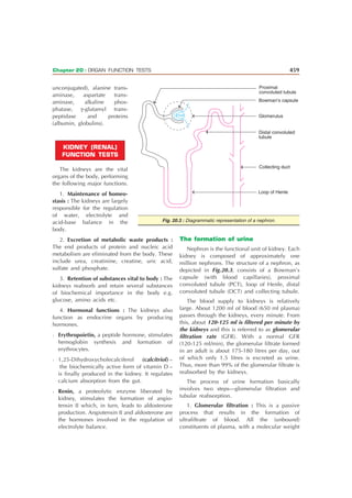
The formation of urine
Nephron is the functional unit of kidney. Each
kidney is composed of approximately one
million nephrons. The structure of a nephron, as
depicted in Fig.20.3, consists of a Bowman’s
capsule (with blood capillaries), proximal
convoluted tubule (PCT), loop of Henle, distal
convoluted tubule (DCT) and collecting tubule.
The blood supply to kidneys is relatively
large. About 1200 ml of blood (650 ml plasma)
passes through the kidneys, every minute. From
this, about 120-125 ml is filtered per minute by
the kidneys and this is referred to as glomerular
filtration rate (GFR). With a normal GFR
(120-125 ml/min), the glomerular filtrate formed
in an adult is about 175-180 litres per day, out
of which only 1.5 litres is excreted as urine.
Thus, more than 99% of the glomerular filtrate is
reabsorbed by the kidneys.
The process of urine formation basically
involves two steps—glomerular filtration and
tubular reabsorption.
1. Glomerular filtration : This is a passive
process that results in the formation of
ultrafiltrate of blood. All the (unbound)
constituents of plasma, with a molecular weight
Proximal
convoluted tubule
Bowman’s capsule
Glomerulus
Distal convoluted
tubule
Collecting duct
Loop of Henle
Fig. 20.3 : Diagrammatic representation of a nephron.
2. BIOCHEMISTRY
460
less than about 70,000, are passed into the
filtrate. Therefore, the glomerular filtrate is
almost similar in composition to plasma.
2. Tubular reabsorption : The renal tubules
(PCT, DCT and collecting tubules) retain water
and most of the soluble constituents of the
glomerular filtrate by reabsorption. This may
occur either by passive or active process. The
excreted urine has an entirely different
composition compared to glomerular filtrate
from which it is derived. The normal
composition of urine is given elsewhere (Refer
inside backcover).
Renal threshold substances
There are certain substances in the blood
whose excretion in urine is dependent on their
concentration. Such substances are referred to as
renal threshold substances. At the normal
concentration in the blood, they are completely
reabsorbed by the kidneys, with a result that their
excretion in urine is almost negligible.
The renal threshold of a substance is defined
as its concentration in blood (or plasma) beyond
which it is excreted into urine. The renal
threshold for glucose is 180 mg/dl; for ketone
bodies 3 mg/dl; for calcium 10 mg/dl and for
bicarbonate 30 mEq/l. While calculating the renal
threshold of a particular compound, it is assumed
that both the kidneys are optimally functioning,
without any abnormality. But this is not always
true—in which case the renal threshold is altered.
For instance, renal glycosuria is associated with
reduced threshold for glucose due to its
diminished tubular reabsorption.
The term tubular maximum (Tm) is used to
indicate the maximum capacity of the kidneys to
absorb a particular substance. For instance,
tubular maximum for glucose (TmG) is 350
mg/min.
Tests to assess renal function
In view of the important and sensitive
functions the kidney performs (described
already), it is essential that the abnormalities
(renal damages), if any, must be detected at the
earliest. Several tests are employed in the
laboratory to assess kidney (renal) function. It
must, however, be remembered that about two-
thirds of the renal tissue must be functionally
damaged to show any abnormality by these tests.
The kidney function tests may be divided into
four groups.
1. Glomerular function tests : All the
clearance tests (inulin, creatinine, urea) are
included in this group.
2. Tubular function tests : Urine concen-
tration or dilution test, urine acidfication test.
3. Analysis of blood/serum : Estimation of
blood urea, serum creatinine, protein and
electrolyte are often useful to assess renal
function.
4. Urine examination : Simple routine exami-
nation of urine for volume, pH, specific gravity,
osmolality and presence of certain abnormal
constituents (proteins, blood, ketone bodies,
glucose etc.) also helps, of course to a limited
degree, to assess kidney functioning.
Some of the important renal function tests are
discussed in the following pages.
CLEARANCE TESTS
The clearance tests, measuring the glomerular
filtration rate (GFR) are the most useful in
assessing the renal function. The excretion of a
substance can be expressed quantitatively by
using the concept of clearance.
Clearance, in general, is defined as the
volume of plasma that would be completely
cleared of a substance per minute. In other
words, clearance of a substance refers to the
milliliters of plasma which contains the amount
of that substance excreted by kidney per
minute. Clearance (C), expressed as ml/minute,
can be calculated by using the formula
U u V
C = ———
P
where U = Concentration of the substance in
urine.
V = Volume of urine in ml excreted per
minute.
P = Concentration of the substance in
plasma.
3. Chapter 20 : ORGAN FUNCTION TESTS 461
Care should be taken to express the concen-
trations of plasma and urine in the same units
(mmol/l or mg/dl).
The clearance of a given substance is
determined by its mode of excretion. The
maximum rate at which the plasma can be
cleared of any substance is equal to the GFR.
This can be easily calculated by measuring the
clearance of a plasma compound which is freely
filtered by the glomerulus and is neither
absorbed nor secreted in the tubule. Inulin (a
plant carbohydrate, composed of fructose units)
and 51Cr-EDTA satisfy this criteria. Inulin is
intravenously administered to measure GFR.
In practice, however, measurement of
clearance for the substances already present in
the blood is preferred. The two compounds,
namely creatinine and urea, are commonly
employed for this purpose. Creatinine clearance
(~145 ml/min) is marginally higher than the GFR
as it is secreted by the tubules. On the other
hand, urea clearance (~75 ml/min) is less than
the GFR, since it is partially reabsorbed by the
tubules.
Diodrast (diiodopyridone acetic acid) is used
as a contrast medium to take urinary tract X-rays.
Diodrast and para amino hippuric acid (PAH)
are peculiar substances as they are entirely
excreted by a single passage of blood through
the kidneys. It is partly filtered by the glomerulus
and mostly excreted by the tubules. PAH has a
clearance of about 700 ml/min (or 1,200 ml, if
expressed as blood). Thus clearance of PAH
represents the renal plasma flow.
Creatinine clearance test
Creatinine is an excretory product derived
from creatine phosphate (largely present in
muscle). The excretion of creatinine is rather
constant and is not influenced by body
metabolism or dietary factors. As already stated,
creatinine is filtered by the glomeruli and only
marginally secreted by the tubules. The value of
creatinine clearance is close to GFR, hence its
measurement is a sensitive and good approach
to assess the renal glomerular function.
Creatinine clearance may be defined as the
volume (ml) of plasma that would be completely
cleared of creatinine per minute.
Procedure : In the traditional method,
creatinine content of a 24 hr urine collection
and the plasma concentration in this period are
estimated. The creatinine clearance (C) can be
calculated as follows :
U u V
C = ———
P
where U = Urine concentration of creatinine
V = Urine output in ml/min (24 hr urine
volume divided by 24 u 60)
P = Plasma concentration of creatinine.
As already stated, creatinine concentration in
urine and plasma should be expressed in the
same units (mg/dl or mmol/l).
Modified procedure : Instead of a 24 hr urine
collection, the procedure is modified to collect
urine for 1 hr, after giving water. The volume of
urine is recorded. Creatinine contents in plasma
and urine are estimated. The creatinine
clearance can be calculated by using the formula
referred above.
Reference values : The normal range of
creatinine clearance is around 120-145 ml/min.
These values are slightly lower in women. In
recent years, creatinine clearance is expressed in
terms of body surface area.
Diagnostic importance : A decrease in
creatinine clearance value (< 75% normal)
serves as sensitive indicator of a decreased GFR,
due to renal damage. This test is useful for an
early detection of impairment in kidney function,
often before the clinical manifestations are seen.
Urea clearance test
Urea is the end product of protein
metabolism. After being filtered by the glomeruli,
it is partially reabsorbed by the renal tubules.
Hence, urea clearance is less than the GFR and,
further, it is influenced by the protein content of
the diet. For these reasons, urea clearance is not
as sensitive as creatinine clearance for assessing
renal function. Despite this fact, several
laboratories traditionally use this test.
4. BIOCHEMISTRY
462
Urea clearance is defined as the volume (ml)
of plasma that would be completely cleared of
urea per minute. It is calculated by the formula
U u V
Cm = ———
P
where Cm = Maximum urea clearance
U = Urea concentration in urine
(mg/ml)
V = Urine excreted per minute in ml
P = Urea concentration in plasma
(mg/ml).
The above calculation is applicable if the
output of urine is more than 2 ml per minute.
This is referred to as maximum urea clearance
and the normal value is around 75 ml/min.
Standard urea clearance : It is observed that
the urea clearance drastically changes when the
volume of urine is less than 2 ml/min. This is
known as standard urea clearance (Cs) and the
normal value is around 54 ml/min. It is
calculated by a modified formula
Cs
U u V
P
Diagnostic importance : A urea clearance
value below 75% of the normal is viewed
seriously, since it is an indicator of renal
damage. Blood urea level as such is found to
increase only when the clearance falls below
50% normal. As already stated, creatinine
clearance is a better indicator of renal function.
Urine concentration test
This is a test to assess the renal tubular
function. It is a simple test and involves the
accurate measurement of specific gravity which
depends on the concentration of solutes in urine.
A specific gravity of 1.020 in the early morning
urine sample is considered to be normal.
Several measures are employed to
concentrate urine and measure the specific
gravity. These include overnight water
deprivation and administration of antidiuretic
hormone. If the specific gravity of urine is above
1.020 for at least one of the samples collected,
the tubular function is considered to be normal.
Osmolality and specific gravity : The
osmolality of urine is variable. In normal
individuals, it may range from 500-1,200
milliosmoles/kg. The plasma osmolality is around
300 milliosmoles/kg. The normal ratio of the
osmolality between urine and plasma is around
2-4. It is found that the urine (without any
protein or high molecular weight substance) with
an osmolality of 800 mosm/kg has a specific
gravity of 1.020. Therefore, measurement of
urine osmolality will also help to assess tubular
function.
Analysis of blood (or serum)
Estimation of serum creatinine and blood
urea are often used to assess the overall kidney
function, although these tests are less sensitive
than the clearance tests. Serum creatinine is a
better indicator than urea in this regard.
The diagnostic importance of urea and crea-
tinine estimations are discussed elsewhere (Refer
Chapter 15).
The relationship between GFR and serum
creatinine levels is depicted in Fig.20.4. It is
observed that the GFR must fall to about 50% of
its normal value before a significant increase in
serum creatinine occurs. Therefore, a normal
0 20 40 60 80 100 120 140 160
1
2
3
4
5
6
7
8
9
10
Serum
creatinine
(mg/dl)
GFR (ml/min)
Fig. 20.4 : The relationship between glomerular filtration
rate (GFR) and serum creatinine concentration.
5. Chapter 20 : ORGAN FUNCTION TESTS 463
serum creatinine level does not necessarily mean
that all is well with the kidney. It is estimated
that a loss of 50% of the functions of nephrons
leads to (approximate) doubling of serum
creatinine concentration.
Cystatin C is a protein marker of kidney
function (serum reference range 0.8-1.2 Pg/dl),
and is more sensitive than creatinine. Even minor
changes in GFR in the early stages of chronic
kidney diseases are associated with increased
cystatin C.
Urine examination
The routine urine examination is undoubtedly
a guiding factor for renal function. The volume
of urine excreted, its pH, specific gravity,
osmolality, the concentration of abnormal
constituents (such as proteins, ketone bodies,
glucose and blood) may help to have some
preliminary knowledge of kidney function. More
information on urine laboratory tests is given in
the appendix.
Choice of renal function tests
In general, the assessment of kidney function
starts with the routine urine examination,
followed by serum creatinine and/or blood urea
estimations and, finally, the specific tests to
measure the tubular and glomerular functions
(clearance tests).
GASTRIC FUNCTION TESTS
The stomach is a major organ of digestion
and performs the following functions
1. Stomach is a reservoir of ingested
foodstuffs.
2. It has a great churning ability which
promotes digestion.
3. Stomach elaborates HCI and proteases
(pepsin) which are responsible for the initiation
of digestive process.
4. The products obtained in the stomach
(peptides, amino acids) stimulate the release of
pancreatic juice and bile.
Secretion of gastric HCI
The parietal (oxyntic) cells of gastric glands
produce HCI. The pH in the gastric lumen is as
low as 0.8 (against the blood pH 7.4). Therefore,
the protons are transported against the
concentration gradient by an active process.
A unique enzyme—namely K+ activated
ATPase—present in the parietal cells is
connected with the mechanism of HCI secretion
(Fig.20.5). The process involves an exchange of
H+ ions (of the parietal cells) for K+ ions (of the
lumen). This is coupled with the consumption of
energy, supplied by ATP. The H+ are
continuously generated in the parietal cells by
the dissociation of carbonic acid which, in turn,
is produced from CO2. The bicarbonate ions
(HCO3
–), liberated from the carbonic acid
(H2CO3) dissociation, enter the blood in
exchange for Cl– ions. The latter diffuse into the
gastric lumen to form HCI. Gastrin—a peptide
hormone of gastrointestinal tract—stimulates HCI
secretion.
Following a meal, there is a slight elevation
in the plasma bicarbonate concentration which
is linked to the gastric HCI secretion. This is
referred to as alkaline tide.
Fig. 20.5 : Mechanism of HCl secretion
( —represents K+
activated ATPase).
Lumen Blood
Cl
–
H
+
Cl
–
HCl
H
+
K
+
K
+
ATP
ADP + Pi
Cl
–
HCO3
–
Parietal cell
HCO3
–
H2CO3
CO2 + H2O
Carbonic
anhydrase