
Empfohlen
Weitere ähnliche Inhalte
Was ist angesagt?
Was ist angesagt? (20)
Ähnlich wie Human Digestive Anatomy
Ähnlich wie Human Digestive Anatomy (20)
Kürzlich hochgeladen
Kürzlich hochgeladen (20)
Human Digestive Anatomy
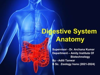
- 1. Digestive System Anatomy Supervisor - Dr. Archana Kumar Department – Amity Institute Of Biotechnology By - Aditi Tanwar B Sc. Zoology hons (2021-2024)
- 2. With this presentation we will learn to develop an understanding of the relationships between the structures and functions of the human digestive system and digestive processes which include the processes of digestion include six activities: ingestion, propulsion, mechanical or physical digestion, chemical digestion, absorption, and defecation. OBJECTIVES:- • Be able to describe the major function or functions of each organ in the digestive system. • Be able to know how the process of chemical digestion. • Know the illness related to elimination of waste and diseases related to nutrition deficiency.
- 3. General Terminologies • NUTRITION- the process of taking in and using food. • NUTRIENTS- substances in food that are used as energy sources of the body • DIGESTIVE SYSTEM- the organs and glands in the body that are responsible for digestion • • DIGESTION- the breakdown of food to smaller molecules
- 4. TWO TYPES OF DIGESTIVE SYSTEM • INCOMPLETE DIGESTIVE SYSTEM- common among invertebrates, there is only a single opening for the ingestion of food. • COMPLETE DIGESTIVE SYSTEM- common among nematodes, annelids, mollusks, echinoderms and vertebrates, this type has digestive tubes with 2 openings.
- 6. Human Digestive System The human digestive system consists of the alimentary canal and the associated glands. The alimentary canal begins with an anterior opening – the mouth, and it opens out posteriorly through the anus. The mouth leads to the buccal cavity or oral cavity. The oral cavity has a number of teeth and a muscular tongue. Each tooth is embedded in a socket of jaw bone. This type of attachment is called thecodont. Majority of mammals including human being forms two sets of teeth during their life, a set of temporary milk or deciduous teeth replaced by a set of permanent or adult teeth. This type of dentition is called diphyodont. An adult human has 32 permanent teeth which are of four different types (Heterodont dentition), namely, incisors (I), canine (C), premolars (PM) and molars (M).
- 7. The hard chewing surface of the teeth, made up of enamel, helps in the mastication of food. The tongue is a freely movable muscular organ attached to the floor of the oral cavity by the frenulum. The upper surface of the tongue has small projections called papillae, some of which bear taste buds.
- 8. 1. Food processing begins in the mouth. 2. The food is then swallowed and moved through pharynx into the esophagus. 3. Then food is mechanically and enzymatically digested in the stomach. 4. Most enzymatic digestion takes place in the small intestine. 5. The large intestine then eliminates wastes leading to the opening for the elimination of waste called anus.
- 9. TWO PHASES OF DIGESTION MECHANICAL PHASE - involves the breaking up of food into small pieces, pushing the food down the food tube, and mixing it with digestive juices. CHEMICAL PHASE- involves the further breaking up of the larger molecules of food into smaller molecules by the action of digestive enzymes.
- 10. Process Of Digestion Mechanical Digestion- starts in the mouth (mastication) where four kinds of teeth tear the food into pieces. Chemical Digestion- our mouth has salivary glands that secrete saliva. This saliva contains the enzyme called salivary amylase which catalyzes maltose into glucose. The pharynx and esophagus conduct food to the stomach. After being chewed, the food is swallowed through the pharynx extending to the oesophagus.
- 11. Digestion Of Food In Stomach The oesophagus is a thin, long tube which extends posteriorly passing through the neck, thorax and diaphragm and leads to a ‘J’ shaped bag like structure called stomach. A muscular sphincter (gastro-oesophageal) regulates the opening of oesophagus into the stomach. The stomach, located in the upper left portion of the abdominal cavity, has four major parts – a cardiac portion into which the oesophagus opens, a fundic region, body (main central region) and a pyloric portion which opens into the first part of small intestine. Each of the openings, the cardiac and the pyloric, has a sphincter muscle that keeps the neighboring region closed, except when food is passing through. In this manner, food is enclosed by the stomach until ready for digestion.
- 12. The stomach has the ability to expand or contract depending upon the amount of food contained within it. When contracted, the interior walls form numerous folds (rugae), which disappear when the walls are distended. The thick mucous-membrane lining of the walls is densely packed with small gastric glands; these secrete a mixture of enzymes and hydrochloric acid that partly digest proteins and fats. Upon entry of food, they relax briefly, then begin to contract. Periodic contractions churn and knead food into a semifluid mixture called chyme; rhythmical pumping (peristaltic) waves move food toward the pylorus and small intestine.
- 14. Small intestine is 5-6m long it is distinguishable into three regions, a ‘C’ shaped duodenum, a long coiled middle portion jejunum and a highly coiled ileum. Ileum opens into the large intestine. It consists of caecum, colon and rectum. Caecum is a small blind sac which hosts some symbiotic micro-organisms. A narrow finger-like tubular projection, the vermiform appendix which is a vestigial organ, arises from the caecum. The caecum opens into the colon. The colon is divided into four parts – an ascending, a transverse, descending part and a sigmoid colon. The descending part opens into the rectum which opens out through the anus.
- 15. Absorption in the small intestines Once broken down the nutrients are absorbed by the inner walls of the small intestine into the blood stream. The nutrients are rendered small enough so that they may pass, or "be transported", across the epithelial cells of the gastrointestinal tract. The nutrients are absorbed by processes of simple/passive diffusion, facilitated diffusion, primary active transport, or secondary active transport. The small intestine is good for absorption since it has a large inner surface area. This is formed many tiny finger-like structures of tissue called villi. The individual epithelial cells also have finger-like projections, which are called known as microvilli. For transport, nutrients commonly rely upon: Lipids – undergo passive or simple diffusion Short-chain fatty acids – diffusion Amino acids – primary active transport Glucose – secondary active transport Fructose – facilitated diffusion
- 16. The job of large intestine is to absorb water, minerals, and some of the remaining nutrients from your food. It will change the leftover waste into a bowel movement. This is also called stool. Rectum stores the stool until we feel the need to have a bowel movement. Muscles of rectum then push the stool through anus and out of body.
- 17. Liver Liver is the largest internal organ of the body. It secrets bile which emulsifies fat and stored in gall bladder then ejected into the small intestine. It converts excess glucose to glycogen and stores it. Converts excess amino acids into fatty acids and urea.
- 18. Pancreas The pancreas is a compound (both exocrine and endocrine) elongated organ situated between the limbs of the ‘C’ shaped duodenum. The exocrine portion secretes an alkaline pancreatic juice containing enzymes The endocrine portion secretes hormones, insulin and glucagon The enzymes secreted by the pancreas are called trypsin and chymotrypsin.
- 19. 1. Bile from the liver and digestive enzymes from the pancreas released into the duodenum. 2. These two then act on the chyme. 3. Then enzymes produced by the epithelial cells lining duodenum catalyze the final steps in the digestion of the major types of nutrients. Chyme moves through the digestive tract by peristalsis and motions of villi. Enzymes digest chyme as follows:- • Carbohydrates are digested to monosaccharide or simple sugars. • Proteins are digested to amino acids • Fats are digested to fatty acids and monoacylglycerols
- 20. Some hormones that regulate digestion
- 21. Absorption • It is the process by which substances are taken in by the cells. • Absorption takes place mainly through the villi of the small intestine. • Digested food in the form of amino acids, simple sugar, fatty acids and glycerol diffuse into the cells of the villi.
- 22. Process Of Absorption • Most of the digested food diffuse into capillaries and reach the blood, while fatty acids and glycerol diffuse into the lacteals and reach another circulating fluid called lymph. • Through diffusion, digested food reaches the blood and lymph and undergoes a process called circulation. • The circulating fluids distribute the digested food to all the cells of the body. • Undigested food passes through the large intestine where absorption of some water, minerals and certain drugs takes place.
- 23. 1. Vomiting: It is the ejection of stomach contents through the mouth. This reflex action is controlled by the vomit center in the medulla. A feeling of nausea precedes vomiting. 2. Jaundice: The liver is affected, skin and eyes turn yellow due to the deposit of bile pigments. 3. Diarrhea: The abnormal frequency of bowel movement and increased liquidity of the fecal discharge is known as diarrhea. It reduces the absorption of food. 4. Constipation: In constipation, the faeces are retained within the colon as the bowel movements occur irregularly. 5. Indigestion: In this condition, the food is not properly digested leading to a feeling of fullness. The causes of indigestion are inadequate enzyme secretion, anxiety, food poisoning, over eating, and spicy food.
- 24. Protein-energy malnutrition (PEM) • Marasmus is produced by a simultaneous deficiency of proteins and calories. It is found in infants less than a year in age, if mother’s milk is replaced too early by other foods which are poor in both proteins and caloric value. This often happens if the mother has second pregnancy or childbirth when the older infant is still too young. In Marasmus, protein deficiency impairs growth and replacement of tissue proteins; extreme emaciation of the body and thinning of limbs results, the skin becomes dry, thin and wrinkled. Growth rate and body weight decline considerably. Even growth and development of brain and mental faculties are impaired.
- 25. Protein-energy malnutrition (PEM) • Kwashiorkor is produced by protein deficiency unaccompanied by calorie deficiency. It results from the replacement of mother’s milk by a high calorie low protein diet in a child more than one year in age. Like marasmus, kwashiorkor shows wasting of muscles, thinning of limbs, failure of growth and brain development. But unlike marasmus, some fat is still left under the skin; moreover, extensive oedema and swelling of body parts are seen.
- 26. Bibliography • Hall, John E. (2011). "General Principles of Gastrointestinal Function". Guyton and Hal Textbook of Medical Physiology (12th ed.). Saunders Elsevier. p. 755. ISBN 978- 1416045748. • Saladin, Kenneth (2011). Human Anatomy. McGraw Hill. p. 659. ISBN 9780071222075. • Saladin, Kenneth (2011). Human Anatomy. McGraw Hill. pp. 621– 622. ISBN 9780071222075. • Britannica Concise Encyclopaedia. Encyclopaedia Britannica, Inc. 2007. ISBN 978- 1593392932. • Wood, Jackie D. (2009), "Gastrointestinal Physiology", in Rhoades, Rodney A.; Bell, David R. (eds.), Medical Physiology: Principles for Clinical Medicine (3rd ed.), Philadelphia, PA: Lippincott Williams & Wilkins, pp. 463–496 • Maton, Anthea (1993-01-01). Human Biology and Health. Prentice Hall 1993. ISBN 978- 0-13-981176-0