1. The Role of GABA in Visual Processing in the Retina
BAKER AS1
. SKINNER BM1
. DECKER ML1
. MALCHOW RP2
. AND KREITZER MA1
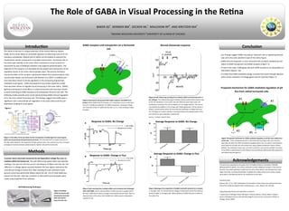
Introduction
The retina of the eye is a unique extension of the Central Nervous System
(CNS), and its study allows an accessible approach to observing many of its tre-
mendous complexities. Retinal cells of catfish can be isolated to examine the
fundamental cellular components of synaptic transmission. Horizontal cells of
the retina span laterally in the outer retina contribute to visual contrast en-
hancement by way of feedback inhibition onto adjacent photoreceptors. The
objective of this research is to elucidate the key players and mechanisms of the
regulatory events in this outer retinal synaptic layer. The primary excitatory
neurotransmitter at this synapse is glutamate release from photoreceptors onto
second order bipolar and horizontal cells (Kreitzer et al 2007). In addition pro-
tons have been shown to be key regulators in the processing of glutamate-
mediated visual signals. GABA and dopamine (see similar dopamine study)
have also been shown to regulate visual processing in the outer retina. GABA’s
(gamma-aminobutyric acid) effects in retinal horizontal cells have been shown
to work by binding to GABA receptors and transporters found on the cells. The
following studies provide early results demonstrating GABA-induced regulation
of H+ flux from retinal horizontal cells. The findings suggest that GABA plays a
significant role in extracellular pH regulation in the outer retina and thus pH-
dependent shaping of visual signals.
(Figure 1) The retina of the eye allows for the transduction of visible light into neural signals,
which can be processed and perceived by the brain. The horizontal cell is a unique structure of
the CNS, which allows for the production of high contrast vision. This contrast occurs by a complex
exchange of chemicals between photoreceptors, bipolar cells and horizontal cells.
Methods
A proton select electrode measured the pH-dependent voltage flux near an
isolated catfish horizontal cell. The self-referencing system takes two separate
readings, one near the cell and one at a set distance of 30μm from the cell. The
difference in voltage signals acquired between the two regions represents the
cell-induced change in proton flux. After standing flux was achieved a back-
ground control was performed 200μm above the cell. 1ml of 1mM GABA was
placed into the dish. Data was compiled on Microsoft Excel and graphs were
made using GraphPad Prism software.
(Figure 4) Self-referencing recording from isolated catfish rod horizontal cell in 2
mL of Ringer. The initial positive signal indicates that the extracellular solution next
to the cell membrane is more acidic than the reference point (30µm from cell
membrane). R indicates the control addition of 1 mL Ringer solution. The next ar-
row indicates the addition of 1mL ringer with 100µm glutamate. The bar above in-
dicates a control period where the electrode was withdrawn to 400µm from the
cell. The figure as a whole indicates a working method and a depolarization of the
cell membrane upon application of glutamate.
(source – Kreitzer research labs)
Conclusion
Our findings suggest GABA may play an important role in regulating extracel-
lular pH in the outer plexiform layer of the retina Figure.
GABA-induced responses in cone horizontal cells are likely mediated by acti-
vation of GABA transporters and GABA receptors (Figure 3).
In half of the trials, challenging cells with GABA resulted in an extracellular al-
kalinization (Figures 5b)
It is likely that GABA-mediated change in proton-flux works through depolari-
zation-driven activation of voltage-gated calcium channels (Figure 7).
(figure 7)Proposed mechanism for GABA-mediated regulation of pH flux from retinal hori-
zontal cells. If the cell depolarizes (#1) to threshold level (see figure 5b), Ca2+ channels
open (#2) and allow the influx of positively charged calcium ions. As calcium concentrations
build inside the cell (#3), the cell counters this using a plasma membrane calcium ATPase
(PMCA). This antiporter moves Ca2+ out of the cell and H+ ions into the cell (#4). The action
of the PMCA is hypothesized to contribute to the extracellular alkylinization (#5) observed in
self-referencing (see figure 4)
Acknowledgements
This work was supported by two grants from the National Science Foundation; 0924383
(MAK) & 0924372 (RPM). The authors would like to thank additional members of the Kreitzer
Lab who have added significant insight into this work: Ethan Naylor, Luke Montgomery, Sonja
Vogel, Tara fuller, and Karisa Burkholder In addition the authors thank Jason Jacoby, A gradu-
ate student in the Malchow lab for his contribution to this work.
Literature Cited:
Kreitzer, M.A., ET AL. 2007, Modulation of Extracellular Proton Fluxes from Retinal Horizontal
Cells of the Catfish By Depolarization and Glutamate. J. Gen. Physiol 130: 169-182.
Supporting Departments and institutions of Authors:
1 Department of Biology, Indiana Wesleyan University, Marion, Indiana 46953: 2 Depart-
ments of Biological Sciences and ophthalmology & visual sciences, University of Illinois at
Chicago, Illinois, 60607.
GABA receptors and transporters on a Horizontal
Cell
Normal Glutamate response
Proposed mechanism for GABA-mediated regulation of pH
flux from retinal horizontal cells.
(Figure 5) pH recording from isolated catfish cone horizontal cell challenged
with 1mM GABA. Fig A is representation of what occurred in roughly half of
the trials. GABA did not cause a change in extracellular proton levels. Fig B is a
Representation of the other half of GABA trial. GABA caused an alkalization
outside the cell.
Figure 1
Self-Referencing Technique
A
B
A
B
(Figure 3) Horizontal cells possess both GABA receptors and GABA trans-
porters. When GABA binds its receptor, a Cl- conductance occurs in the direc-
tion of Cl- equilibrium potential. For GABA transporters, cotransport allows
the movement of Na+:Cl-:GABA into the cell in a 2:1:1 ratio resulting in depo-
larization.
(Figure 2) Isolated
catfish horizontal cells
showing the electrode
both near and far
away from the cell.
#2
#3 #4
#5
#1
#1
1
INDIANA WESLEYAN UNIVERSITY 2
UNIVERSITY OF ILLINIOS AT CHICAGO
(Figure 6)Averages from population of GABA trials (with standard error shown).
A. Average trials for cell that did not change in extracellular proton flux when ex-
posed to GABA. B. Averaged cells. Where addition of GABA did have an effect on
the proton flux.
Response to GABA: No Change
Response to GABA: Change in Flux
Average Response to GABA: No Change
Average Response to GABA: Change in Flux