

Empfohlen
Empfohlen
Weitere ähnliche Inhalte
Was ist angesagt?
Andere mochten auch
Andere mochten auch (8)
Ähnlich wie Web sep09lr
Ähnlich wie Web sep09lr (20)
Mehr von Abu-Hussein Muhamad
Mehr von Abu-Hussein Muhamad (20)
Kürzlich hochgeladen
Kürzlich hochgeladen (20)
Web sep09lr
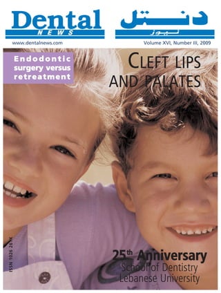
- 1. www.dentalnews.com Volume XVI, Number III, 2009 Endodontic surgery versus retreatment CLEFT LIPS AND PALATES ISSN 1026 261X 25 Anniversary th School of Dentistry Lebanese University
- 3. The nano-optimized composite system Tetric EvoCeram Tetric EvoFlow ® ® MORE THAN A COMPOSITE M O R E T H A N A F L O WA B L E Nano-optimized mouldable Nano-optimized flowable ceramic composite Ideally complements Tetric EvoCeram www.ivoclarvivadent.com Ivoclar Vivadent AG Bendererstr. 2 | FL 9494 Schaan | Liechtenstein | Tel.: +423 / 235 35 35 | Fax: +423 / 235 33 60
- 4. Modulmix and Hydrorise, the brand new impressions system for high precision results Zhermack, leading company in the technology of A-silicones, introduces a unique impression system delivering outstanding quality, accurate results and easy handling. The combination between Modulmix and Hydrorise assures the following great performances: excellent flow properties - the hyper hydrophilic behaviour provides the most accurate impressions ever time saving - thanks to high speed dispensing for rapid tray-filling less waste - thanks to the new patented dynamic/static Tel. +39 - 0425 597611 - Fax +39 - 0425 597642 mixing tip comm.expo@zhermack.com - www.zhermack.com
- 5. CONTENTS Vo l u m e X V I , N u m b e r I I I , 2 0 0 9 EDITORIAL TEAM Alfred Naaman, Nada Naaman, Jihad Fakhoury, Dona Raad, Antoine Saadé, Lina Chamseddine, 13 Adopting minimum intervention Tarek Kotob, Mohammed Rifai, Bilal Koleilat, in dentistry Mohammad H. Al-Jammaz Dr. Steffen Mickenautsch COORDINATOR Lina Jadaa ART DEPARTMENT Krystel Kouyoumdjis 20 Cleft lips and palates SUBSCRIPTION Micheline Assaf, Nariman Nehmeh Dr. Abu-Hussein Muhamad ADVERTISING Josiane Younes PHOTOGRAPHY Albert Saykali TRANSLATION Gisèle Wakim, Marielle Khoury 28 Lumineers DIRECTOR Tony Dib Dr. David Silber ISSN 1026-261X DENTAL NEWS – Sami Solh Ave., G. Younis Bldg. POB: 116-5515 Beirut, Lebanon. Tel: 961-3-30 30 48 Fax: 961-1-38 46 57 Email: info@dentalnews.com Website: www.dentalnews.com INTERNATIONAL REVIEW BOARD Pr. M.A. Bassiouny BDS, DMD, MSc, Ph.D. Director International Program, Temple University, Philadelphia, USA. 36 Endodontic surgery versus retreatment Pr. N.F. Bissada D.D.S., M.S.D Professor and Chairman, Department of Periodontics, Case Western Reserve Dr. Richard Mounce University, USA. Pr. Jean-Louis Brouillet D.C.D, D.S.O. Chairman, Department of Restorative Dentistry, Aix-Marseille II, France. 54 25th Anniversary - School of Dentistry Pierre Colon D.C.D., D.S.O. Maître de conférence des universités, Paris, France. Lebanese University Dr. Jean-Claude Franquin, Directeur de l’Unité de Recherche ER116, Marseille, France. Pr. Gilles Koubi D.C.D., D.S.O. Department of Restorative Dentistry, Aix-Marseille II, France. Pr. Guido Goracci. University LA SAPIENZA, School of Medicine & Dentistry, Roma, Italia. Dr. Olivier Hue, Faculté de chirurgie dentaire de Paris VII, rue Garancière, Paris, France. Brian J. Millar BDS, FDSRCS, Ph.D. Guy’s, King’s, and St. Thomas’ College School of Medecine & Dentistry, London, UK. Pr. Dr. Klaus Ott, Director of the Clinics of Westfälischen Wilhelms-University, Münster, Germany. Wilhelm-Joseph Pertot DEA, Maître de conférence, Aix-Marseille II, France. Pr. James L. Gutmann, Professor and Director, Graduate Endodontics, Baylor College of Dentistry, Dallas, Texas, USA. Pr. Dr. Alfred Renk, Bayerische Julius-Maximilians-University, Würzburg, Germany. Dr. Philippe Roche-Poggi DEA. Maître de conférence des universités, Aix-Marseille II, France. Michel Sixou D.C.D., D.E.A. Department of Priodontology, Toulouse, France. 3 Contents Pr. M. Sharawy B.D.S., Ph.D. Professor and Director, Department of Oral biology, Medical College of Georgia, Augusta, Georgia, USA. DENTAL NEWS IS A QUARTERLY MAGAZINE DISTRIBUTED MAINLY IN THE MIDDLE EAST & NORTH AFRICA IN COLLABORATION WITH THE COUNCIL OF DENTAL SOCIETIES FOR THE GCC. Statements and opinions expressed in the articles and communications herein are those of the author(s) and not necessarily those of the Editor(s) or publisher. No part of this magazine may be reproduced in any form, either electronic or 72 Product Review mechanical, without the express written permission of the publisher.
- 8. PantOs DG XP Ceph P R O F E S S I O N A L T E C H N O L O G Y The latest technologies for digital imaging in PantOs DG xp Ceph, featuring two specific imaging detectors for panoramic and cephalometric radiology. Digital Cephalometric radiology is on the same system in addition to multiprojection panoramic. The one-shot digital Ceph features a 10x10 inch detection area and instant imaging on computer with a short exposure. A solid and reliable solution with fixed digital sensors. No slow scanning, no risk of movement, no image distortion. More precise results, quickly. igita D l 1-SHOT 1-SHOT C eph Blue X Imaging Srl Via Idiomi 1/8-33 • 20090 Assago • Italy Ph. +39 02 4571 2171 • Fax +39 02 4570 3385 e-mail: bluex@bluex.it • www.bluex.it Have a nice imaging
- 9. A-dec 300 stylish functional innovative A healthy new choice for dentistry Find out why the A-dec 300 is a great choice for your practice. For details visit www.a-dec300.com or contact your local authorised A-dec dealer . A-dec 300 www.a-dec300.com A-dec A-dec Inc. 2601 Crestview Drive, Newberg, Oregon 97132 USA Tel: +44 2476 322089 Fax: +44 2476 345106 Web: www.a-dec.com
- 11. Seal-Tight Dry air on demand. It will blow you away. Disposable air/water syringe tips. • Disposable. For maximum infection protection. • Saves Time. Dramatically reduces time needed for reprocessing autoclavable tips. • Flexible. Can be bent to a 90° angle without affecting water flow and improving ergonomics. • Dry Air. The yellow rubber seal acts as a new O-ring ensuring dry air every time. Cleanic® The universal prophy paste is now available in tubes. With Perlite technology. • Universal prophy paste with integrated cleaning variability. • Single application: cleaning and polishing in a single application (ca. 15 seconds/3 teeth). • Perlite technology: high cleaning power converting into gentle polishing action thanks to Perlite technology (dynamic behaviour of Perlite-particles). • Gentle: the prophy paste is gentle to the tooth structure while maintaining a high efficiency. Oral Health Examination Kit Sterile. Single-use. Convenient. Diagnostic examination kit. • Ready-to-use. Minimises tray set-up time. • Hygienic. Eliminates cross-contamination issues. Simply use once and discard. • Convenient. Excellent for routine use, especially during busy surgery times, staff shortages, autoclave breakdowns, home visits and for high-risk patients. • Time-saving. Reduces time spent sterilising and handling sharps. • Cost-effective. Increases productivity and reduces inventory. Flexible Clearance Tabs™ Ideal for surgery use • Give you an easy and accurate way to make direct intra-oral measurements of occlusal and interproximal tooth preparation clearance. • Each colour coded tab is used like a feeler gauge to check clearance dimension. • Available in a convenient dispenser box designed especially for the dental surgery. • Each tab is for single use only. KerrHawe SA P.O. Box 268 6934 Bioggio Switzerland Freephone: 00800 41 05 05 05 Fax: ++41 91 610 05 14 www.KerrHawe.com Your practice is our inspiration.™
- 13. INTERNATIONAL CALENDAR September 23–26, 2009 The Beirut International Dental Meeting 2009 November 6 - 7, 2009 Congress Palace Dbayeh 1st Dental – Facial Cosmetic International Conference Contact: Dr. Antoine Karam, President of the Lebanese Dental Association Where Science Meets Art of Beauty Email: lda@lda.org.lb Jumeirah Beach Hotel UAE Website: www.LDA.org.lb Email: info@cappmea.com Website: http://www.cappmea.com October 14–16, 2009 The 17th Scientific International Conference of Syrian Dental Association, November 10 - 12, 2009 Damascus Ommayad Palace for conferences, Damascus Ebla Hotel 1st Dubai International Implant Summit Contact: Syrian Dental Association, Damascus, Syria, POB: 11104 Crowne Plaza Dubai - UAE Tel: 963 11 222 1446 – Fax: 963 11 222 48 45 Email: matios.tcholakian@index.ae Email: syrdent@scs-net.org Website: http://www.diis.ae Website: http://www.syr-sda.com November 10 - 12, 2009 October 15 - 17, 2009 DENTISTRY 2009 37th International Expodental Rome ADNEC, Abu Dhabi – UAE Pavilions 7-8-9 of the new Fiera Roma Exhibition Center Email: dentistry@iirme.com Email: pressoffice@expodental.it Website: http://www.dentistryme.com Website: http:// www.expodental.it November 11 - 13, 2009 October 21 - 23, 2009 Egyptian Dental Association – 14th International Dental Congress The 4th Riyadh International Pharmacy Meeting - 4th RIDPM Problem solving in Dentistry Riyadh, Saudi Arabia from October 21st to 23rd, 2009 [02-04/10/1430]. Intercontinental Hotel - Cairo - City Stars Email: meeting@riyadh.edu.sa Email: eda@internetegypt.com.eg Website: http://riyadh.edu.sa/meeting Website: http;//www.eda-egypt.org October 27 - 29, 2009 November 29 – December 2nd, 2009 The 5th Bahrain Dental Society Conference 2009 Greater New York Dental Meeting 27- 29 October 2009 Tel: 212-398-6922 Email: bahds@batelco.com.bh Fax: 212-398-6934 Website: http://www.bahrain-dental.com Email: info@gnydm.com Website: http://www.gnydm.com November 3 - 6, 2009 1st Pan Arab Endodontic conference March 9 - March 11, 2010 LandMark Hotel (RadissonSAS) Amman, Jordan AEEDC Dubai Dr. Ibrahim Abu Tahun Dubai International Convention & Exhibition Centre Email: ihtahun@yahoo.com Email: index@emirates.net.ae Website: http://www.jda.org.jo/endo Website: http://www.aeedc.ae ADVERTISING INDEX ACTEON 71 - A-DEC 7 - BELMONT 27 - BEYOND 25 - BIEN AIR 23 - BIOMET 3I 54 - BLUE X 6 - CASTELLINI 21 - CAVEX 35 - COLGATE 41 - COLTENE WHALEDENT 53 - DISCUS DENTAL 75 - DR. WILD 4, 5 - DUERR 17 - GC 31 - GSK C2, 51 - HU-FRIEDY 59 - INTENSIV 49 - INVISALINE 26 - IVOCLAR – VIVADENT 1, C4 - KAVO C3 - KAVO – GENDEX 55 - KERR 9, 19 - KETTENBACH 34 - KOMET 78 - LEONE 10 - MECTRON 42 - METASYS 33 - MICRO MEGA 67 - MOCOM 63 - MORITA 29 - NISSIN 39 - NOBEL BIOCARE 37 - NSK 80 - SIRONA 15 - SOREDEX 8 - STRAUMANN 69 - SULTAN 12 - VITA 47 - VOCO 65 - W&H 61 - ZHERMACK 2 - ZIMMER 43 www.dentalnews.com 11 w w w. d e n t a l n e w s . c o m T e l : 9 6 1 - 3 - 3 0 3 0 4 8 F a x : 9 6 1 - 1 - 3 8 4 6 5 7 Email: info@dentalnews.com Volume XVI, Number III, 2009 GET YOUR ISSUE ONLINE DENTAL NEWS, VOLUME XVI, NUMBER III, 2009
- 14. If you like Genie, you’ll ™ love these Sultan products… Silgimix™ Alginate Replacement Impression Material Quicker, better impressions—without the mess. It’s the dust-free alternative to alginate materials. Genie™ Bite A bite registration that sets, rock hard, in 60 or 90 seconds. Comes in easy-to-use automix cartridges. 3-Way™ Disposable Impression Trays Single use—ideal for accurate impressions, counter impressions and bite registrations. Genie’s™ superior flow… for incredible impressions, every time. Genie™ Magic Mix To capture the very best impressions, it’s critical that your impression Genie is available in material exhibits superior “flow”—the ability to capture all details, including 380 ml cartridges for automix machines. those past the prepared margin. Flow is the single most important feature Get 5% more material of your impression material, because the better the flow, the better you’ll than foil bag systems. capture important details. For flow that stands above the rest, choose Genie™. An independent study confirms it: when compared to leading brands of impression material, Genie™ proved to have superior flow.* Isn’t it time you got the best performance for the best price? Genie™ * Study available upon request. To order Genie™ contact your dental supply , dealer or visit www.sultanhc.com/genie. www.sultanhealthcare.com
- 15. OPERATIVE DENTISTRY Adopting minimum intervention in dentistry: Diffusion, bias and the role of scientific evidence Dr S Mickenautsch, BDS, PhD Division of Public Oral Health, University of the Witwatersrand, 7 York Rd, 2193 Parktown/Johannesburg, South Africa Email: neem@global.co.za Abstract provider to advise healthy patients about their risks regarding Minimum Intervention (MI) in dentistry aims to empower possible future ailments11. Such risks may be due to aspects related patients through information, skills, and motivation to take to a patient's lifestyle or to other factors with the potential to charge of their own oral health and consequently require only have an impact upon health12. These aspects are then assessed minimum intervention from the dental profession. Although MI to determine the basis on which addressing the identified risk in dentistry has until now focused mainly on caries-related topics, factors with targeted prevention is possible13. it follows the 3-step philosophy of disease risk assessment, early Patients with manifest disease are helped by as early as possible disease detection and, if required, minimally invasive treatment. identification of such manifestation14-16. As disease at an early This philosophy is applicable to any type of disease. The subsequent stage is often relatively contained, treatment can consequently benefit of MI is its focus on disease causes and ultra-conservative, be simple, very conservative and minimally invasive1. patient-friendly treatment. Successful diffusion of MI depends Laboratory findings, clinical considerations and protocols, materials on substantiation of its beneficial claims through low-bias evidence. and technologies for all three steps of MI in dentistry have been 13 Adopting minimum intervention in dentistry Such evidence provides the first step for a wider adoption which, reported elsewhere3-6,17. Patients benefit from MI because of its furthermore, depends on complex factors related to adopter focus on the cause of disease instead of on merely addressing behavior. disease symptoms7. A further benefit for patients is its patient- friendly nature, due to its minimally invasive treatment options. Introduction MI procedures are considered to be atraumatic, since patients Since the beginning of this millennium information about the experience less discomfort and pain than traditional treatment procedures and benefits of minimum intervention, an innovative, options incur8. Experience and expectation of pain and discomfort modern healthcare approach for dentistry, has been increasingly during dental treatment has been associated with dental fear18. disseminated1-8. As with any innovation, wide adoption of minimum A study investigating the dental fear levels of children and adults intervention by the dental profession is reliant upon factors related during atraumatic restorative treatment (ART), in comparison to to the process of diffusion9. This paper aims to contribute to the those receiving traditional restorative treatment using high-speed discussion of this topic by highlighting the role, which both bias drilling, found patients treated with ART to be significantly less and scientific evidence can play in this process. fearful than the others19. Patients with low levels of dental fear are more cooperative during treatment than those with high fear Minimum intervention levels20. Positive patient attitude and cooperation resulting from Minimum Intervention (MI) in dentistry aims to empower reduction of fear during treatment sessions may further benefit patients, through information, skills and motivation, to take the healthcare provider, as a direct correlation between dental charge of their own oral health in order to require only minimum fear and operator stress in daily dental practice has been intervention from the dental profession (Hien Ngo, National observed21. University of Singapore; oral communication, September 2004). The MI benefits for patients, attributable to addressing causes Although the focus of MI in dentistry has so far been on caries- of disease and to the reduced discomfort, and the benefits for related topics10, the approach follows the 3-step philosophy of healthcare providers, resulting from stress reduction through disease risk assessment; early disease detection and possible reduced patient fear and consequent higher patient coopera- minimally invasive subsequent treatment. Such philosophy is tion, have been stated as reasons for adopting MI into daily applicable to any type of disease2. MI enables the healthcare dental practice. DENTAL NEWS, VOLUME XVI, NUMBER III, 2009
- 16. OPERATIVE DENTISTRY Diffusion of innovation attrition bias (Table 1) 24. Despite its stated benefits the still new philosophy of MI faces, Bias may affect studies by causing either an over-or under estimation as most innovations commonly do, the process of diffusion. of the treatment effect of an investigated clinical procedure. This Rogers9 (2003) defined “innovation” as an idea, practice or may lead to a situation where a new ineffective treatment procedure object that is perceived as new, and “diffusion” as the process is presented as effective or an effective treatment is presented as through which innovation spreads. Diffusion comprises (i) innovation ineffective. The overestimation of a treatment effect through itself; (ii) the type and availability of channels through which the bias has been observed to be the most common25, thus providing innovation is communicated to others; (iii) time and (iv) the the rationale for late adopters to doubt superiority claims at the prevailing social system9. onset. Schulz et al. (1995) reported a 41% treatment effect over- The social system constitutes the community of potential adopters estimation due to selection bias alone26. Such overestimation of innovation, categorized as follows: the innovators themselves, would mean that a study comparing the treatment effect of a early adaptors, early majority, late majority and laggards9. Rogers new clinical procedure against a standard one would report a (2003) estimated the percentage distribution of these groups as Risk ratio (RR) of 0.82 while the true RR would only be 1.13. The being 2.5%, 3.5%, 34%, 34% and 16%, respectively9. Except term “Risk” (R) describes the number of patients having an for the innovators themselves, these adopter groups' responses event (e.g. remaining ill after treatment) (nill) divided by the total to innovation can vary between adoption, non-adoption or number of patients treated (ntotal)27. rejection22. An innovation is considered self-sustaining once it R = nill : ntotal has been accepted by 10-20% of all potential adopters9. As well If the effect of treatment with a new procedure is compared as adoption of an effective innovation, rejection and resistance with the effect of a conventional, standard procedure, a “Risk against such an innovation are possible. ratio” (RR) can be calculated by dividing the patient Risk of remaining ill after treatment with the new procedure (Rnew) by Research bias the patient Risk of remaining ill after treatment with the standard One of the factors governing the response to an innovation by procedure (Rold)28. potential adopters is insecurity concerning uncertainties about RR = Rnew : Rold the advantages of new ideas, practices or objects as compared The so calculated RR indicates whether treatment with the new to those of current ones22. Doubts regarding claims of superiori- procedure, in comparison to treatment with the standard procedure, ty of, for example, new products or clinical procedures are justi- increases or decreases the risk (or chance) that patients may 14 Adopting minimum intervention in dentistry fied if these are based on studies containing high degrees of bias remain ill28 . A presented RR of 0.82 would imply that the new or systematic error. Bias has been defined as “any process at any procedure has reduced the chance of remaining ill for 18% of stage of inference tending to produce results that differ system- patients. (A risk ratio of 1.00 would indicate no difference in risk atically from the true values”23. The most important types of bias between the two procedures.) However, in a case of a 41% in clinical studies are selection-, performance-, detection-and overestimation through bias, a real RR of 1.13 would mean that Table 1. Types of bias in clinical trials Bias Description Selection bias New clinical procedures are usually tested in clinical trials consisting of 2 groups of patients: One group, forming the control group, is treated with a conventional, most commonly used procedure being considered as “currently accepted standard of care”. A second group (test group) is treated with the new procedure. At the end of the study the success (or failure) rates of both procedures are compared. Selection bias occurs when patients are selected into the 2 groups with known or unknown different characteristics. For example, if patients in the test group have conditions, which favor the success of treatment and which are lacking in patients of the control group then the new clinical procedure cannot be credited with the treatment success43. Performance bias Similar to selection bias, performance bias leads to wrong study results if the characteristics of patients in one group of a clinical study support or hinder the treatment effect of a clinical procedure. However, unlike in selection bias, performance bias is induced through active intervention, e.g. through additional treatment during the study in preference to one group only44. Detection bias Detection bias is created if the outcomes of both test-and control group are assessed differently. In other words, if the outcome of one group is assessed more favorably then the other44. Attrition bias Attrition bias occurs when patients allocated to either test-or control group are excluded from the outcomes assessment. For example, if patients in the control group are excluded for whom the standard clinical procedure lead to a treatment success. In such case the overall success rate of the standard treatment would be com- parable lower than the new clinical procedure, thus falsely indicating that the later is superior24. DENTAL NEWS, VOLUME XVI, NUMBER III, 2009
- 18. OPERATIVE DENTISTRY Table 3 - Evidence hierarchy the new procedure has in fact increased by 13% the chance of Study Design patients' remaining ill. If such new clinical procedure were to be Highest evidence Large randomised trials with clear results adopted into daily practice on the basis of the biased overesti- value / lowest bias Small randomised results with unclear results mated results, then 13 out of 100 patients treated with the new COHORT studies procedure would have been worse off than they would have Case-control studies been if treated with the standard procedure. Case series and reports Negative experiences of early adopters of an apparently ineffective innovation, as shown in the example above, would in time lead to Lowest evidence Expert reports value / highest bias its rejection. Early adopters have been described as interacting more frequently with peers than late adopters9. Therefore, negative expe- tion38. Nevertheless, diffusion of innovation is more likely if the riences of an innovation by early adopters would be communicated evidence supporting it is regarded as being strong38,39. to other adopter groups and this would prevent further diffusion. Furthermore, it has been observed that clinicians do recognize a In that case, the critical mass of 10-20% of adopters29 would not hierarchy of evidence and most frequently regard randomized be reached and the innovation would thus remain unsustainable. control trials (RCT) as the “gold standard”38. Locock et al. (1999) described RCTs as providing the only form of evidence that may Evidence and diffusion convince clinicians to adopt change40. Therefore strong evidence To avoid negative feedback from early adopters during the diffu- is an important prerequisite for achieving wider adoption of an sion process, an innovation needs to be based on low-bias innovation. Once strong positive evidence regarding an innova- research because high internal validity of research provides the tion is available, further aspects of diffusion need to be consid- prerequisite for the successful generalization and adoption of ered. These are related to complex factors of adopter behavior. the innovation24 . Bias reduction in clinical studies focused on According to Morris et al. (1989), they may include past educa- treatment is realized through a range of interventions (Table 2) tional and professional experiences, work environment and pro- to be considered while planning and conducting a study24,29,30. fessional and personal aspirations41. Fitzgerald et al. (2002) add In addition, it has been acknowledged that various study designs further considerations related to whether the innovation threat- contain various degrees of bias31-33. For that reason an 'evidence ens the established skill base and, consequently, the status and hierarchy' of study designs has been established (Table 3) 31-33. professional position of potential adopters, and to the impact of It also has been recommended that once a study is conducted, financial incentives which may facilitate or inhibit adoption of an 16 Adopting minimum intervention in dentistry its reporting should follow guidelines in order to assure recognition innovation42. The latter may be further reinforced by perceptions of study quality34. Such guidelines include the CONSORT statement of potential adopters as to whether the innovation offers advan- for randomized control trials35 and the STROBE statement for tages that the current methods do not22. observational studies, such as Cohort and case-control studies36. Studies with low bias are identified through systematic reviews, MI Evidence using explicit, systematic methods designed to limit bias and the The need for strong (low-bias) evidence as an important prerequisite chance effects37. Where possible the results of the identified for wide adoption of innovation38-40 applies also to MI. The Cochrane studies are statistically combined, using META analysis and thus library (online: www.cochrane.org) and Midentistry's compendium providing more precise estimates of healthcare effects37. database (online: www.midentistry.com/compendium.html) are Despite the value of low-bias evidence, it has been shown that known sources for evidence generated through systematic on its own this is not sufficient to facilitate diffusion of innova- reviews and META analysis and cover aspects of disease risk Table 2. Bias-reducing interventions assessment; early disease detection and minimally invasive treatment. The compendium database follows Cochrane recommendations Bias Intervention and guidelines regarding the conduct of systematic reviews and Selection bias (a) Selection of study subjects using a random allocation sequence META analysis but focuses exclusively on MI topics, including disease (b) Concealment of allocation sequence from investi- treatment and etiology, prognosis and diagnosis. gators24 Conclusions Performance bias Blinding (masking) of study subjects and care providers as Minimum intervention (MI) in dentistry focuses on causes of dis- to the differences per test-or control group24 ease and allows for ultra¬conservative treatment that is more Detection bias Blinding (masking) of study assessors as to the differences patient-friendly than traditional dentistry. Successful diffusion of per test-or control group24 MI requires substantiation of its beneficial claims through low- bias evidence. Such evidence provides the first step for a wider Attrition bias Inclusion of all randomized study subjects into the adoption, which furthermore depends on complex factors of analysis regardless of their adherence to the study protocol, thus following “intention-to-treat” principle29,30 adopter behavior. DENTAL NEWS, VOLUME XVI, NUMBER III, 2009
- 20. OPERATIVE DENTISTRY REFERENCES 1. Frencken JE, Holmgren CJ. ART: A Minimal Intervention Approach To Manage 25. Chalmers TC, Matta RJ, Smith H Jr, Kunzler AM. Evidence Favoring The Use Of Dental Caries. Dent Update 2004; 31: 295-301. Anticoagulants In The Hospital Phase Of Acute Myocardial Infarction. N Engl J Med 2. Mickenautsch S. An Introduction to Minimum Intervention Dentistry. Singapore 1977; 297: 1091-6. Dent J 2005; 27: 1-6. 26. Schulz KF, Chalmers I, Hayes RJ, Altman DG. Empirical Evidence Of Bias: 3. Mount GJ, Ngo H. Minimal Intervention: A New Concept For Operative Dentistry. Dimensions Of Methodological Quality Associated With Estimates Of Treatment Quintessence Int 2000; 31: 527-33. Effects In Controlled Trials. J Am Med Assoc 1995; 273: 408-12. 4. Mount GJ, Ngo H. Minimal Intervention: Early Lesions. Quintessence Int 2000; 27. The Cochrane collaboration. Cochrane Handbook For Systematic Reviews Of 31: 535-46. Interventions. Updated version 4.2.6; 2006. p. 102-103. 5. Mount GJ, Ngo H. Minimal Intervention: Advanced Lesions. Quintessence Int 28. The Cochrane collaboration. Cochrane Handbook For Systematic Reviews Of 2000; 31: 621-9. Interventions. Updated version 4.2.6; 2006. p. 103-105. 6. Murdoch-Kinch CA, McLean ME. Minimally Invasive Dentistry. J Am Dent Assoc. 29. May GS, Demets DL, Friedman LM, Furberg C, Passamani E. The Randomized 2003; 134: 87-95. Clinical Trial: Bias In Analysis. Circulation. 1981; 64: 669-73. 7. Tyas MJ, Anusavice KJ, Frencken JE, Mount GJ. Minimal Intervention Dentistry - 30. Sackett DL, Gent M. Controversy In Counting And Attributing Events In Clinical A Review. Int Dent J 2000; 50: 1-12. Trials. N Engl J Med 1979; 301: 1410-2. 8. Whitehouse J. Minimally Invasive Dentistry. Clinical Applications. Dent Today 31. Cook DJ, Guyatt GH, Laupacis A, Sackett DL. Rules Of Evidence And Clinical 2004; 23: 56-61. Recommendations On The Use Of Antithrombotic Agents. Chest 1992; 102: 305S- 9. Rogers EM. Diffusion Of Innovation. 5th ed. New York: Free Press 2003. 311S. 10. World Dental Federation. Minimal Intervention In The Management Of Dental 32. Sackett D. Rules Of Evidence And Clinical Recommendations Can J Cardiol Caries. FDI policy statement; 2002. 1993; 9: 487-9. 11. Sonbul H, Al-Otaibi M, Birkhed D. Risk Profile Of Adults With Several Dental 33. Woolf SH, Battista RN, Anderson GM, Logan AG, Wang E. Assessing The Restorations Using The Cariogram Model. Acta Odontol Scand 2008; 66: 351-7. Clinical Effectiveness Of Preventive Manoeuvres: Analytic Principles And Systematic 12. Walsh LJ. Lifestyle impacts on oral health. In: Mount GJ, Hume WR, editors. Methods In Reviewing Evidence And Developing Clinical Practice Preservation And Restoration Of Tooth Structure. Brighton: Knowledge books and Recommendations. A Report By The Canadian Task Force On The Periodic Health software; 2005. p. 83-109. Examination. J Clin Epidemiol 1990; 43: 891-905. 13. Ngo HC, Gaffney S. Risk Assessment In The Diagnosis And Management Of 34. Moher D, Simera I, Schulz KF, Hoey J, Altman DG. Helping Editors, Peer Caries. In: Mount GJ, Hume WR editors. Preservation and restoration of tooth Reviewers And Authors Improve The Clarity, Completeness And Transparency Of structure. Brighton: Knowledge books and software; 2005. p. 61-82. Reporting Health Research. BMC Medicine 2008; 6:13. 14. Angmar-Månsson B, ten Bosch JJ. Quantitative Light-Induced Fluorescence 35. Moher D, Schulz KF, Altman DG. The CONSORT statement: Revised (QLF): A Method For Assessment Of Incipient Caries Lesions. Dentomaxillofac Recommendations For Improving The Quality Of Reports Of Parallel-Group Radiol 2001; 30: 298-307 Randomised Trials. Lancet 2001; 357: 1191-4. 15. Mendes FM, Nicolau J, Duarte DA. Evaluation Of The Effectiveness Of Laser 36. von Elm E, Altman DG, Egger M, Pocock SJ, Gøtzsche PC, Vandenbroucke JP; Fluorescence In Monitoring In Vitro Remineralization Of Incipient Caries Lesions In STROBE Initiative. The Strengthening the Reporting of Observational Studies in Primary Teeth. Caries Res 2003; 37: 442-4. Epidemiology (STROBE) statement: guidelines for reporting observational studies. 16. Mendes FM, Sigueira WL, Mazzitelli JF, Pinheiro SL, Bengtson AL. Performance Bull World Health Organ 2007; 85: 867-72. of DIAGNOdent For Detection And Quantification Of Smooth -Surface Caries In 37. The Cochrane collaboration. Cochrane Handbook For Systematic Reviews Of Primary Teeth. J Dent 2005; 33: 79-84. Interventions. Updated version 4.2.6; 2006. p. 15. 17. Kitasako Y, Nakajima M, Foxton RM, Aoki K, Pereira PNR, Tagami J. 38. Dopson S, Fitzgerald L, Ferlie E, Gabbay J, Locock. No Magic Targets! Changing Physiological Remineralization Of Artificial Demineralized Dentine Beneath Glass Clinical Practice To Become More Evidence Based. Health Care Manage Rev 2002; Ionomer Cements With And Without Bacterial Contamination In Vivo. Oper Dent 27: 35-47. 2003; 28: 274-80. 39. Dopson S, Gabbay J, Locock L, Chambers D. Evaluation of the PACE pro- 18 Adopting minimum intervention in dentistry 18. Vassend O. Anxiety, Pain And Discomfort Associated With Dental Treatment. gramme: Final report. Southampton: Templeton College, University of Oxford and Behav Res Thu 1993; 31: 659-66. Wessex Institute for Health Research and Development, University of 19. Mickenautsch S, Frencken JE, van't Hof M. Atraumatic Restorative Treatment Southampton, 1999. And Dental Anxiety In Outpatients Attending Public Oral Health Clinics In South 40. Locock L, Chambers D, Surender R, Dopson S, Gabbay J. Evaluation Of The Africa. J Public Health Dent 2007; 67: 179-84. Welsh Clinical Effectiveness Initiative National Demonstration Projects: Final Report. 20. Yamada MKM, Tanabe Y, Sano T, Noda T. Cooperation During Dental Southampton: Templeton College, University of Oxford and Wessex Institute for Treatment; The Children's Fear Survey Schedule In Japanese Children. Int J Paediatr Health Research and Development, University of Southampton, 1999. Dent 2002; 12: 404-9. 41. Morris A, Vito A, Bomba M, Bentley J. The Impact Of A Quality Assessment 21. Moore R, Brødsgaard I. Dentists' Perceived Stress And Its Relation To Program On The Practice Behaviour Of General Practitioners: A Follow Up Study. J Perceptions About Anxious Patients. Community Dent Oral Epidemiol 2001; 29: Am Dent Assoc 1989; 119:705-9. 73-80. 42. Fitzgerald L, Ferlie E, Wood M, Hawkins C. Interlocking Interactions, The 22. Parashos P, Messer HH. The Diffusion Of Innovation In Dentistry: A Review Diffusion Of Innovations In Health Care. Human Relations 2002; 55: Using Rotary Nickel-Titanium Technology As An Example. Oral Surg Oral Med Oral 14: 29-49. Pathol Oral Radiol Endod 2006; 101: 395-401. 43. Altman DG, Bland JM. Statistic notes. Treatment Allocation In Controlled Trials: 23. Murphy EA. The Logic Of Medicine. Baltimore: Johns Hopkins University Press, Why Randomize? Br Med J 1999; 318: 1209. 1976. 44. Noseworthy JH, Ebers GC, Vandervoorst MK, Farquhar RF, Yetsir E, Roberts R. 24. Jüni P, Altman DG, Egger M. Assessing The Quality Of Controlled Clinical Trials. The Impact Of Blinding On The Result Of A Randomized, Placebo-Controlled Br Med J 2001; 323: 42-6. Multiple Sclerosis Clinical Trial. Neurology 1994; 44: 16-20. DENTAL NEWS, VOLUME XVI, NUMBER III, 2009
- 21. OptiDam™ The first rubber dam with 3-dimensional shape. Making dental work fast and efficient. • OptiDam creates a dry and clean operating field, enabling safe dental procedures. • OptiDam isolates all soft tissue for perfect accessibility: The patient’s tongue no longer needs to be restrained. And the patient’s cheeks, lips and gums no longer interfere with your work. • OptiDam establishes a non-contaminated field – a basis for durable clinical access: Moisture-sensitive materials can be used correctly. The area being worked on is kept completely dry. • OptiDam offers optimum protection for both patient and dental staff: Your patients are protected against aspiration or ingestion of foreign objects. Airborne debris is reduced. SoftClamp™ Universal Rubber Dam Clamp. The gentle alternative to metal clamps • Secure. Unique design with grip-tight coating on the jaws minimizes rotation, ensuring secure, solid retention. • Safe. No sharp edges. Evenly distributed clamping force. Minimizes risk of harm to soft tissue, tooth structure or delicate restorations. • Versatile. Provides a secure, stable fit for varying molar tooth anatomies and sizes. • Compatible. Accommodates all types of rubber dam forceps. Prevents forceps tips from penetrating through the clamp, which could affect the gingiva. • Autoclavable. Well-suited for multiple use. Fixafloss® The new fixation of rubber dams. First multifunctional floss for the gentle fixation of rubber dams. • Innovative: first waxed dental floss with conical clamping element included (silicone). • Gentle: for gentle rubber dam fixation without clamp in the anterior area. • Universal: depending on the anatomical situation also indicated for posterior teeth rubber dam fixation. KerrHawe SA P.O. Box 268 6934 Bioggio Switzerland Freephone: 00800 41 05 05 05 Fax: ++41 91 610 05 14 www.KerrHawe.com Your practice is our inspiration.™
- 22. PEDIATRIC DENTISTRY Cleft Lips and Palates; The Roles Of Specialists DR. ABU-HUSSEIN MUHAMAD - DDS, MScD Limited to Pediatric Dentistry Athens-GREECE Abu-hus@hotmail.com left lips and cleft palates are among the most common Role of the Geneticist C of birth defects and if left untreated can lead to serious medical and concurrent speech and language problems. However, while the consequences of cleft lips and palates can be severe and long-lasting, these can be averted by Consultation with a geneticist is crucial in order to do a DNA test of the child with Apert's as well as the parents of the child. Per the Mountain States Genetics Regional Collaborative Center's website (accessed August 11, 2007) in Apert's syndrome, there medical intervention, especially if it is done as early as possible. is bicoronal synostosis, midface retrusion, and symmetric syndactyly This paper explores the various options for surgical, medical, (webbing of the digits) in both the hands and feet. The cranio- dental, and speech and audiological management of cleft of the facial dysostosis syndromes are inheritable. Per Carinci et al (2005 ), secondary palates in children with Apert's syndrome and the the gene mutation for each syndrome has been identified. ways in which these interventions can help children with these Apert's syndrome is inherited in an autosomal dominant fashion, particular birth defects. meaning that there is a 50% likelihood of recurrence of the syn- We should begin this discussion by establishing what a cleft palate is, drome in the offspring of the affected individual. Often, the parents Cleft Lips and Palates;The Roles Of Specialists in medical terms. Per the Cleft Palate Foundation website (accessed of these children are not affected, and the gene mutation arises August 17, 2007) a cleft palate occurs when the palatal plates spontaneously during development. However, the severity of of an individual (which lie in the roof of the mouth) for various the syndrome may vary from one generation to the next. The reasons fail to come together or “close” during the second month researchers state that the gene encoding for FGFR2 is mutated, of fetal development. From an early age and up to adulthood, and this results in a wide spectrum in the expression of phenotype the skull, face, jaws, hands and feet undergo frequent surgical and making anomalous development more complicated. correction. These operations require planning and coordination Furthermore, Apert's may not have been detected in a parent, amongst various specialists, including a neurosurgeon, a craniofacial until a child with more remarkable Apert's traits was born. plastic surgeon, an anesthesiologist, a maxillofacial oral surgeon, Therefore DNA testing is of the utmost importance to give par- an orthodontist, a dentist, an orthopedist and an orthopedic surgeon. ents the data to make informed decisions about the odds of Continuing care throughout the first twelve years of a child with bearing another child with Apert's or other genetically inherited Apert's life (as well as past the first twelve years) is facilitated by disorders. Lastly, it is of great importance to determine the co- a social worker, psychologist, audiologist and speech language occurrence of other congenital and genetic anomalies that may 20 pathologist. In addition to the planning and performance of affect the progression of the child's development. many operations at specialist clinics, each child and his or her family should have regular contact with the craniofacial team. Role of the Speech Language Pathologist There will often be meetings with several members of the mul- Therapy and follow-up care is coordinated with the speech language tidisciplinary craniofacial team as well as other medical profession- pathologist (SLP). Per Shipster et al (2002), a cohort of ten children als: the occupational therapist, the pediatrician, the nutritionist, with Apert's syndrome was studied and a thorough analysis of the guidance counselor, the psychologist and the physiothera- their speech and language characteristics was done. They often pist. For the purposes of this research paper there will be a have hyponasal resonance due to an under-developed midface, focused discussion of six medical professionals' roles on the small nose, and excessively long soft palate. If there is a cleft craniofacial team in the first twelve years of a child with Apert's palate, they may also have hypernasal resonance. Articulation of life. These professionals are: geneticist, speech language pathol- speech sounds is often distorted due to the malocclusion and high ogist, audiologist, plastic surgeon, dentist and orthodontist. arched palate. Impaired hearing or a general developmental DENTAL NEWS, VOLUME XVI, NUMBER III, 2009
- 24. PEDIATRIC DENTISTRY delay will also affect speech and language development. Otoloaryngologist) are: low-set ears, microtia, macrotia, posteriorly Individuals with Apert's Syndrome often require glasses to correct rotated external ears, ossicular fixation, wide cochlear aqueduct, near- or long-sightedness and thus the Speech Language Pathologist and abnormal surface configuration of the pinna. Regular audi- must bear this in mind when doing drills using written media. ological testing is done according to schedule in order to assess The researchers state that there are specific areas requiring therapy ongoing changes in the child's hearing. based on their examination and standardized testing of these children. Resonance and voice are at issue due to marked nasal Role of the Plastic Surgeon obstruction, affecting the nasality of words produced by the chil- Per the Children's Craniofacial Association website (accessed dren. Diplophonia and wet voice quality were apparent in the August 19, 2007) the physical characteristics of Apert's include cohort's voice and resonance. As a result therapy must focus on defects of the skull, eyes, and face. The skull is: short from back nasality and voice therapy as well. Testing of the cohort deter- to front, wide on the sides. The eyes are: bulging, eyelids tilt mined severely impaired receptive and expressive language skills downward abnormally at the sides. The face is: mid-face has a in nearly 50% of the group. This involves an approach to ther- sunken-in appearance, the upper jaw slopes backward, lower apy which must address these issues to strengthen receptive and teeth project in front of the upper teeth. Early surgery relieves consequently receptive abilities. Attention was delayed by 2;0 to the pressure on the brain and eyes by allowing the bone plates 3;0 chronological years. The cohort displayed single channel of the skull to be detached from one another. Even in severe attention control that lags behind normal children. This means cases of Apert's syndrome a significant cosmetic and functional that therapy must focus on eye contact, pragmatics and posture improvement is possible and a decreased risk of optic difficulties issues. There were delays in the phonetic and phonological skills or blindness secondary to orbital hypoplasia can be achieved. of these children. Phonetic errors mainly involved blade production Per Paravatty et al (1999) plastic surgery procedures include of alveolar consonants as well as some lateralization of alveolar release of the prematurely fused sutures; the traditional surgery fricative and affricates. While these issues mostly would be involves advancing the frontal bones, correcting the bulging eyes helped by alveolar ridge surgery, in the meantime therapy may and upper facial deformities including the retrusion or hypoplasia focus on proper articulation of these phonemes. Phonologically of the midface. Depending on the severity of Apert's syndrome problems involved stopping of fricatives and affricates, final con- and the associated congenital abnormalities, other operations sonant deletion, voicing of voiceless consonants, and fronting of such as rhinoplasty - plastic surgery of the nose, genioplasty - velars and palatoalveolar sounds. These are all major areas for plastic surgery of the chin, eye muscle surgery to correct strabis- Cleft Lips and Palates;The Roles Of Specialists work on the part of the SLP. It is important to note that areas of mus or eyelid surgery to correct the abnormal downward tilt, therapy for the cohort members during earlier years of life were: and surgical separation of the fingers and/or toes are performed Portage and Makaton signing (which was relinquished to instead according to a staged treatment plan. To give the brain space to work on spoken language). grow and to improve the shape of the head, the fused bones are subjected to early surgery, often when the child is six months old. Role of the Audiologist Corrections of the midface and jaws are currently not undertaken Per Rajenderkumar, Bamiou and Simimanna (2005), the major until adolescence, when all the permanent teeth are in place. It concern related to audiological treatment of Apert's is the risk of is also important to note that plastic surgery of the hand in hearing impairment caused by repeated infections in the middle Apert's syndrome often has to be started early, to allow the child ear. However, the researchers point to the significant debate to develop a grip. regarding the efficacy of repeated pressure equalization tube Per the Mountain States Genetics Regional Collaborative insertion vs. the efficacy of amplification to ensure hearing ability. Center's website (accessed August 11, 2007), the fingers are all Otoloaryngologists will handle the middle ear infections by separated and shaped, using skin and possibly also bone transplants. 22 inserting pressure equalization tubes into the eardrum to equalize This may enable the child to have three to five fingers on each middle-ear air pressure and drain liquid. The audiologist will hand. After such surgery, the child will require training under the check the ears for placement of the tubes as they often fall out supervision of an occupational therapist to develop grip and of place. coordination; the Speech Language Pathologist will work on Per the researchers, some individuals may require hearing aids, as a similar issues of tone and articulation related to muscle strength hearing impairment will affect speech and language development. in the speech mechanism. They argue based on their data from a cohort of seventy cases Per Sadove, Van Aalst, and Culp (2004) the plastic surgeon is that hearing aids are more effective than tube insertion in the involved in early repair of the cleft lip in the first few months of long run. Hearing impairments caused by sensorineural damage life and works with the dentist and orthodontist to manage (the inability of the nervous system to mediate sound impulses) appliances to close the cleft in the secondary palate. The surgeon are uncommon. Further impairments of hearing that are monitored also is involved with surgery to the hands and feet to create digits. and managed by the audiologist (with consultation with a Regarding the secondary palate surgery, there are a number of DENTAL NEWS, VOLUME XVI, NUMBER III, 2009
- 26. PEDIATRIC DENTISTRY approaches. These include the von Langenbeck repair, the Veau- school age. The purpose of this device is to bring the maxilla in Wardill-Kilner palatoplasty, two-flap palatoplasty, vomer flap surgery, alignment with the rest of the head, with consideration of the Z-furlow (Z-plasty), and four-flap palatoplasty surgeries. It is mandible and dentition in the process of the orthopedic treatment. important to note that the researchers report the discussion There is debate as to the proper timing for orthopedics and the between two schools of thought on repair timing. One is to efficacy of using extra-oral pin-retained appliances versus passive repair early in life to accommodate the onset of speech at 1 year appliances. Per the researchers 54% of craniofacial centers use of age, vs. delaying repairs to allow for maxillo-facial growth neonatal maxillary orthopedics. with a complete transverse facial growth at 5 years of age. The The second process involves orthodontic treatment of the decid- current approach stated by the researchers, is to do the soft uous dentition stage, which the researchers state has a direct palate repair at 0;3 to 0;6 and the secondary hard palate by 1;6 CA. correlation with the patency of circummaxillary sutures. This occurs in the latter period of 5;0 to 7;0 years. It is significant to Role of the Dentist reiterate the research of Kaloust, Ishii, and Vargervik (1997), Dental treatment is necessary in the case of Apert's syndrome. bearing in mind once again that there is a 0.96 year delay in Per Kaloust, Ishii, and Vargervik (1997) the oral cavity of these dentition of Apert's vs. normal children's dentition. Treatments children is characterized by supernumary teeth, missing teeth, are needed for the lack of deciduous dentition in the area of the impaction and crowding, and delayed eruption. The maxilla is alveolar cleft, and these treatments may include a face mask to affected and the mandible has an abnormal shape and size. protract growth. Treatment to manage crossbite includes equili- The dentist works closely with the orthodontist to time adjustments bration for occlusal interference. to the oral cavity and dentition. The researchers point to two sig- The third process is in the mixed dentition period in the 9;0 to nificant findings: a delay of 0.96 years vs. a normal timeline of 11;0 years age range. This is concurrent with alveolar bone dentition, as well as a marked slowdown in dental maturity that grafting, 6 months prior to graft insertion with fixed appliances slows more notably with age. This means that the dentist must placed on the maxillary arch. The researchers explain that this time their involvement with the patient along these parameters. eliminates crossbite and other unfavorable consequences of mal- They will work on extraction of supernumary teeth and impaction positioned incisors, and helps with dental aesthetics. and crowding, but the work is done at different ages as normal Eruption of the canine adjacent to the cleft of the secondary children. The dentist also works closely with the orthodontist palate is of importance as this will control the timing for further and plastic surgeon to assess dentition all the while that process- orthodontic treatment. The researchers point to evidence that Cleft Lips and Palates;The Roles Of Specialists es are worked on for closing the alveolar ridge after age 5;0 as the canines erupt in synchronicity with bone graft placement. well as palatal closure and velopharngeal port surgeries. The fourth process is in the permanent dentition stage, anywhere Per Shipster et al (2002), there are a remarkable number of from 10;0 to 13;0 years of age or even older. In this period there cases that present with Class III malocclusion, specifically Class III is a determination whether orthognathic surgery is indicated. incisor relationship, anterior bite and bilateral posterior crossbite. The researchers state that there is a high percentage of patients The degree of incisor crowding and irregularity is variable among that require this surgery vs. the general population, but that it is cases. The dentist considers how difficult it is for the child to not needed in more than 10% of the Apert's and cleft palate maintain good oral hygiene owing both to crowded teeth and to patients. This is particularly relavent in Apert's patients as they restrictions in fine motor skills. An electric toothbrush may be a have a high incidence of Class III skeletal issues and thus the useful aid. Frequent appointments with the dentist and/or a den- orthodontist carefully exams the other evidence from the cranio- tal hygienist are important and, as there is an increased risk of facial team in order to determine candidacy for treatment. caries, preventive fluoride treatment should be given. Conclusion 24 Role of the Orthodontist There is clearly a need for further and more controlled research Orthodontists play a significant role in the treatment of a child on the disciplines involved in the craniofacial teams. There is a with Apert's syndrome. Per Kuijpers-Jagtman (2006) there are need for larger cohorts to gather more data specific to Apert's four distinct processes that the orthodontist will participate in up syndrome as this will give better evidence about treatment efficacy to and beyond the child's twelfth year of life. The orthodontist and treatment outcomes. The craniofacial teams should be will confer with the dentist and plastic surgeon that care for a advised to produce studies that have to do strictly with Apert's child with Apert's. This is to determine proper timing for the syndrome so that the body of research regarding genetics in par- implementation of orthodontic treatment. ticular so that models for future treatment can be perfected even The first process begins from 0;0 to 7;0 years, after the initial more for the benefit of these patients. treatment plan is devised with the craniofacial team. This is the period during which the orthodontist constructs neonatal maxil- lary orthopedics for the child ages infant through elementary DENTAL NEWS, VOLUME XVI, NUMBER III, 2009
- 28. PEDIATRIC DENTISTRY REFERENCES Cleft Palate Foundation (2007). About Cleft Lip and Palate. Retrieved August 13, from a Genetics Consultation. Retrieved August 11, 2007, from 2007, from http://www.cleftline.org/parents/about_cleft_lip_and_palate. http://www.mostgene.org/dir/expect.htm. Carinci, F., Pezzetti, F., Locci, P., Becchetti, E., Carls, F., Avantaggiato, A., Becchetti, Paravatty, R., Ahsan, A., Sebastian, B., Pai, K., Dayal, P. (1999). Apert syndrome: A A., Carinci, P., Baroni, T., & Bodo, M. (2005). Apert and Crouzon's Syndromes: case report with discussion of craniofacial features. Quintessence International, Clinical Findings, Genes and Extracellular Matrix. The Journal of Craniofacial 30(6), p. 423-426. Surgery, 16(3), p. 361-368. Rajenderkumar, D., Bamiou, D., & Sirimanna, T. (2005). Management of hearing Children's Craniofacial Association (2007). A guide to understanding Apert's Syndrome. loss in Apert syndrome. The Journal of Laryngology and Otology, 119, p. 385-390. Retrieved August 19, 2007, from http://www.ccakids.com/Syndrome/Apert.PDF. Sadove, A., Van Aalst, J., & Culp, J. (2004). Cleft palate repair: art and issues. Kaloust, S., Ishii, K., & Vargervik, K. (1997). Dental development in Apert Clinics in Plastic Surgery, 31, p. 231-241. Syndrome. Cleft Palate-Craniofacial Journal, 34(2), p. 117-121. Shipster, C., Hearst, D., Dockrell, J., Kilby, E., & Hayward, R. (2002) Speech and language Kuijpers-Jagtman, A. (2006) The orthodontist, an essential partner in CLP treatment. skills and cognitive functioning in children with Apert syndrome: a pilot study. B-ENT, 2(4), p. 57-62. International Journal of Language and Communication Disorders. 37(3), p. 325-343. Mountain States Genetics Regional Collaborative Center (2007). What to Expect Cleft Lips and Palates;The Roles Of Specialists 26 DENTAL NEWS, VOLUME XVI, NUMBER III, 2009