Lung tumor
•
28 gefällt mir•14,264 views

A lung tumor is an abnormal rate of cell division or cell death in lung tissue or in the airways that lead to the lungs.

Empfohlen
Weitere ähnliche Inhalte
Was ist angesagt?
Was ist angesagt? (20)
Ähnlich wie Lung tumor
Ähnlich wie Lung tumor (20)
Mehr von Abhay Rajpoot
Mehr von Abhay Rajpoot (20)
Kürzlich hochgeladen
Kürzlich hochgeladen (20)
Lung tumor
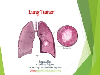
- 1. Lung Tumor Prepared by Mr.Abhay Rajpoot HOD (Dep. of Medical Surgical) abhayrajpoot5591@gmail.com
- 2. INTRODUCTION A lung tumor is an abnormal rate of cell division or cell death in lung tissue or in the airways that lead to the lungs. Lung tumor is a type of cancer that begins in the lungs. The lungs are two spongy organs in your chest that take in oxygen when you inhale and release carbon dioxide when you exhale.
- 4. DEFINITION A benign lung tumor is an abnormal growth of tissue that serves no purpose and is found not to be cancerous. Benign lung tumor may grow from many different structures in the lung.
- 5. INCIDENCE It is the leading cause of death among all racial groups in US. Accounting for 31% of all ca deaths in men & 27% of deaths in women. In 2005 more than 1,68,000 people died from lung ca in US; an estimated 1,84000 new cases were diagnosed in the same year.
- 6. RISK FACTORS: • Smoking. The risk of lung cancer increases with the number of cigarettes you smoke each day and the number of years you have smoked. Quitting at any age can significantly lower your risk of developing lung cancer. • Exposure to secondhand smoke. Even if you don't smoke, your risk of lung cancer increases if you're exposed to secondhand smoke. • Previous radiation therapy. If you've undergone radiation therapy to the chest for another type of cancer, you may have an increased risk of developing lung cancer. • Exposure to asbestos and other carcinogens. Workplace exposure to asbestos and other substances known to cause cancer — such as arsenic, chromium and nickel — can increase your risk of developing lung cancer, especially if you're a smoker. • Family history of lung cancer. People with a parent, sibling or child with lung cancer have an increased risk of the disease.
- 7. ETIOLOGY: Cigarette smoking which contain 43 known chemical carcinogens & ca promoters is most significant cause of ca. Genetic abnormality chromosome 3 with loss of genetic material. Alteration of tumor suppressor gene.
- 8. PATHOPHYSIOLOGY: Due to etiological factors Damage of bronchial epithelial cells Mutation of bronchial epithelial cells Epithelial cells become neoplastic
- 9. TYPES OF LUNG CA Acc. To cell type tumor can divide into : s. no. Cell type & prevalence Presentation & associated manifestation spread 1 Small-cell carcinoma (20-25%) Central lesion with hilar mass common, early meditational involvement,, no cavitations, SIADH, Cushing syndrome Aggressive tumor, more than 40% of clients have distant metastasis at time of presentation 2 Adenocarcinoma (20-40%) Peripheral mass involving; few local symptoms Early metastasis to CNS skeleton, & adrenal glands 3 Squamus cell carcinoma (30-32%) Central lesion located in large bronchi; clients presents with cough, dyspnea, atelectasis & wheezing Spread by local invasion 4 Large cell carcinoma (10-15%) Usually peripheral lesion that is larger than that associated with adenocarcinoma & tends to cavitate; gynecomastia Early metastasis
- 10. CLINICAL MANIFESTATION: • A new cough that doesn't go away • Coughing up blood, even a small amount • Shortness of breath • Chest pain • Hoarseness • Losing weight without trying • Bone pain • Headache
- 11. DIAGNOSTIC EVALUATION: Chest x-ray: usually provide the Ist evidence of lung cancer. It may be used as a screening tool for lung ca. Sputum specimen: is sent for cytologic examination. The sputum sample is collected on arising in the morning. If malignant cells are found in the sputum more invasive examinations are required. Bronchoscopy: done to visualize & obtain tissue for biopsy from the tumor. CT-scan: it is used to evaluate & localize tumors in the lung parenchyma & pleura. CT scanning can also detect distant tumor metastasis & evaluate tumor response to treatment. Cytologic examination: cells or tissues for cytologic examination & biopsy may be obtained by aspirating fluid from a pleural effusion, percutaneous needle biopsy & lymph node biopsy.
- 12. CONTI… CBC, liver function test & serum electrolytes: Including ca are obtained to evaluate for evidence of metastatic disease or paraneoplstic syndromes. Tuberculin test is performed to rule out TB as a cause of symptoms. Pulmonary function test: may be performed prior to the initiation of treatment if the client has manifestations of respiratory insufficiency (e.g. dyspnea, low oxygen saturation level).
- 13. MANAGEMENT: Chemotherapy: Used in combination, chemotherapeutic drugs to be attached at different parts of the cell cycle & in different ways, increasing the effectiveness of therapy. Chemotherapy drugs that commonly used are- Vance Alkaloids (Vinblastine), Doxorubicin, Taxanes (Docetaxel), Plantin analogus (Cisplantin, & Carboplantin).
- 14. CONTI… Radiation therapy: It is used alone or in combination with surgery & chemotherapy. Goals- Treatment- prior to surgery, R/T may be used to debulk tumors. Palliative- (symptom relief) it may also be used to relieve manifestation such as cough, hemoptysis & dyspnea from bronchial obstruction. R/T may be delivered by external beam radiation to the primary tumor site or by intraluminal radiation or brachytherapy.
- 15. SURGICAL MANAGEMENT The goal of surgery is to remove all involved tissue while preserving as much as functional lung as possible. s. no. procedure description Used for 1 Laser bronchoscopy Bronchoscopy guided laser used to resect tumor Tumors localized in a main bronchus 2 mediastinoscopy Visualization of mediastinum using an endoscope passed through a suprasternal incision Evaluation & biopsy of a meditational tumors & lymph nodes 3 thoracotomy Incision into the chest wall Access the lung & thoracic cavity for surgery
- 16. 4 Wedge resection Removal of an individual bronchovascular segment of a lobe Peripheral lung tumor with no evidence of extension to chest wall or metastasis 5 Segmental resection Resection of a section of a major bronchus with reconstruction of remaining normal bronchus Small lesion of major bronchus 6 Sleeve resection Resection of a section of a major bronchus with reconstruction of remaining normal bronchus Small lesion of a major bronchus 7 lobectomy Removal of a single lung lobe Tumor confined to a single lobe 8 pneumonecto my Removal of an entire lung Tumor widespread throughout the lung, involving the main bronchus or fixed to the hilum
- 17. COMPLICATION • Shortness of breath. People with lung cancer can experience shortness of breath if cancer grows to block the major airways.. • Coughing up blood. Lung cancer can cause bleeding in the airway, which can cause you to cough up blood (hemoptysis). • Pain. Advanced lung cancer that spreads to the lining of a lung or to another area of the body, such as a bone, can cause pain. • Fluid in the chest (pleural effusion). Lung cancer can cause fluid to accumulate in the space that surrounds the affected lung in the chest cavity (pleural space). Fluid accumulating in the chest can cause shortness of breath. • Cancer that spreads to other parts of the body (metastasis).
- 18. Nursing management: Ineffective breathing pattern r/t tumor & treatment of tumor. Activity intolerance r/t resectional lung surgery & inoperable lung ca. Acute pain r/t surgical procedure or terminal stage of ca. Anticipatory grieving r/t advanced diagnosis of lung ca.
- 19. THANK YOU