
Empfohlen
Weitere ähnliche Inhalte
Was ist angesagt?
Was ist angesagt? (20)
Ähnlich wie Long-term potentiation.ppt
Ähnlich wie Long-term potentiation.ppt (20)
Kürzlich hochgeladen
Kürzlich hochgeladen (20)
Long-term potentiation.ppt
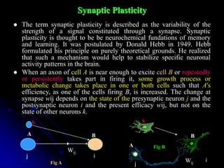
- 1. Synaptic Plasticity The term synaptic plasticity is described as the variability of the strength of a signal constituted through a synapse. Synaptic plasticity is thought to be be neurochemical fundations of memory and learning. It was postulated by Donald Hebb in 1949. Hebb formulated his principle on purely theoretical grounds. He realized that such a mechanism would help to stabilize specific neuronal activity patterns in the brain. When an axon of cell A is near enough to excite cell B or repeatedly or persistently takes part in firing it, some growth process or metabolic change takes place in one or both cells such that A's efficiency, as one of the cells firing B, is increased. The change at synapse wij depends on the state of the presynaptic neuron j and the postsynaptic neuron i and the present efficacy wij, but not on the state of other neurons k. j i A B Wij A B j i Wij j i Wij k k Fig A Fig B
- 2. Long-term potentiation Definition In neuroscience, long-term potentiation (LTP) is the strengthening (or potentiation) of the connection between two nerve cells. This connection lasts for an extended period of time (minutes to hours in vitro and hours to days and months in vivo). LTP can be induced experimentally by applying a sequence of short, high-frequency stimulations to nerve cell synapses. The phenomenon was discovered in the mammalian hippocampus by Terje Lømo in 1966 and is commonly regarded as the cellular basis of memory.
- 3. History Santiago Ramón y Cajal proposed that memories might be stored in the connections between nerve cells. By the turn of the 19th century, neurobiologists had good reason to believe that memories were generally not the product of new nerve cell growth. Scientists generally believed that the number of neurons in the adult brain (roughly 1011) did not increase significantly with age. With this realization came the need to explain how memories were created in the absence of new cell growth.
- 4. History Among the first neuroscientists to suggest that learning was not the product of new cell growth was the Spanish anatomist Santiago Ramón y Cajal. In 1894 Santiago Ramón y Cajal proposed that memories might be formed by strengthening the connections between existing neurons to improve the effectiveness of their communication. Hebbian theory, introduced by Donald Hebb in 1949, echoed Ramón y Cajal's ideas, and further proposed that cells may grow new connections between each other to enhance their ability to communicate:
- 5. History When an axon of cell A is near enough to excite a cell B and repeatedly or persistently takes part in firing it, some growth process or metabolic change takes place in one or both cells such that A's efficiency, as one of the cells firing B, is increased. (Hebb, The organization of behavior) Similarly, memories may be forgotten through the weakening or loss of connections. For example, a man might be startled by the sound of a car alarm outside. Sensory cells in the ear record the sound and send it to the brain where it activates neurons that control the man's muscles. But as the blaring alarm continues, those connections are weakened so that the alarm no longer causes the man to be startled. These theories about memory formation were unfortunately foresighted. Neuroscientists were simply not yet equipped with the neurophysiological techniques necessary for elucidating the biological underpinnings of learning in animals. These skills would not come until the latter half of the 20th century, at about the same time as the discovery of long-term potentiation.
- 6. Discovery of long-term potentiation 1. LTP was first discovered in the rabbit hippocampus. In humans the hippocampus is located in the medial temporal lobe. 2. LTP was first observed by Terje Lømo in 1966 in the Oslo, Norway, laboratory of Per Andersen (12740104). There, Lømo conducted a series of neurophysiological experiments exploring the role of the hippocampus in the rabbit short-term memory. Targeting the synapses between granule cells of the perforant pathway and those of the dentate gyrus, Lømo elicited excitatory postsynaptic potentials (EPSPs) from dentate gyrus cells by stimulating the perforant pathway. He observed that a high-frequency train of stimulation produced larger, prolonged EPSPs compared to the responses evoked by a single stimulation. This phenomenon was soon dubbed "long-term potentiation". 3. Timothy Bliss, who joined the Andersen laboratory in 1968, collaborated with Lømo in 1973 to publish the first characterization of LTP in rabbit hippocampus.
- 7. Types of LTP Since its original discovery in the rabbit hippocampus, LTP has been observed in a variety of other neural structures, including the cerebral cortex, cerebellum, amygdala, and many others. The underlying mechanisms of LTP are generally conserved across these different regions, but there are subtle differences in LTP's precise molecular machinery between sites. Very broadly, there are two types of LTP: 1. Associative and 2. Nonassociative. At rest, the NMDA receptor is blocked by magnesium, preventing the flow of calcium into the postsynaptic cell.
- 8. Associative LTP Associative LTP is the molecular analog of associative learning (e.g. classical conditioning). It is the strengthening of the connection between two neurons that have been simultaneously active. To detect the simultaneous activity of the pre- and postsynaptic cells, associative LTP requires a so- called coincidence detector. In many parts of the brain where associative LTP is observed, the NMDA receptor (NMDAR) fills the role of coincidence detector. At rest, the NMDAR's calcium channel is blocked by magnesium; the blockade is relieved only after strong postsynaptic depolarization. The calcium channel is also ligand-gated, so that it only opens when presynaptically- released glutamate binds the receptor. When the NMDAR opens, calcium floods the postsynaptic cell triggering associative LTP.
- 9. Associative LTP NMDAR-dependent LTP has been demonstrated in the hippocampus, particularly in the Schaffer collaterals and perforant pathway, and several other brain regions including parts of the amygdala (9403688) and cerebral cortex (2446147). There are several types of associative LTP that do not depend on NMDA receptors. NMDAR-independent LTP has been observed, for example, in the amygdala, where it depends instead on voltage-gated calcium channels.
- 10. Nonassociative LTP Nonassociative LTP is brought about by the repeated application of one stimulus (whereas in associative LTP there are at least two stimuli). At nonassociative synapses, such as those involved in habituation and sensitization, persistent stimulation of the synapse triggers an influx of calcium into the postsynaptic cell. As in associative LTP, calcium signals the beginning of long-term potentiation, but the precise mechanisms of nonassociative LTP are still unknown.
- 11. Properties of LTP NMDA receptor-dependent LTP classically exhibits four main properties: Rapid induction, Cooperativity, Associativity, and Input specificity:
- 12. Properties of LTP Induction: LTP can be rapidly induced by applying one or more brief tetanic stimuli to a presynaptic cell. (A tetanic stimulus is a high- frequency sequence of individual stimulati.) Cooperativity: LTP can be induced either by strong tetanic stimulation of a single pathway, or cooperatively via the weaker stimulation of many. It is explained by the presence of a stimulus threshold that must be reached in order to induce LTP. Associativity: refers to the observation that when weak stimulation of a single pathway is insufficient for the induction of LTP, simultaneous strong stimulation of another pathway will induce LTP at both pathways. There is some evidence that associativity and cooperativity share the same underlying cellular mechanism (Synaptic tagging). Input specific: Once induced, LTP at one synapse is not propagated to adjacent synapses; rather LTP is input specific.
- 13. Properties of LTP Rapid induction: LTP can be rapidly induced by applying one or more brief tetanic stimuli to a presynaptic cell. (A tetanic stimulus is a high-frequency sequence of individual stimuli.) A neuron is impaled by an intracellular electrode to record the membrane potential while presynaptic fibers are stimulated by means of a second extracellular electrode. Small pulses are applied to the presynaptic fibers in order measure the strength of the postsynaptic response. The amplitude of the test pulse is chosen so that the stimulation evokes a postsynaptic potential, but no action potentials. No action potential
- 14. Properties of LTP In a second step, the input fibers are strongly stimulated by a sequence of high frequency pulses so as to evoke postsynaptic firing (Fig. B). Rapid induction: LTP
- 15. Properties of LTP After that the strength of the postsynaptic response to small pulses is tested again and a significantly increased amplitude of postsynaptic potentials is found (Fig. C). This change in the synaptic strength persists over many hours and is thus called long-term potentiation. Rapid induction: LTP
- 16. Properties of LTP Cooperativity: LTP can be induced either by of a single pathway, or cooperatively via the weaker stimulation of many. It is explained by the presence of a stimulus threshold that must be reached in order to induce LTP. Strong tetanic stimulation LTP
- 17. Properties of LTP Cooperativity: LTP can be induced either by of a single pathway, or cooperatively via the weaker stimulation of many. It is explained by the presence of a stimulus threshold that must be reached in order to induce LTP. Many weaker stimulation LTP
- 18. Properties of LTP Associativity: refers to the observation that when weak stimulation of a single pathway is insufficient for the induction of LTP, simultaneous strong stimulation of another pathway will induce LTP at both pathways. There is some evidence that associativity and cooperativity share the same underlying cellular mechanism (Synaptic tagging). Weak stimulation Simultaneous strong stimulation LTP
- 19. Properties of LTP Once induced, LTP at one synapse is not propagated to adjacent synapses; rather LTP is input specific. Synapse input-specific LTP Basal activity (no LTP) LTP
- 20. Phases of LTP LTP is often divided into two phases: 1. An early protein synthesis-independent phase (E-LTP) that lasts between one and five hours, and 2. A late protein synthesis-dependent phase (L-LTP) that lasts from days to months. Broadly, E-LTP produces short-lived synaptic facilitation by making existing postsynaptic glutamate receptors (e.g. AMPA receptors) more sensitive to glutamate. Conversely, L-LTP results in a pronounced facilitation of the postsynaptic response largely through the synthesis of new proteins. These proteins include glutamate receptors (e.g. AMPAR), transcription factors, and structural proteins that enhance existing synapses and form new connections. There is also considerable evidence that late LTP prompts the postsynaptic synthesis of a retrograde messenger that diffuses to the presynaptic cell increasing the probability of neurotransmitter vesicle release on subsequent stimuli.
- 22. Early LTP E-LTP can be induced experimentally by applying a few trains of tetanic stimulation to the connection between two neurons. Through normal synaptic transmission, this stimulation causes the release of neurotransmitters, particularly glutamate, from the presynaptic terminal onto the postsynaptic cell membrane, where they bind to neurotransmitter receptors embedded in the postsynaptic membrane. Though a single presentation of the stimulus is usually not sufficient to induce LTP, repeated presentations cause the postsynaptic cell to be progressively depolarized. In NMDAR-dependent synapses, this progressive depolarization relieves the magnesium blockade of the NMDA receptor. When the next stimulus is applied, glutamate binds the NMDA receptor and calcium floods the postsynaptic cell, rapidly increasing the intracellular concentration of calcium. It is this rapid rise in calcium concentration that induces E-LTP.
- 23. ELTP Beyond calcium's critical role in the induction of E-LTP, Few downstream molecular events leading to the expression and maintenance of E-LTP are known with certainty. E-LTP induction depends upon the activity of several protein kinases, including calcium/calmodulin-dependent protein kinase II (CaMKII), PKC, PKA, MAPK, and tyrosine kinases. Postsynaptically, the ELTP is expressed primarily through the enhancement of receptor/channel sensitivity. In NMDA-dependent LTP in the CA1 hippocampus, the endogenous calcium chelator calmodulin rapidly binds calcium as a result of NMDAR opening. The calcium-calmodulin complex directly activates CaMKII which 1) phosphorylates voltage-gated potassium channels increasing their excitability; 2) enhances the activity of existing AMPA receptors; and 3) phosphorylates intracellular AMPARs and activates Syn GAP (a Ras GTPase activating protein) and the MAPK cascade, facilitating the insertion of AMPARs into the postsynaptic membrane.
- 24. E-LTP PKA serves a role similar to that of CaMKII, but PKA's effects are more broad. PKA's activity is enhanced during LTP induction by elevated levels of cAMP as a result of calcium's activation of adenylyl cyclase. Like CaMKII, PKA phosphorylates voltage-dependent potassium channels and also calcium channels enhancing their excitability to future stimuli. Additionally, PKA phosphorylates intracellular AMPAR stores, facilitating their insertion postsynaptically. PKA may also enhance AMPAR delivery via activation of the MAPK cascade. While LTP is induced postsynaptically, it is partially expressed presynaptically. One hypothesis of presynaptic facilitation is that enhanced CaMKII activity during early LTP gives rise to CaMKII autophosphorylation and constitutive activation. Persistent CaMKII activity then stimulates NO synthase, leading to the enhanced production of the putative retrograde messenger, NO. Since NO is a diffusable gas, it freely diffuses across the synaptic cleft to the presynaptic cell leading to a chain of molecular events that facilitate the presynaptic response to subsequent stimuli.
- 25. The late phase of LTP is dependent upon gene expression and protein synthesis, mediated largely by CREB-1. Late LTP can be experimentally induced by a series of three or more trains of tetanic stimulation spaced roughly 10 minutes apart. Unlike early LTP, late LTP requires gene transcription and protein synthesis (3401749), making it an attractive candidate for the molecular analog of long-term memory. The synthesis of gene products is driven by kinases which in turn activate transcription factors that mediate gene expression. camp response element binding protein-1 (CREB-1) is thought to be the primary transcription factor in the cascade of gene expression that leads to prolonged structural changes to the synapse enhancing its strength. CREB-1 is both necessary (2141668) and sufficient for late LTP. It is active in its phosphorylated form and induces the transcription of so- called immediate-early genes, including c-fos and c-jun. Ultimately, the products of CREB-1-mediated transcription and protein synthesis give rise to new building materials for the synaptic connection between pre- and postsynaptic cell. Late LTP
- 26. During L-LTP, constitutively active CaMKII activates a related kinase, CaMKIV. Additionally, enhanced Ca2+ levels during late LTP increase cAMP synthesis via adenylyl cyclase-1, further activating PKA and resulting in the phosphorylation and activation of MAPK (10964936). Facilitated by cAMP, both CaMKII and CaMKIV translocate to the cell nucleus along with PKA and MAPK (mediated by PKA), where they phosphorylate CREB-1. There is also some evidence that L-LTP is mediated in part by NO. In particular, NO may activate guanylate cyclase, leading to the production of cyclic GMP and activation of protein kinase G (PKG), which phosphorylates CREB-1. PKG may also cause the release of Ca2+ from ryanodine receptor-gated intracellular stores, increasing the Ca2+ concentration which activates other previously mentioned kinase cascades to further activate CREB-1. Late LTP
- 27. LTP and behavioral memory In neuroscience, the Morris water maze is a behavioral procedure designed to test the spatial memory of rats and other small mammals. It was developed by neuroscientist Richard Morris in 1981, and is commonly used today to explore the role of the hippocampus in the formation of memories about space. In the typical paradigm, a rat is placed into a small pool of opaque water which contains a escape platform hidden a few millimeters below the water surface. Visual cues, such as colored shapes, are placed around the pool in plain sight of the rat. When released, the rat swims around the pool in search of an exit while various parameters are recorded, including the time spent in each quadrant of the pool, the time taken to reach the platform (latency), and total distance traveled. The rat's escape from the water reinforces its desire to quickly find the platform, and on subsequent trials (with the platform in the same position) the rat is able to locate the platform more rapidly. This improvement in performance occurs because the rat has learned where the hidden platform is located relative to the salient visual cues.
- 29. LTP and behavioral memory Richard Morris provided some of the first evidence that LTP was indeed required for the formation of memories. He tested the spatial memory of two groups of rats, One whose hippocampi were bathed in the NMDA receptor blocker APV (APV (also called AP5) is a selective NMDA receptor (NMDAR) antagonist that competitively inhibits the active site of NMDAR. Its chemical name is 2-amino-5-phosphonovalerate), and The other acting as a control group. Both groups were then subjected to the Morris water maze, in which rats were placed into a pool of water and tested on how quickly they could locate a platform hidden beneath the water's surface.
- 30. LTP and behavioral memory Rats in the control group were able to locate the platform and escape from the pool, whereas the ability of APV-treated rats to complete the task was significantly impaired. Water maze Water Platform Platform Water
- 31. LTP and behavioral memory Moreover, when slices of the hippocampus were taken from both groups of rats, LTP was easily induced in controls, but could not be induced in the brains of APV-treated rats. This provided some evidence that the NMDA receptor — and thus LTP — was somehow involved with at least some types of learning and memory. LTP Control APV-treated
- 32. LTP and behavioral memory Similarly, Susumu Tonegawa has demonstrated that a specific region of the hippocampus, namely CA1, is crucial to the formation of spatial memories. So-called place cells located in this region are responsible for creating "place fields" of the rat's environment, which may be roughly equated with maps of the rat's surroundings. The accuracy of these maps determines how well a rat learns about its environment, and thus how well it can navigate about it. Tonegawa found that by impairing the NMDA receptor, specifically by genetically removing the NMDAR1 subunit in the CA1 region, the place fields generated were substantially less specific than those of controls. That is, rats produced faulty spatial maps when their NMDA receptors were impaired. As expected, these rats performed very poorly on spatial tasks compared to controls, providing more support to the notion that LTP is the underlying mechanism of spatial learning.
- 33. LTP and behavioral memory Enhanced NMDA receptor activity in the hippocampus has also been shown to produce enhanced LTP and an overall improvement in spatial learning. Trangenic mice with enhanced NMDA receptor function by overexpressing the NR2B subunit in the hippocampus. These mice, nicknamed "Doogie mice" after the precocious doctor Doogie Howser, had larger long-term potentiation and excelled at spatial learning tasks, once again suggesting LTP's involvement in the formation of hippocampal-dependent memories.