
Empfohlen
Empfohlen
Weitere ähnliche Inhalte
Was ist angesagt?
Was ist angesagt? (20)
Ähnlich wie Regional conditions of upper limb
Ähnlich wie Regional conditions of upper limb (20)
Mehr von Aaishwaryaa Rai
Kürzlich hochgeladen
Kürzlich hochgeladen (20)
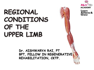
Regional conditions of upper limb
- 1. Dr. AISHWARYA RAI, PT BPT, FELLOW IN REGENERATIVE REHABILITATION, CKTP.
- 6. •The shoulder joint is a synovial joint of the ball and socket variety. •Structurally, it is a weak joint because the glenoid cavity is too small and shallow to hold the head of the humerus in place •The shoulder joints get excessive mobility at the cost of its own stability, since both are not feasible to the same degree. •BONES FORMING THE SHOULDER COMPLEX 1. Clavicle 2. Humerus 3. Scapula 4. Sternum • Due to the compromised mobility of the joint a few structures that provide stability to the joint are: 1. The rotator cuff 2. Ligaments 3. Glenoid Labrum 4. Long head of Biceps brachii muscle SHOULDER JOINT
- 7. Fig: SHOULDER JOINT COMPLEX
- 8. ELBOW JOINT The elbow joint is a synovial joint between the lower end of humerus and the upper ends of radius and ulna bones. Articular Surfaces: Upper: The capitulum and trochlea of the humerus. Lower: (i) Upper surface of the head of the radius articulates with the capitulum, (ii) trochlear notch of the ulna articulates with the trochlea of the humerus. The elbow joint is continuous with the superior radioulnar joint. The humeroradial, the humeroulnar and the superior radioulnar joints are together known as cubital articulations.
- 9. FIG: ELBOW JOINT FIG: RADIO-ULNAR JOINTS
- 10. WRIST JOINT THE WRIST JOINT IS A SYNOVIAL JOINT OF ELLIPSOID VARIETY BETWEEN LOWER END OF RADIUS AND THREE LATERAL BONES OF PROXIMAL ROW OF CARPUS. Articular Surfaces Upper: 1.Inferior surface of the lower end of the radius 2.Articular disc of the inferior radioulnar joint Lower: 1.Scaphoid 2.Lunate 3.Triquetral bones.
- 11. Fig: wrist joint
- 12. SHOULDER CONDITIONS Rotator Cuff Lesions. Adhesive Capsulitis Deltoid Contracture Bursitis
- 14. ROTATOR CUFF LESIONSThis includes both rotator cuff tears and impingement syndrome. Impingement Syndrome: Commonly associated with supraspinatus tendon, where the supraspinatus tendon gets impinged below the coracoacromial arch. The major site of compression is anterior to the angle of the acromion. Hence, the proper term is anterior impingement syndrome or painful arc syndrome. The pathology of subacromial impingment generally relates to a chronic repetitive mechanical process in which the conjoint tendon of the rotator cuff muscles undergoes repetitive compression and micro trauma as it passes under the coraco-acromial arch. As the arm is flexed, abducted or rotated the subacromial space width changes and the cuff becomes increasingly compressed.
- 15. In this syndrome, the arc of shoulder abduction elevation is painful from about 45–160 degrees, but this is painless up to the initial 45 degrees as well as in the terminal range from about 160 degrees to full elevation. The passive ROM is pain free. The cause is mechanical – the tender structure get nipped in between the tuberosity of the humerus and the acromion process or coracoacromial ligament.
- 16. NEER’S STAGES OF IMPINGEMENT Stage 1 is characterized by edema and tendinitis and is more typical in patients under 25 years old. Stage 2 is characterized by chronic inflammation, thickening and fibrosis of the impinged tendon due to repeated insults. This further decreases the size of the suprahumeral space. Stage 2 is more typical in patients between 25 and 40 years old and patients who have had a history of episodes of shoulder pain. Stage 3 is characterized by tendon degeneration, rupture, and arthritis. Patients with this stage of impingement are usually over 40 years old and have a prolonged history of shoulder problems. Often, at this stage, there will be radiographic evidence and a high likelihood of partial or full thickness rotator cuff tears.
- 17. CLASSIFICATION: PRIMARY: Impingement occurs beneath the coracoacromial arch and is due to the subacromial overloading. SECONDARY: Occurs due to relative decrease in the subacromial arch and is due to microinstability of the GH joint or ST joint. POSTERIOR(INTERNAL): In this the supraspinatus and infrapinatus tendons are pinched between the posterior and superior aspects of the glenoid when the arms are in an elevated and externally rotated position. It is usually seen in athletes involved in more of overhead activities like throwers, swimmers, etc.
- 18. KEY SYMPTOMS OF IMPINGEMENT Toothache like Antero-lateral shoulder region. Pain sleeping on the affected side Stiffness Catching of the shoulder during use Pain on active and passive range of motion Local tenderness Nocturnal pain
- 21. TREATMENT: If conservative method fails even after three months, then surgical method is opted for Arthroscopic repair in small and partial tears. Open methods in major tears. Depending upon the etiological factors, the following surgical techniques are described: Excision of adhesions and manipulation of shoulder, excision of calcium deposits, repair of incomplete tear, acromioplasty, acromionectomy for more disabling pain with normal range of movements, direct suture for complete rupture of rotator cuff, rotation and transposition of flap, free graft, etc. Results are good in 85-90 percent.
- 22. Deltoid contracture/fibrosis is a condition where intramuscular fibrous bands within lead to contractures and stiffness of the shoulder joint. It is seen in people of all ages, but it has been reported primarily in children. It is often associated with congenital/developmental defects, a genetic disposition or application of intramuscular injections. The fibrosis could develop by any of the following Direct injury to the muscle by the needle toxicity of the drug. Volume of injection causing ischemia of selected portion Local edema Fibrotic compression Damage to vessels Fibrotic compartmentalization Muscle ischemia Entrapment neuropathy DELTOID CONTRACTURE
- 24. Fig; Arms Resting in abduction Fig: Winging of scapula
- 25. a. Abduction contracture of the left shoulder with winging of the scapula. b. The winged scapula disappears with horizontal abduction of the shoulder. c. anterior protrusion of the left humeral head and winging of the scapula.
- 26. Fig: Deltoid contracture along with scoliotic changes
- 27. Patient presents with a history of restriction of shoulder movement affecting the activities of daily living. The patient would typically complaint of inability to pull the arm fully down to the side of the body or across. A complaint of pain near the near the shoulder and neck may be present. On examination, one may notice a dimple over the deltoid skin. Palpation would reveal a fibrous palpable band. Patient should be enquired for the history of injections, similar contracture in other parts of the body and a family history of similar contractures. Depending on the part of the deltoid involved, the limb of the patient may be: oAbducted – Middle portion oFlexed and abducted – Anterior portion oExtended and abducted – Posterior Portion In severe cases involving anterior or posterior parts, the subluxation/dislocation of the humeral head may occur. Examination should look for high riding scapula(Sprengel deformity) or other abnormalities like scoliosis or chest wall abnormality. Motion of neck, glenohumeral joint and scapula should be assessed. Winging of scapula and freedom of scapular motion should be noted. Look for evidence of contractures elsewhere in both the upper and the lower extremities.
- 28. Diagnosis: X-ray, CT Scan, MRI. May reveal flattening of the head of the humerus and drooping of acromion due to constant contracture. With increased contracture, inferior border of the scapula rotates medially, causing the winging of the scapula. In severe abduction contracture, secondary scoliosis may occur. Treatment: The surgical treatment should be considered if Contracture > 25 degrees; Age> 5 years; Progressively increasing; Painful. Fibrous bands are released or resected. The arm is immobilized across the body in adducted position for about two weeks after the surgery. After this patient is put on physical therapy and movements are started.
- 29. ADHESIVE CAPSULITISIt is defined as a clinical syndrome characterized by painful restriction (Figs 30.1A and B) of both active and passive shoulder movements due to causes within the shoulder joint or remote (other parts of the body). Patient Presentation: The hallmark of adhesive capsulitis is pain and decreased motion. Needless to say, there are many conditions which cause either pain or decreased motion or both; adhesive capsulitis can appear as an isolated condition or accompanying another shoulder condition. Patients with adhesive capsulitis present with gradual unilateral shoulder pain that is often diffuse and worse at night. They also report an insidious onset of increasing stiffness. Some patients may “remember” a trivial trauma inciting the process, though this is likely a faulty attribution.
- 31. Fig: The capsule, outlined in blue, holds the humeral head against the glenoid Fig: Decreased ROM in an adhesive capsulitis affected patient
- 32. Clinically, isolated adhesive capsulitis has 3 distinct phases, namely: the painful phase, the stiff phase, and the resolution phases. The Painful (or “freezing”) Phase begins gradually, with no known precipitant. This phase, which lasts weeks to months, is characterized by diffuse, disabling pain that is worse at night. The Stiff Phase is noted by marked stiffness that limits range of motion in multiple plains, interfering with activities of daily living. Pain is less intense at this point. This phase can last a year or longer. Most patients enter a Resolution (“thawing”) Phase during which motion improves, though often some limitations of range of motion compared to the contralateral shoulder remain. On physical exam, patients with adhesive capsulitis have significantly reduced active and passive range of motion in two or more planes (see Figure 3 for normal ranges of motion). External rotation and abduction are the most commonly affected movements. Patients also have difficulty internally rotating, and report problems scratching their hand on their back or hooking their bra from behind.
- 33. Fig: An arthrogram showing a reduced volume of contrast material within the shoulder joint. The red line outlines the border of the normal border of the capsule
- 34. BURSITIS Bursae are membranous sacs lined by synovial membrane and sited so that they prevent friction or wear and tear of muscles and tendons as they pass over the bone. It is of two types: True bursa: eg. Found subacromially, etc. Adventitious bursa: Occur in response to friction over tendon and can occur anywhere.
- 35. Bursitis is the inflammation of these bursa. The inflammation arises because of excessive pressure, friction or gouty deposit. Irritative bursitis: This is the commoner of the two types. It is caused by excessive pressure or friction, occasionally due to a gouty deposit. Inflammation of the bursa results in the effusion of a clear fluid within the bursal sac. With prolonged inflammation, the sac gets thickened and may cause pressure erosion on the adjacent bone. Infective bursitis: Uncommonly, a bursa may get infected by a pyogenic or tubercular infection. TREATMENT: Rest Icing NSAIDs Corticosteroids If infective- antibiotics and surgical drainage. Surgical: Excision of the bursa (rarely done)
- 36. Fig: subacromial bursitis Fig: Application of corticosteroid injections
- 37. Fig: Prepatellar bursitis Fig: Olecranon bursitis
- 39. Lateral Epicondylitis is a common clinical entity characterized by pain and tenderness at the common origin of the extensor group muscles of the forearm, usually as a result of a specific strain, overuse, or a direct bang. It is considered a cumulative trauma injury that occurs over time from repeated use of the muscles of the arm and forearm, leading to small tears of the tendons (Tendonitis). CAUSES Epicondylitis: This is due to single or multiple tears in the common extensor origin, periostitis, angiofibroblastic proliferation of extensor carpi radialis brevis (ECRB), etc. Inflammation of adventitious bursa: Between the common extensor origin and radio humeral joint. Calcified deposits: Within the common extensor tendon. Painful annular ligament: It is due to hypertrophy of synovial fringe between the radial head and the capitulum’s. Pain of neurological origin: e.g. cervical spine affection, radial nerve entrapment, etc LATERAL EPICONDYLITIS
- 40. STAGES Stage I: There is acute inflammation but no angioblastic invasion. The patient complains of pain during activity. Stage II: This is the stage of chronic inflammation. There is some angioblastic invasion. The patient complains of pain both during activity and at rest. Stage III: Chronic inflammation with extensive angioblastic invasion. The patient complains pain at rest, night pains, and pain during daily activities. PATIENT PRESENTATION Patient complains of pain on the outer aspect of the elbow and has difficulty in gripping objects and lifting them. Sportspersons will have difficulty in extending the elbow. Eventually the pain may become so constant and severe so as to stop the patient from further playing or interferes with activities of daily living, such as carrying a briefcase, wringing wet clothes or even holding a cup of tea. Grip becomes weak. Morning stiffness may be felt.
- 42. TREATMENT Conservative treatment: The treatment consists of rest and trying to avoid the movements that cause pain. NSAIDs and a tennis elbow splint are used for pain relief. Local injection of hydrocortisone with local anaesthetic solution relieves pain in majority of cases. Surgical treatment: The extensor muscles are stripped from their origin, i.e., lateral epicondyle, and are allowed to fall back. An above- elbow slab with elbow in 90 degrees flexion is given for a period of 10 days postoperatively. The elbow is then mobilized.
- 43. FIG: KINESIOTAPING FOR TENNIS ELBOW
- 44. This condition is not as common as the tennis elbow. The site of tenderness being the medial epicondyle of the humerus and the seat of inflammation is the common origin of the flexor group of muscles. The muscles of the common flexor group are strained or torn from the medial epicondyle of humerus. Pain is produced by extension of the elbow, supination and valgus strain. It is called Golfer elbow because it is thought to be caused when a golfer hits the ground instead of the ball, thus producing a valgus strain to the elbow thereby straining the flexor group of muscles. TREATMENT: A steroid injection is given locally into the muscle belly at its origin. The physiotherapeutic management programme proceeds on the same lines as for lateral epicondylitis MEDIAL EPICONDYLITIS
- 45. FIG: KINESIOTAPING FOR GOLFER’S ELBOW
- 47. HAND & WRIST CONDITIONS De Quervain’s Disease Carpal Tunnel Syndrome Tenosynovitis Tenovaginitis Wrist Ganglion Duputyren’s Contracture
- 48. TENOSYNOVITI SFollowing an infection or an injury, the synovial lining of the tendon sheath responds by secreting excessive synovial fluid. This condition is called tenosynovitis. When it is traumatic, it affects the tendons of the abductor pollicis longus and extensor pollicis brevis, which form the radial boundary of the anatomical snuff box. It may present as irritative or infective tenosynovitis. CLINICAL PRESENTATION Severe Pain Crepitus on palpation No adhesions or limitations of ROM *Usually irritative occurs at wrist and ankle *Infective occurs due to low grade organisms or TB.
- 50. TREATMENT Conservative: Restriction of movement- use of splint, bandaging, POP cast Iceing Antibiotics Physical Therapy Surgery: Splitting of the fibrous sheath, which will require pre-op and post-op PT.
- 51. TENOVAGINITI S The tendon sheath rather than fibrous sheath is affected. In tenovaginitis, the difference in the pathology is that instead of secreting abnormal amount of synovial fluid, the tendon gets thickened and fibrosed. This limits the movement of the tendon within its sheath. TRIGGER FINGERS AND THUMB It is a stenosing tenovaginitis, in which the sheath of a flexor tendon thickens, apparently spontaneously, to entrap the tendon. CLINICAL FEATURES Initially, the only symptom is pain at the base of the affected finger, especially on trying to passively extend the finger. As the sheath further thickens, the contained tendon gets swollen proximal to it. The swollen segment of the tendon does not enter the sheath when an attempt is made to straighten the finger from the flexed position. This is called ‘locking of finger’. This locking can be overcome either by a strong effort in which case the finger extends with a snap-like trigger of a pistol or by extending the finger passively with other hand.
- 53. TREATMENT In early stages, local ultrasonic therapy provides relief. In a long standing problem, a local injection of hydrocortisone relieves the pain. In some cases, splitting of the tight tendon sheath may be required.
- 55. DE QUERVAINS DISEASEIt is also called as stenosing tenosynovitis of the first dorsal compartment of the wrist involving the abductor pollicis longus and extensor pollicis brevis tendons. This is a condition characterised by pain and swelling over the radial styloid process. It results from inflammation of the common sheath of abductor pollicis longus and extensor pollicis brevis tendons. On examination, the tenderness is localised to the radial styloid process. Pain is aggravated by adducting the thumb across the palm and forcing ulnar deviation and on asking the patient to perform radial deviation againstresistance (Finkelstein's test). There may be a palpable thickening of the sheath.
- 58. TREATMENT Conservative: Drug therapy to combat inflammation. Restriction of movements by applying a small hand splint in functional position with immobilization of the thumb. During early stage, ice massage, and at the later stage, ultrasound or any deep heating modality helps in reducing inflammation and pain. Local corticosteroid infiltration may be effective in some. Surgical intervention: in the form of splitting the lateral wall of the tendon sheath may be necessary in non responding cases.
- 59. CARPAL TUNNEL SYNDROME This is a syndrome characterised by the compression of the median nerve as it passes beneath the flexor retinaculum through the carpal tunnel. CARPAL TUNNEL Carpal tunnel is the overcrowded fibro-osseous canal formed between three carpal bones (scaphoid, trapezoid and hamate) and the transverse carpal ligament. The median nerve and the nine tendons of the digital flexors pass through this 2–2.5 cm tunnel. Therefore, any bony or soft- tissue pathology to it precipitates compressive forces over the median nerve passing through it.
- 60. ETIOLOGY Occupational overuse of wrist joint, e.g., computer keyboards Malunited Colles’ fracture, rheumatoid or osteoarthritis Ganglia or haematoma at the wrist Connective tissue disorders (amyloidosis) Endocrine disorders like DM, hypothyroidism, menopause Metabolic causes like gout Growth hormone abnormality (acromegaly) Pregnancy CLINICAL FEATURES Pain and discomfort at the wrist during movements or even at rest. Burning, aching, warmth, paraesthesia in the hand and wrist in the distribution of the median nerve (tingling, numbness or burning sensation); relieved by shaking hands. Vasomotor symptoms like swelling, cold, dry and shiny skin. Motor weakness of opponens, flexor pollicis brevis results in difficulty in making circle with thumb and index finger. Bottle sign – poor contact of thumb and index finger due to the paralysis of abductor pollicis brevis, causing difficulties in gripping a cylindrical object, e.g., bottle or cylinder. Loss of true opposition. Late stage muscular atrophy – thenar muscles of the hand
- 62. TREATMENT Conservative Treatment of the relevant causative factor which has precipitated the syndrome. A simple splint that blocks movements at the wrist is adequate to avoid compressive stretches to the carpal tunnel. Nonsteroidal anti-inflammatory drug (NSAID) agents Local ultrasonic exposure Pain-free relaxed passive or speedy active movements are encouraged in the pain-free range by removing the splint. Cryotherapy, TENS over the palmar aspect of the wrist may be tried. Local corticosteroid injection may be given only after confirming the absence of sensory deficit. Surgical: Surgical release of the median nerve may have to be undertaken by the division of the carpal flexor retinaculum and/or transverse carpal ligament either by open surgery or by endoscopic surgery.
- 64. It is the commonest cystic swelling on the dorsum of the wrist. It results from mucoid degeneration of the tendon sheath or the joint capsule. Ordinarily, there are no symptoms other than the swelling itself. Sometimes, a mild discomfort or pain is experienced. The cyst may sometimes be so tense as to resemble a solid tumour of the tendon sheath. Often the cyst is multi-loculated. Aspiration of the cyst is performed and an injection of hylase given. If the cyst recurs, excision may be required. WRIST GANGLION= bible bumps
- 67. DUPUTYREN’S CONTRACTURE This is a condition characterised by a flexion deformity of one or more fingers due to a thickening and shortening of the palmar aponeurosis. The cause is unknown, but a hereditary predisposition has been established. There is an increased incidence of the disorder among cirrhotic patients and in epileptics on sodium hydantoin. CLINICAL FEATURES In early stages, thickening of the palmar aponeurosis is felt at the bases of ring and little fingers. Later, a flexion deformity of the fingers develops. Dupuytren's contracture can be differentiated from a similar deformity due to contracture of the flexor tendons; in the former only the MP and PIP joints are flexed, unlike the latter where the DIP joints are also flexed.
- 70. Surgery. This is the most common treatment used for advanced cases. It may be done when you have limited use of your hand. During Dupuytren's contracture surgery, the surgeon makes a cut (incision) in your hand and takes out the thickened tissue. This can improve the mobility of your fingers. Some people have contractures return. They may need surgery again. Steroid shot (injection). If a lump is painful, a steroid injection may help ease the pain. In some cases, it may stop your condition from getting worse. You may need repeated injections. Radiation therapy. This treatment is not as common in the U.S. Low energy X-rays are directed at the nodules. This works best in the early stage of the disease. It can soften the nodules and help keep contractions from happening. Enzyme injection. This is a newer, less invasive procedure done by specially trained surgeons. Your doctor injects a medicine into the area to numb the hand. Then the enzyme is injected into the lump of tissue. Over several hours, the enzyme breaks down and dissolves the tough bands. This lets the fingers straighten when the cord is snapped by the surgeon, usually the next day. Needle aponeurotomy. This is another newer, less invasive procedure. Medicine is injected into the area to numb the hand.
- 71. REFERENCES B D Chaurasia’s Human Anatomy, Regional and Applied Dissection and Clinical; Volume 1; Upper Limb and Thorax Essential Orthopedics (Including Clinical Methods); Fifth Edition; J Maheshwari. Textbook of Orthopedics, Fourth Edition; John Ebnezar. Orthopedics and Applied Physiotherapy; Jayant Joshi Prakash Kotwal. Orthopedics by Dr. C Hutchinson Kibler WB, Sciascia A, Wilkes T. Scapular dyskinesis and its relation to shoulder injury. J Am Acad Orthop Surg. 2012 Jun. 20(6):364-72. Chen WJ, Wu CC, Lin YH, Shih CH. Treatment of deltoid contracture in adults by distal release of the deltoid. Clin Orthop Relat Res. 2000 Sep. (378):136-42. Ngoc HN. Fibrous deltoid muscle in Vietnamese children. J Pediatr Orthop B. 2007 Sep. 16(5):337-44. Hang YS, Miller JW. Abduction contracture of the shoulder. A report of two patients. Acta Orthop Scand. 1978 Apr. 49(2):154-7. .