2. INTRODUCTION
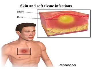
• Skin and soft tissue infection : invasion of organism through skin or
organism reach the skin from blood
• Pyogenic infections : pus produced
• Pus : localized deep tissue , surface ( pahrynx) , musosa of bladder ,
meninges and any part of body
3. Clinical types (SSTIs)
• 1) PRIMARY LESION: due to damage of trauma or disease
• 2) SECONDARY LESION: As a result of any primary infections
4. • Area of infection
• Skin
• Subcutaneous tissue
• Fasciae
• Muscles
8. FASCIA AND MUSCLES
SKIN LESION ETIOLOGICAL AGENT
Necrotizing fasciitis S.PYOGENES
PYOMYOSITIS S.PYOGENES
S.AUREUS
MYONECROSIS CLOSTRIDIAL MYONECROSIS
9. Erysipelas
• Common in very young, old, debilitated patients
• Symptoms:fever, shivering, chills, fatigue, headaches, v
omiting (48 hrs of infection)
• Well demarcated, painful, erythematous indurated
plaques, Blisters & ulceration
• More severe infections: lymphedema
• Face, legs
• Cause: Streptococcus pyogenes , Streptococcus
agalactiae (newborns)
10. Impetigo
• A contagious superficial infection of the skin
• common in children
• involves : face, around the mouth and nose
.
• spread by direct contact
Symptoms: red sores to yellowish-brown crust
• Prevention :good hygiene and hand washing .
Staphylococci or β-haemolytic streptococci
11. • It has two forms
• 1) non-bullous (crusted):
Streptococcus pyogenes
"honey-crust" lesions
• 2) Bullous impetigo:
• Staphylococcus aureus, rupture of the
bullae
• "varnish-like" crust
13. Folliculitis
• Infections of the superficial part of the single
hair follicle
• Itchy or tender papules and pustules.
• Small pustules often pierced by a hair
• arms, legs, buttocks, genitals, chest, back,
head, and face (except the lips, palms of the
hands, and soles of the feet)
• Staphylococcus aureus
15. Boils (furuncles)
• Staph. Infections of the deeper part of hair
follicle
• most common on the face, neck, armpit,
buttocks, and thighs
• Symptoms:
- Swelling, redness, and pus-filled bump under
the skin
- A white or yellow center or tip
16. CARBUNCLE
• Deep staph. Infection of several
adjacent hair follicle
• Collection of boil at one side (more
larger than single boil)
• neck, back, thighs
• In diabetics & debilitated
• • Treatment
– Antibiotics,
– Surgical incision
17. Ecthyma
• By both streptococci and staphylococci
• Ulcer forms under a crusted surface of the
infection
• Heals with scarring
• predisposing factors :Poor hygiene and
malnutrition , Minor injuries
18. • Treatment-
– Improved hygiene and nutrition
– Antibiotics
(phenoxymethylpenicillin and flucloxacillin)
19. Cellulitis
• Infection of normal skin flora or exogenous Bacteria
S. aureus and ß-haemolytic streptococci)
• Skin on the lower legs (face, arms and other areas)
• Trauma and Ulceration
• Infection can spread to blood stream
• Bacteremia /septicemia
• Associated with fever and lymphadenopathy
• Affected skin appears swollen , red, typically painful ,
and warm to the touch
20. Clinical features
• Acute localised pain
• Oedema
•
• Lymphangitis & lymphadenitis
• Fever, Malaise, Leucocytosis
• progressing proximally from the affected area
• More serious staphylococcus infection called methicillin-resistant
Staphylococcus aureus (MRSA)
23. • Investigations
• Swabs taken from relevant sites (from leading edge or aspirating blisters)
• Gram stain and Blood cultures
•
Serological-
– antistreptolysin O titre (ASOT)
– antiDNAse B titre (ADB)
25. Necrotizing fasciitis/flesh-eating
disease
• Infection that results in the death of parts of the
body's soft tissue
• limbs and perineum.
• Symptoms : red or purple skin in the affected
area, severe pain, fever, and vomiting
• Mode of infection : break in the skin such as a cut
or burn
• Risk factors : poor immune function such
as diabetes or cancer, obesity, alcoholism, intrave
nous drug use, and peripheral artery disease
26. • Classification : 4 types (types of bacteria infecting the soft tissue)
1) Type I infection: most common type (70-80 % cases) , abdominal or groin
areas ,
• Staphylococcus aureus, Streptococcus pyogenes, and enterococci ,
Escherichia coli, Pseudomonas aeruginosa, and anaerobes,
(Bacteroides and Clostridium species [ Clostridium perfringens, Clostridium
septicum, and Clostridium sordellii] )
• Trauma is not the cause of such infections ( Previous history of abscess
infection or gut perforation)
27. Clostridium perfringens
alpha-
toxin
theta-
toxin
excessive
platelet
aggregation
blocks blood vessels and deprives
the vital organs of oxygen supply
C
R
E
A
T
E
acidic, oxygen-deficient
environment for the
proliferation of bacteria
once alpha-toxin absorbed by soft tissues
inhibit the migration of white blood cells from blood vessels into the
soft tissue, thus impairing phagocyte function
destruction of red blood cells
in blood vessels, damage to the
integrity of the blood vessels,
and suppression of heart
function
28. • 2) Type 2 : 20 to 30% of cases ,
• Streptococcus pyogenes bacteria
• young, healthy adults with a history of injury
• 3) Type III infection: Vibrio vulnificus
• 4) Type iv : related to fungal infection
29.
30. • Clinical Features:
• Severe pain at the site of initial
infection
• • Tissue necrosis.
• • spreading erythema
• • pain
• • Fever ,Tachycardia
32. Staphylococcal scalded skin
syndrome
• Exfoliate or epidermolytic toxin.
• Skin looks like (scalded or burned)
• Risk factors : any age (children under 5 years),
Weak immune system, chronic kidney disease or
kidney failure
• SYMPTOMS :
-Fussiness (irritability)
-Tiredness
-Fever
-Redness of the skin
-Fluid-filled blisters
- Top layer of skin may peel away
33. Hidradenitis suppurativa
• Chronic infection of obstructed sweat
gland
• Staphylococcus aureus and
streptococcus anginosus group
• Multiple tender swellings (Enlarging and
discharging pus)
34. Pyomyositis
• S. aureus & Streptococcus infection of the
skeletal muscles
• Pus forming in muscle layer
• Symptoms: Fever, Sepsis, Localized
inflammation , Muscle pain
• Predisposing factors: Immunodeficiency,
Trauma and malnutrition
35. Gas Gangrene
(Clostridial myonecrosis)
• Clostridium perfringens
• Extensive tissue destruction
• gas production by fermentative action of
bacteria.
• Swollen reddish-black
• foul smelling tissue with crepitus.
36. LABORATORY DIAGNOSIS OF
Skin and soft tissue infections
• 1) SPECIMEN COLLECTION :
• Pus : wound by sterile swab
• Pus from abscess :by incision and needle aspiration
• Vesicle or bulla fluid : needle aspiration
• Subcutaneous infection: base of lesion or biopsy of deep tissues
• Skin scrapping : hair and nail clipping (fungal infections)
37. • 2) Microscopy :
• Gram staining : morphology of causative agent
• KOH mount : fungal suspected
• Tzanck smear : HSV and varicella virus
38. • 3) Culture :
• Aerobic bacteria : BA and MA for overnight at 37 o c
• Atypical mycobacterium : LJ media
• Dermatophytes : SDA
• Anaerobic organisms : RCM and BHI
•
39. • 4) Identification : colony morphology , culture smear and
biochemical reactions
• 5) AST : Based on type of organism