
Empfohlen
Weitere ähnliche Inhalte
Kürzlich hochgeladen
Kürzlich hochgeladen (20)
Empfohlen
Empfohlen (20)
20150217 R Brush Case - how to treat Peri-Implantitis
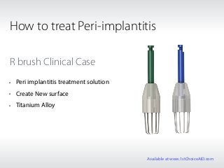
- 1. R brush Clinical Case • Peri implantitis treatment solution • Create New surface • Titanium Alloy How to treat Peri-implantitis Available at www.1stChoiceAID.com
- 2. Patient Information Patient: 51 years old male Medical status: healthy Dental history: implants placed on #25, 26 & 27 Chief complaint: "I have some discomfort around the left maxillary area." Diagnosis 3 Brånemark type external implants were placed in the left maxillary posterior area with sinus graft (left). 8 years later, an advanced chronic periimplantis with severe bone loss around the implant was found in the middle implant area (right). Available at www.1stChoiceAID.com
- 3. The 3 unit SCRP (Screw & Cement Retained Prosthesis) was removed through the screw holes. Soft tissue around the bridge looked fine. Available at www.1stChoiceAID.com
- 4. A severe defect (8mm vertical defect and no buccal and lingual wall) was seen. How can the contaminated rough surface be cleaned and decontaminated mechanically and chemically? Is that possible the contaminated implant surface be reosseointegrated? Available at www.1stChoiceAID.com
- 5. A regular size of R-Brush (Neobiotech, Korea) was used to decontaminate the contaminated rough surface mechanically. Available at www.1stChoiceAID.com
- 6. Before using the R-Brush, the original screw from the prosthesis was inserted to a protection cap. Available at www.1stChoiceAID.com
- 7. The Protection cap was connected to the screw hole to prevent the bristle to go into the screw hole. Available at www.1stChoiceAID.com
- 8. A regular size of R-Brush was connected to a 1:1 contra-angle and rotated using a speed of around 8000 rpm(or2000rpm) with copious water or chlorhexidine solution. 30 to 60 seconds were taken to clean one thread. It took about 5 minutes to clean 8 threads. Available at www.1stChoiceAID.com
- 9. A clean surface like a machine surface was seen. It is known that the R-Brush could eliminate the original surface and create a new rough surface which is not like machine surface but like new rough surface having Ra: 1-1.2. Available at www.1stChoiceAID.com
- 10. An allogenicgraft (RegenOss) was used to graft the large defect. The implant itself could be a space maintainer. Available at www.1stChoiceAID.com
- 11. A collagen membrane was used to cover the graft material. Available at www.1stChoiceAID.com
- 12. It was submerged and sutured with a supramid suture material. Available at www.1stChoiceAID.com
- 13. A radiograph right after the surgery. Available at www.1stChoiceAID.com
- 14. A remarkable clinical result was found 4 month after the surgery. A complete hard bone formation around the defect area was seen. Available at www.1stChoiceAID.com
- 15. Radiographic view 4 month after the surgery. Available at www.1stChoiceAID.com
- 16. A remarkable clinical result was found 4 month after the surgery. A complete hard bone formation around the defect area was seen. Available at www.1stChoiceAID.com
- 17. Buccal view at 4 months after delivery of the old SCRP. The gingiva looks healthy. Available at www.1stChoiceAID.com
- 18. Periapical radiograph shows the result of regeneration of the bone around the periimplantitis area 4 months after the surgery. Available at www.1stChoiceAID.com
- 19. 8 months follow-up. Available at www.1stChoiceAID.com
- 20. Again, before and after the treatment. This treatment option may be one of the options for treating periimplantitis. Available at www.1stChoiceAID.com