Joints_Skeleton system.pptx
•
2 gefällt mir•1,784 views

A part of the body where two bones fit together and are able to bend.

Empfohlen
Weitere ähnliche Inhalte
Was ist angesagt?
Was ist angesagt? (20)
Ähnlich wie Joints_Skeleton system.pptx
Ähnlich wie Joints_Skeleton system.pptx (20)
Mehr von ABHIJIT BHOYAR
Mehr von ABHIJIT BHOYAR (20)
Kürzlich hochgeladen
Kürzlich hochgeladen (20)
Joints_Skeleton system.pptx
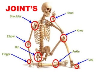
- 1. JOINT’S
- 2. A joint is a point where two bones make contact.
- 3. Classification of Joints Fibrous Cartilaginous Synovial A. Structural Classification A.
- 5. C. Regional Classification Skull Type Vertebral Type Limb Type IMMOVABLE SLIGHTLY IMMOVABLE FREELY IMMOVABLE
- 6. FIBROUS JOINTS • Fibrous joints are defined as the joints in which the bones are connected by fibrous tissue. • They are called fixed or immovable joints as they do not allow any movement between the bones. • They do not have a joint cavity, and the fibrous tissue which connects the bones is made up of collage fibres.
- 7. These can be grouped in the following three subtype 1. Sutures are immobile joints in the cranium. The plate-like bones of the skull are slightly mobile at birth because of the connective tissue between them, termed fontanelles.
- 8. Conti.. • This initial flexibility allows the infant’s head to get through the birth canal at delivery and permits the enlargement of the brain after birth. • As the skull enlarges, the fontanelles reduce to a narrow layer of fibrous connective tissue that suture the bony plates together. Eventually, cranial sutures ossify- the two adjacent plates fuse to form one bone (termed synostosis).
- 9. 2. Gomphoses . Gomphoses are the immobile joints between the teeth and their sockets in the mandible and maxillae. The periodontal ligament is the fibrous tissue that connects the tooth to the socket.
- 10. Syndesmoses 3. Syndesmoses are slightly movable joints (amphiarthroses). In syndesmosis joints, the two bones are held together by an interosseous membrane. Eg Middle Tibiofibular Joint, a fibrous joint formed by the interosseus membrane connecting the shafts of the tibia and the fibula
- 11. Cartilaginous joints • Cartilaginous joints are a type of joint where the bones are entirely joined by cartilage, either hyaline cartilage or fibrocartilage. • These joints generally allow more movement than fibrous joints but less movement than synovial joints.
- 12. 1.Primary cartilaginous joints • Primary cartilaginous joints: These cartilaginous joints are composed entirely of hyaline cartilage and are known as synchondroses. Most exist between ossification centres of developing bones and are absent in the mature skeleton, but a few persist in adults. eg First Sternocostal Joint, between first rib and manubrium (all other sternocostal joints are plane synovial joints); Growth plates.
- 13. 2. secondary cartilaginous joint • The secondary cartilaginous joint, also known as symphysis, may involve either hyaline or fibrocartilage. These joints are slightly mobile (amphiarthroses). eg The pubic symphysis: Intervertebral discs
- 14. Synovial joint • The primary purpose of the synovial joint is to prevent friction between the articulating bones of the joint cavity. • While all synovial joints are diarthroses, the extent of movement varies among different subtypes and is often limited by the ligaments that connect the bones. • Nearly all joints of the limbs and most joints of the body fall into this class
- 15. Synovial joints are the most common type of joint in the body • These joints are termed diarthroses, meaning they are freely mobile.
- 16. Conti.. • A key structural characteristic for a synovial joint that is not seen at fibrous or cartilaginous joints is the presence of a joint cavity. • The joint cavity contains synovial fluid, secreted by the synovial membrane (synovium), which lines the articular capsule. • This fluid-filled space is the site at which the articulating surfaces of the bones contact each other.
- 17. Conti.. • Hyaline cartilage forms the articular cartilage, covering the entire articulating surface of each bone. • The articular cartilage and the synovial membrane are continuous. A few synovial joints of the body have a fibrocartilage structure located between the articulating bones. • This is called an articular disc, which is generally small and oval-shaped, or a meniscus, which is larger and C-shaped.
- 18. • Synovial joints are often further classified by the type of movements they permit. • There are six such classifications: hinge (elbow), saddle (carpometacarpal joint), planar (acromioclavicular joint), pivot (atlantoaxial joint), condyloid (metacarpophalangeal joint), and ball and socket (hip joint).
- 20. Features of all Synovial Joints • Articular capsule with synovial membrane • Synovial cavity containing synovial fluid • Hyaline articular cartilage: acts like a Teflon coating over the bone surface, allowing the articulating bones to move smoothly against each other without damaging the underlying bone tissue.
- 21. Additional features within some Synovial Joints • Fibrocartilage structure located between the articulating bones: Articular disc, which is generally small and oval-shaped, or a Meniscus, which is larger and C-shaped. These structures can serve several functions, depending on the specific joint. Can serve to smooth the movements between the articulating bones eg at the temporomandibular joint. • Intrinsic ligament: fused to or incorporated into the wall of the articular capsule
- 22. • Intracapsular ligament: located inside of the articular capsule. • Intra-capsular tendons eg. popliteus tendon within the knee joint • Intra-articular tendons eg. long head of biceps tendon within the shoulder joint
- 23. • Knee joint. In a Synovial joint, the ends of bones are encased in smooth cartilage. Together, they are protected by a joint capsule lined with a synovial membrane that produces synovial fluid. The capsule and fluid protect the cartilage, muscles, and connective tissues.
- 24. The six types of synovial joints are: 1. Plane Joints: Multiaxial joint, the articular surfaces are essentially flat, and they allow only short nonaxial gliding movements. Examples are the gliding joints introduced earlier—the intercarpal and intertarsal joints, and the joints between vertebral articular processes. Gliding does not involve rotation around any axis, and gliding joints are the only examples of nonaxial plane joints
- 25. Hinge Joints: Uniaxial Joint, the cylindrical end of one bone conforms to a trough-shaped surface on another. Uniaxial hinge joints permit flexion and extension only, typified by bending and straightening the elbow and interphalangeal joints. 2
- 26. 3. Pivot Joints: Uniaxial Joint, the rounded end of one bone conforms to a “sleeve” ring composed of bone of another. The only movement allowed is uniaxial rotation of one bone around its own long axis. An example is the joint between the atlas and dens of the axis, which allows you to move your head from side to side to indicate “no.” Another is the proximal radioulnar joint, where the head of the radius rotates within a ringlike ligament secured to the ulna.
- 27. 4.
- 28. The examples of saddle joints in the body are the carpometacarpal joints of the thumbs. 5. Movements allowed by these joints are clearly demonstrated by twiddling your thumbs.
- 29. These joints are multiaxial and the most freely moving synovial joints. 6..
- 30. Nerve supply of synovial joint • Sensory and autonomic fibers innervate synovial joints: • The autonomic nerves are vasomotor in function, controlling the dilation or constriction of blood vessels. • The sensory nerves of the articular capsule and ligaments (articular nerves) provide proprioceptive feedback from Ruffini endings and Pacinian corpuscles. Proprioception of the joint permits reflex control of posture, locomotion, and movement. Free nerve endings convey pain sensation that is diffuse and poorly localized. The articular cartilage has no nerve supply.
- 31. Blood Supply • Synovial joints receive vascular supply through a rich anastomosis of arteries extending from either side of the joint ie the periarticular plexus. Some vessels penetrate the fibrous capsule to form a rich plexus deeper in the synovial membrane.
- 32. Clinical Anatomy • Arthritis – inflammation that causes stiffness and pain in the joints eg rheumatoid arthritis or gout, or degeneration (osteoarthritis) • Bursitis – inflammation of the bursae (fluid-filled sacs that cushion and pad bones) • Tendonitis – inflammation, irritation and swelling of a tendon that is attached to the joint. • Injury – including strain or sprain of a ligament or nearby tendon or muscle, or bone fracture
- 33. • Dislocation of joint • Sprain • Neuropathic joint