2. Ahmedabad
Sub-Medical surgical nursing
Topic- Cancer esophagus
Submitted to-Mr. P. Yonatansir
Submitted by-MrsHeena Mehta
Sr no Content Page no
1 identification
2 history
3 Physical examination
4 investigation
5 Disease condition
6 defination
7 pathophysiology
8 management
9 Nursing diagnosis
10 Health teaching
11 Bibliography
3. IDENTIFICATION DATA
PATIENT’S NAME: ChetangiriDevgiriGoswami
Indoor . NO: F 59456
AGE:40 years
SEX:Male
DATE OF ADMISSION`: 18-07-2012
DR’S UNIT: Unit-3 Dr.Devenpatel
WARD: cancer male preoperative ward no-3
MARRITAL STATUS: married
RELIGIO: Hindu
EDUCATION: 5thstd .
OCCUPATION:Labour work
ADDRESS:Bavaji no delo,nearsanatanashram,S.G.highway,Ahmedabad.
DIAGNOSIS:Oesophagus carcinoma
HEIGHT: 152Cm
WEIGHT: 54Kg
4. PRESENTING COMPLAINS:
Patient having complained of following:
-Swallowing difficulti
-Nausea
-Vomiting
-Discomfort in chest
-Pain during swallowing
-Body ache
-Mild fever
-Constipation
PRESENT HISTORY:
Chetangiri is asymptomatic before 3month the he gradually developed swallowing
difficulty,painduering swallowing, tighteness in the chest, so went to the nearest private hospital
butsymptoms not relieve than refer to the civil hospital for treatment.
PAST HISTORY:
PAST MEDICAL HISTORY:
Upto 40 years chetangiribhai had not any need for stay in hospitalition for any major illness, he
need symptomatic treatment as per symptoms and relieve the symptoms
PAST SURGICAL HISTORY:
5. Before two month he had done oesophageal biopsy for the swallowing difficulty in the civil
hospital and finally diagnose the oesophageal carcinoma .
DIET HISTORY:
Chetangiri’s family is vegetarian so hisfamily eats vegetarian diet. His wife cooked
all type of vegetarian diet and sometimes he eat raw vegetables with fruit.He drink
very hot tea in the day three to four times.
PERSONAL HISTORY:
Diet : vegetarian & taking all type of small amount diet
Appetite : Decreased
Sleep :disturb
Micturation : No burning micturation
Bowel habit: Abnormal habits
Smoking : 1 pack bidi in day
Alcohol : Some times
Drugs : No
Tobacco : Sometimes
No any other habits
FAMILY HISTORY:
In his family no any family members have history of any Hypertension, Diabetes mellitus,
Ischemic heart disease, Epilepsy, Asthma, Storks, Arthritis, Cancer or any other disease.
Sr. Name of Family Age in Relationship
Education Occupation
No. Members Year With patient
1 ChetangiriDevgiri 40Yrs. patient 5th Labour worker
Goswami
2 AlkagiriGoswami 35Yrs wife 2nd pass Housewife with
labour work
6. 3 ShivgiriGoswami 25Yrs Son 7th pass
Labour work
4 RajangiriGoswami 24Yrs Son’s wife 7th pass Housewife
5 SangitaGoswami 10Yrs Grand 4thstd -
daughter
6 RamangiriGoswam Yrs Grand son 1st -
i
SOCIOECONOMIC HISTORY :
In his family all family member are labour worker so his family’s income is not good they earn
and eat daily and not store adequet stock for diet .they eat routine diet such as roti,rice ,some
times green vegetables, potetoes more used, once in week they cooked dal. There is no any
adequatefacallity in his house also.
PHYSICAL EXAMINATION
VITAL SIGN
Date Temp ( Pulse Respiration(/min) BP (mm of
F) (/min) Hg)
18-7- 99 F 90/min 20/min 120/74
2012
19-7- 98.4 F 9o/min 22 min 118/64
2012
20-7- 98.6 F 100/min 22 min 116/78
2012
21-7- 98.6F 96/min 24 min 118/74
2012
22-7- 98.4 F 100/min 20 min 120/70
2012
GENERAL OBSERVATION:
Sensorium: She is conscious and well oriented
Foul body odour: no any bad odour from her body
Foul breath : no
Posture : normal
7. Hair: Brown hair, clean no any dandruff.
GENERAL APPERANCE:
Body image: normal
Health: Unhealthy
Activity: less active
MENTAL STATUS:
Consciousness: conscious
Look: weakness, fatigue due to her disease.
Posture
Body curves: normal
Movement: Full movement(if given deep pain than small reflection was done by patient)
Height: 152cm Weight: 52kg
SKIN CONDITION:
Color: pallor
Texture: Rough skin
Temperature: warm
Lesions: no lesions present
HEAD & FACE:
Scalp: clean
Face: pale, fatigue, fear, anxiety
EYES
Eyebrow: normal
Eye lashes: no infection, not open by patient
Eyelids: no any injury or oedema is present
Eye balls: not sunken
Conjunctiva: pale
Sclera: no jaundiced
Pupils: constricted
Vision: react to light
8. EAR:
External ear: no discharge present
Hearing: normal
NOSE:
External nares: Redness present
Nostrils: normal. keeping face mask for proper oxygenation
MOUTH & PHARYNX:
Lips: dry
odour of the mouth: not present
Teeth: normal ,dirty
Mucus membrane: dry
Tongue: pale and moist
NECK:
Lymph node: Not palpable
Thyroid gland: normal
Range of motion: flexion, extension and rotation when done by someone, patient able to
done by own self.
CHEST:
Thorax: expansion
Breath sound: Crab herd with stethoscope
Heart: normal
ABDOMEN:
Observation: no skin rashes and scar
Auscultation: reduced bowel sound
Palpation: no tenderness present
Percussion: not presence of gas, fluid or masses
EXTREMITIES:
Lower extremities: fully movements of lower extremities. mildoedema present
Upper extremities: can move both hands but mild oedema is present
9. Genital and rectum:
No enlarged inguinal lymph nodes, No hemorrhoids, no enlargement of prostate glands.
Bladder & Bowel Pattern: Abnormal
INVESTIGATION
Serum Biochemistry test:
Investigation In patient Normal value
Hemoglobin 14 % gm% 14 – 17 gm %.
RBC 98 mg/dl 153mg/ml
UREA 18.34 mg/dl 15-45mg/dl
WBC 8000/cumm 4000-11000/cumm
S.creat. 0.85mg/dl 0.7-1.5mg/dl
SGPT 48U/L 0-55U/L
S. Alkpo4 68U/L <50-150U/L
S.Billirubin 0.7mg/dl 0.2-1.2mg/dl
BLOOD CHEMISTERY
FASTING 96.0mg/ dl 70-110mg/dl
X-RAY CHEST:
-Both cp angles appear clear
-Heart size &aorta appear within normal limits
-Rest of bony thorax under vision appear normal.
ECG: wnl
Biopsy-Finding
-There is circumferential,ulcerative proliferative growth starting ,extending upto 33cms.
MEDICATION
-Injection amikasine 500gm i/v 12hourly.
-Injection diacloran 1 ampoule i/m sos.
10. - Injection Rantac 1 ampouls i/v 12 hourly.
- Injection Glucose 5% 1 litre i/v slowly.
Maintain intake and output chart daily
Contineus observation of the patient on monitor for any abnormal symptoms.
TPR chart 1 hourly Monitoring continuously for blood pressure, respiration rate, pulse,
and for oxygen saturation.
Care taken of catheter daily
Care taken of all tubes which are inserted
Watched for respiratory failure .
Changed the dressing and adhesive tap at the site of intracath.
DISEASE CONDITION
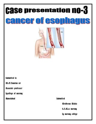
ANATOMY AND PHYSIOLOGY OF OESOPHAGUS-
- The adult esophagus is a 25 cm-long tube and is fixed superiorly at the cricopharyngeus
muscle, which is considered as the upper esophageal sphincter.
- Esophagus courses inferiorly through the posterior mediastinum behind the trachea and the
heart and exits the thorax through the hiatus of the diaphragm.
- The so-called lower esophageal sphincter (LES) is not a true anatomic sphincter, but rather a
functional one.
- Tonic muscular contraction at the lower end of the esophagus creates an action similar to
that of a one-way flutter valve.
- The transition from the normal squamous mucosa of the esophagus to the gastric mucosa at
the esophago-gastric junction occurs abruptly at the level of the diaphragm.
- The venous drainage of the esophagus is important in portal hypertension because it forms
esophageal varices.
- The functions of the esophagus include:
i) Esophagus conducts food and fluids from the pharynx to the stomach and
ii) Prevents reflux of gastric contents into the esophagus.
- These functions require coordinated motor activity including both extrinsic and intrinsic
innervation, myogenic properties and humoral substances.
Clinical features of esophageal dysfunction include:
1. Dysphagia- is the difficulty in swallowing due to mechanical and functional disorders.
2. Heartburn- is the retrosternal burning pain. It is usually due to regurgitation of gastric
contents into lower esophagus.
11. 3. Hematemesis- is the vomiting of blood due to inflammation or ulceration or rupture of
blood vessels.
DEFINITION-
Esophageal cancer (or oesophageal cancer) is malignancy of the esophagus. There are various
subtypes, primarily squamous cell cancer andadenocarcinoma , Squamous cell cancer arises
from the cells that line the upper part of the esophagus. Adenocarcinoma arises from
glandular cells that are present at the junction of the esophagus and stomach
.
CAUSES:
In Book In Patient
No
Barrett's esophagus
Heredity No
Age is another critical factor No
A man with a personal history of cancer No
Lifestyle and Dietary Causes due to obesity No
Tobacco smoking Yes
Human papillomavirus (HPV) Yes
Plummer-Vinson syndrome (anemia and esophageal May be
webbing)
Tylosis and Howel-Evans syndrome No
Achalasia No
PATHOPHYSIOLOGY:
12. The progression of Barrett metaplasia to adenocarcinoma is associated with several changes in
gene structure, gene expression, and protein structure.
The oncosuppressor gene TP53 and various oncogenes, particularly erb -b2, have been studied
as potential markers.
Casson and colleagues identified mutations in the TP53 gene in patients with Barrett
epithelium associated with adenocarcinoma.
alterations in p16 genes and cell cycle abnormalities or aneuploidy appear to be some of the
most important and well-characterized molecular changes.
However, the exact sequence of events in the progression of Barrett esophagus to
adenocarcinoma is not known. Probably multiple molecular pathways interact and are
involved.
o Allelic losses at chromosomes 4q, 5q, 9p, 9q, and 18q and abnormalities of p53, Rb, cyclin
D1, and c-myc have been implicated.
CLINICAL MENIFESTATION:
In Book In Patient
Dysphagia (difficulty swallowing) Present
odynophagia (painful swallowing) Present
Pain behind the sternum or in the Present
epigastrium
coughing and an increased risk of aspiration Not Present
pneumonia.
nausea and vomiting Present
upper airway obstruction Not present
13. superior vena cava syndrome. Not Present
The tumor surface may be fragile and Not Present
bleed, causing hematemesis
lung metastasis could cause shortness of Not Present
breath, pleural effusions
Present
ASSESSMENT & DIAGNOSTIC FINDINGS:
IN BOOK IN PATIENT
- Taking a thorough history - Done
including family history
- Physical examination - Done
- microscopic analysis of the - Done
biopsy
- Laboratory work (cholesterol - Done
levels, glucose )
Biopsies - Done
Computed tomography (CT) - Not Done
Positron emission tomography - Not Done
Esophageal endoscopic ultrasound - Not done
MANAGEMENT:
Esophageal cancer affecting the lower esophageus. Insets show the tumor in more
detail both before and after placement of a stent.
The treatment is determined by the cellular type of cancer (adenocarcinoma or
squamous cell carcinoma vs other types), the stage of the disease, the general condition
of the patient and other diseases present.
On the whole, adequate nutrition needs to be assured, and adequate dental care is
vital.
If the patient cannot swallow at all, an esophageal stent may be inserted to keep the
esophagus patent; stents may also assist in occluding fistulas.
A nasogastric tube may be necessary to continue feeding while treatment for the tumor
is given, and some patients require a gastrostomy (feeding hole in the skin that gives
direct access to the stomach). The latter two are especially important if the patient
tends to aspirate food or saliva into the airways, predisposing for aspiration pneumonia.
14. Esophagectomy is the removal of a segment of the esophagus; as this shortens the
length of the remaining esophagus, some other segment of the digestive tract (typically
the stomach or part of the colon or jejunum) is pulled up to the chest cavity and
interposed.
If the tumor is unresectable or the patient is not fit for surgery, palliative esophageal
stenting can allow the patient to tolerate soft diet.
SURGICAL MANAGEMENT:
The thoracoabdominal approach opens the abdominal and thoracic cavities together.
The two-stage Ivor Lewis (also called Lewis-Tanner) approach involves an initial
laparotomy and construction of a gastric tube, followed by a right thoracotomy to excise
the tumor and create an esophagogastric anastomosis.
The three-stage McKeown approach adds a third incision in the neck to complete the
cervical anastomosis
A fourth method of EMR employs the use of a clear cap and prelooped snare inside the
cap. After insertion, the cap is placed on the lesion and the mucosa containing the lesion
is drawn up inside the cap by aspiration. The mucosa is caught by the snare and
strangulated, and finally resected by electrocautery. This is called the "band and snare"
or "suck and cut" technique.
Although most lesions treated in the esophagus have been early squamous cell cancers,
EMR can also be used to debulk or completely treat polypoid dysplastic or malignant
lesions in Barrett’s esophagus.
Laser therapy is the use of high-intensity light to destroy tumor cells; it affects only the
treated area. This is typically done if the cancer cannot be removed by surgery. The
relief of a blockage can help to reduce dysphagia and pain.
Photodynamic therapy, a type of laser therapy, involves the use of drugs that are
absorbed by cancer cells; when exposed to a special light, the drugs become active and
destroy the cancer cells.
MEDICALMANAGEMENT
Chemotherapy depends on the tumor type, but tends to be cisplatin-based (or
carboplatin or oxaliplatin) every three weeks with fluorouracil (5-FU) either continuously
or every three weeks. In more recent studies, addition of epirubicin was better than
other comparable regimens in advanced nonresectablecancer.Chemotherapy may be
given after surgery (adjuvant, i.e. to reduce risk of recurrence), before surgery
(neoadjuvant) or if surgery is not possible; in this case, cisplatin and 5-FU are used.
Ongoing trials compare various combinations of chemotherapy; the phase II/III REAL-2
15. trial – for example – compares four regimens containing epirubicin and either cisplatin
or oxaliplatin, and either continuously infused fluorouracil or capecitabine.
Radiotherapy is given before, during or after chemotherapy or surgery, and sometimes
on its own to control symptoms. In patients with localised disease but contraindications
to surgery, "radical radiotherapy" may be used with curative intent.
NURSING MANAGEMENT:
- Identify at risk patients, & teach lifestyle modifications to prevent development any
complication.
- Teach patient to control cholesterol levels through dietary reduction of cholesterol intake,
exercise, smoking cessation.
- Note & report findings from history, physical examination, & laboratory results that
indicate hypertension or diabetes, &teach to control blood pressure by taking treatment
in the nearest hospital.
NURSING DIAGNOSIS:
1. .Altered body temperature due to presence of infection.
2. Imbalance nutritional level less than body requirement related to loss of appetite.
3. Activity intolerance related to surgery done.
6 Impaired body image due osurgeory.
7 Alteredself image and confidence due to fegure.
HEALTH TEACHING:
Arrange specific services for patient(e.g. respiratory therapy education, physical therapy
for exercise & breathing)
Explain patient’s reletives about discharge planning.
Give advice about regular medication as per timing.
Explain and demonstrate about chest physiotherapy by doing deep breathing exercise .
Explain and demonstrate about coughing and how to remove cough.
Advice given about good nutritive .
Advide given for prevention of infection management.
Explain about follow up care.
16. BIBLIOGRAPHY:
1. Bennette and Plum; “TEXTBOOK OF MEDITION ; 10thedition, 1996;
W.B. Saunders Company, New York : 1996. PP :
2. Black J.M; “MEDICAL SURGICAL NURSING; 5th edition, 1999
; W.B. Saunders Company, Philadelphia. PP:
3. Brunners&Suddarth’s; “TEXT BOOK OF MEDICAL SURGICAL
NURSING VOL-_1”;10th edition, 2004; Elsevier Publishers, New Delhi,
India. PP:
4. B T Basavanthappa;”TEXT BOOK OF NURSING THEORIES”,Jaypee brothers
Medical Publishers ,New Delhi.
PP: 40-
WEBSITES:
- http://www.wikipedia.com.
- http://www.patho.respiratory disease.org/.com.in
- http://www.google.com.
- http://www.medicine.com.